355
CLINICS 2006;61(4):355-8
Maternité. Hôpital Necker-Enfants Malades, AP-HP, Université de Paris V, Paris, France/ Service de gynécologie. Hôpital Cochin- Saint Vincent de Paul, AP-HP, Université de Paris V, Paris, France.
Clinica Obstétrica, Hospital das Clínicas, São Paulo University Medical School - São Paulo/ SP, Brazil.
Email: [email protected]
LETTER TO THE EDITOR
THREE-DIMENSIONAL ULTRASONOGRAPHIC
DIAGNOSIS OF A CERVICAL PREGNANCY
Rodrigo Ruano, Fabien Reya, Olivier Picone, Nicolas Chopin, Pedro Paulo Pereira, Alexandra Benachi, Marcelo Zugaib
Cervical pregnancy is a rare condition characterized by implantation of a fertilized ovum in the endocervical ca-nal below the interca-nal os level; its incidence is less than 0.1% of all ectopic pregnancies.1 Although predisposing
factors have been described, the cause of cervical pregnancy remains unknown.2 Cervical pregnancy has a high
morbid-ity potential due to massive hemorrhage that can be asso-ciated with it, but mortality is low due to early ultrasonographic diagnosis, using transvaginal examina-tion.2,3 Making a differential diagnosis between cervical
pregnancy and isthmic pregnancy is very important for prognostic reasons, since cervical pregnancy is not com-patible with viable pregnancies, while an isthmic pregnancy can reach viability and term.2 However, making the
cor-rect diagnosis using conventional 2-dimensional ultrasonog-raphy (2D-US) remains a challenge. Magnetic resonance imaging (MRI) has been used in few cases of cervical preg-nancy to improve diagnostic accuracy.4–6 So far, to our
knowledge, 3-dimensional ultrasound (3D-US) has not been described in this situation. The present article presents a case of cervical pregnancy diagnosed by transvaginal 3D-US examination.
CASE REPORT
A 37-year-old woman, gravida 4 para 2, was admitted to our obstetrical emergency unit with clinical signs of threatened abortion at 14 weeks of gestation. Regarding her obstetric history, the patient had 2 previous abortions in the first trimester, performed by curettage. Afterwards, she also had 2 lower-segment caesarean sections at 33 and 34 weeks, both because of severe preeclampsia.
She presented moderate vaginal bleeding associated with lower abdominal pain at the emergency room. Clini-cal examination of the maternal abdomen was difficult
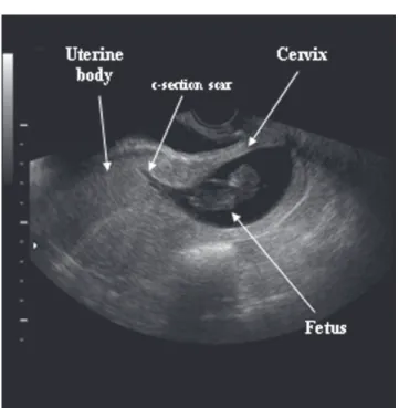
be-cause of maternal obesity. Vaginal examination revealed an anterior position of the cervix, which was extremely thin, with the external os closed. The uterus was retroverted, and speculum examination showed a small amount of cervical bleeding. Conventional transvaginal 2D-US (VOLUSON 730, General-Electric, Zipf, Austria, with a vaginal trans-ducer 3-9 MHz) showed a fetus without heart activity, measuring 38.0 mm, crown-rump length (11 weeks), lo-cated in the cervical portion of the uterus (Figure 1). The cervix was dilated above the external os, and the gestational sac seemed to be attached to the lower scar segment of the previous cesarean sections. It was difficult to precisely iden-tify the endometrial cavity due to the retroverted position of the uterus. At this time, 2 differential diagnoses were proposed: abortion or cervical pregnancy.
To confirm diagnosis, a transvaginal 3D-US (VOLUSON 730, General-Electric, Zipf, Austria, with a 3D vaginal trans-ducer 3-9 MHz) was performed. Because the maternal
356
CLINICS 2006;61(4):355-8 Three-dimensional ultrasonographic diagnosis
Ruano R et al.
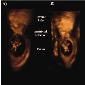
der was partially full, the whole uterus was completely ac-quired, and the volumetric images were stored on a remov-able hard disk. By working on the multiplanar imaging, we were able to evaluate the endometrium and the uterine isth-mus on the coronal section, confirming that the pregnancy was located inside the dilated cervix, which was separated from the uterus body by a constricted isthmus (Figure 2). By adjusting the depth of the volumetric box on the sagittal section (A-plane), it was possible to evaluate the en-dometrium and the isthmus on the coronal section (C-plane), revealing that the endometrial cavity was thin and that the upper limit of the gestational sac was attached to the isth-mic region, which confirmed the diagnosis of cervical preg-nancy. These findings could be clearly observed on rendered 3D sonographic images (Figure 3).
The patient was treated by intramuscular injections of methotrexate (60 mg/kg in 3 doses). After the third injec-tion, uterine voiding occurred with light bleeding. The pre-medication plasma β-HCG level was 884 IU/mL, decreas-ing to less than 10 IU/mL 3 months after the procedure. No maternal complications occurred.
DISCUSSION
This paper presents a case of cervical pregnancy diag-nosed by combining transvaginal 2D-US with 3D-US
ex-aminations. In this case, due to maternal obesity and the posterior position of the uterine body, it was difficult to make the correct diagnosis using only conventional trans-vaginal 2D-US.
The exact etiology of cervical pregnancy is unknown, although there are many predisposing factors, which in-clude endometrial damage after curettage or chronic en-dometritis, leiomyoma, intrauterine devices, in-vitro ferti-lization, and primary embryo anomaly.2
Diagnosis of cervical pregnancy is based on clinical and ultrasonographic findings, but its differentiation from isthmic pregnancy remains a challenge. The main ultrasonographic criteria for diagnosis of cervical pregnancy are as follows: i) gestational sac in the cervix, ii) empty uterine cavity, iii) di-lated cervix, and iv) normal uterine size.7 In the present case,
two initial hypothetic diagnoses were made based on 2D-US: abortion or cervical pregnancy. The diagnosis of cervical preg-nancy could be only confirmed on 3D-US, which gave clear and precise images of the location of the pregnancy.
The great advantage of transvaginal 3D-US over trans-vaginal 2D-US is the possibility of having a coronal sec-tion in the multiplanar imaging.8–9 In the present case, it
was helpful to determine the limits of the gestational sac, allowing the correct diagnosis of cervical pregnancy. This imaging method also allowed a better analysis of the en-dometrial cavity using the coronal section in the multiplanar imaging.
Different therapeutic approaches have been proposed for cervical pregnancy, varying from radical methods to more
Figure 2. Multiplanar imaging with the three orthogonal sections: a) sagittal section, b) transversal section, c) coronal section, and d) reconstructed three-dimensional image. The depth of the sectional plane of the render box was adjusted on the A-plane (sagittal section) in order to evaluate the isthmus on the C-plane (coronal section). Note that, on coronal section and on the rendered 3D image, it is possible to delineate the upper limit of the cervical pregnancy at the constricted isthmus (i). ub: uterine body; i: isthmus; c: cervix.
357
CLINICS 2006;61(4):355-8 Three-dimensional ultrasonographic diagnosis
Ruano R et al.
conservative treatments; the choice of approach depends on the clinical conditions such as blood loss, gestational age, viability of the cervical pregnancy, gestational sac location, and depth of trophoblast invasion. Total abdominal hyster-ectomy is the treatment of choice for cervical pregnancies diagnosed during the second trimester.7 The present
wide-spread use of ultrasonography has allowed early diagnosis of cervical pregnancies (even before clinical manifesta-tions), leading to more conservative management.10–12
Con-servative management techniques include the following: i) cervical curettage with balloon tamponade13–14; ii) ligature
of the hypogastric or cervico-vaginal arteries15–16; iii)
elec-tive angiographic embolization of the uterine artery17–20; and
iv) clinical use of methotrexate.21–22 In the present case,
con-servative management with methotrexate was chosen be-cause the β-HCG level was less than 10,000 IU/mL and fetal heart activity was absent.23 According to previous
re-ports, medical management of cervical pregnancy with methotrexate can prevent the need for hysterectomy in 91% of the cases.24
In conclusion, transvaginal 3D-US may be useful as a complementary imaging method for the correct diagnosis of cervical pregnancy by allowing the correct location of the gestational sac. It can also be useful in obese patients or in those cases with retroverted uterus, allowing a better analysis of the endometrial cavity on coronal planes.
REFERENCES
1. Van de Meerssche M, Verdonk P, Jacquemyn Y, Serreyn R, Gerris J. Cervical pregnancy: three case reports and a review of the literature. Hum Reprod. 1995;10:1850-5.
2. Riethmuller D, Courtois L, MailletR, Schaal JP. Prise en charge des grossesses cervicales et abdominales. J Gynecol Obstet Biol Reprod. 2003;32 (suppl. N 7):3S101-3S108.
3. Guerrier C, Wartarian R, Roblet V, Rohmer E, Le Lirzin R. La grossesse cervicale. Apport de l’échographie au diagnostic et à la prise en charge. Rev Fr Gynecol Obstet. 1995;90:352-9.
4. Jung SE, Byun JY, Lee JM, Choi BG, Hahn ST. Characteristic MR findings of cervical pregnancy. J Magn Reson Imaging. 2001;13:918-22.
5. Itakura A, Okamura M, Ohta T, Mizutani S. Conservative treatment of a second trimester cervicoisthmic pregnancy diagnosed by magnetic resonance imaging. Obstet Gynecol. 2003;101:1149-51.
6. Okamoto Y, Tanaka YO, Nishida M, Tsunoda H, Yoshikawa H, Itai Y. MR Imaging of the Uterine Cervix: Imaging-Pathologic Correlation. Radiographics. 2003;23:425-45.
7. Ushakov FB, Elchalal U, Aceman PJ, Schenker JG. Cervical pregnancy: past and future. Obstet Gynecol Surv. 1997;52:45-59.
358
CLINICS 2006;61(4):355-8 Three-dimensional ultrasonographic diagnosis
Ruano R et al.
9. Jurkovic D, Geipel A, Gruboeck K, Jauniaux E, Natucci M, Campbell S. Three-dimensional ultrasound for the assessment of uterine anatomy and detection of congenital anomalies: a comparison with hysterosalpingography and two-dimensional sonography. Ultrasound Obstet Gynecol. 1995;5:233-7.
10. Bakour SH, Thompson PK, Khan KS. Successful conservative management of cervical ectopic pregnancy with combination of methotrexate, mifepristone, surgical evacuation and tamponade using a double balloon three-way catheter. J Obstet Gynaecol. 2005;25:616-8.
11. El-Matary AM, Ashworth F. Cervical ectopic pregnancy with successful conservative treatment. J Obstet Gynaecol. 2005;25:411-2.
12. Gosakan R, Arutchelvam S, Gergis HH, Emovon E. Medical management of a cervical ectopic pregnancy. J Obstet Gynaecol. 2005;25:82-3.
13. Fylstra DL, Coffey MD. Treatment of cervical pregnancy with cerclage, curettage and balloon tamponade. A report of three cases. J Reprod Med. 2001;46:71-4.
14. Okeahialam MG, Tuffnell DJ, O’Donovan P, Sapherson DA. Cervical pregnancy managed by suction evacuation and balloon tamponade. Eur J Obstet Gynecol Reprod Biol. 1998;79:89-90.
15. Akutagawa N, Nishikawa A, Saito T, Sagae S, Kudo R. Conservative vaginal surgery for cervical pregnancy. Brit J Obstet Gynaecol 2001;108:888-9.
16. Saygli Yilmaz ES, Aydin D, Yilmaz Z. Conservative treatment of cervical pregnancies by evacuation after transvaginal suture ligation of the cervicovaginal branches of uterine arteries. Acta Obstet Gynecol Scand. 2002;106:988-90.
17. Cosin JA, Bean M, Grow D, Wiczyk H. The use of methotrexate and arterial embolization to avoid surgery in case of cervical pregnancy. Fertil Steril. 1997;67:1169-71.
18. Dilbaz S, Atasay B, Bilgic S, Caliskan E, Oral S, Haberal A. A case of conservative management of cervical pregnancy using selective angiographic embolization. Acta Obstet Gynecol Scand. 2001;80:87-9.
19. Su YN, Shih JC, Chiu WH, Lee CN, Cheng WF, Hsieh FJ. Cervical pregnancy: assessment with three dimensional power Doppler imaging and successful management with selective uterine artery embolization. Ultrasound Obstet Gynecol. 1999; 14:284-7.
20. Ryu KY, Kim SR, Cho SH, Song SY. Preoperative uterine artery embolization and evacuation in the management of cervical pregnancy: report of two cases. J Korean Med Sci. 2001;16:801-4.
21. De Greef I, Berteloot P, Timmerman D, Deprest J, Amant F. Viable cervical pregnancy with levonorgestrel containing intrauterine device, treated successfully with methotrexate and mifepristone. Eur J Obstet Gynecol Reprod Biol. 2005;120:233-5.
22. Monteagudo A, Minior VK, Stephenson C, Monda S, Timor-Tritsch IE. Non-surgical management of live ectopic pregnancy with ultrasound-guided local injection: a case series. Ultrasound Obstet Gynecol. 2005;25:282-8.
23. Bai SW, Lee JS, Park JH, Kim JY, Jung KA, Kim SK, et al. Failed methotrexate treatment of cervical pregnancies. J Reprod Med. 2002;47:483-8.