Universidade de Lisboa
Faculdade de Farmácia
Darier disease: molecular study of a
two-generation Portuguese family
Andreia Sofia Rafael de Almeida
Dissertation Report supervised by Professor Isabel Antolin Rivera
Master in Biopharmaceutical Sciences
Abstract
Background Darier Disease is a rare autosomal dominant disorder, which predominantly affects the skin of individuals regardless their gender or ethnicity. Clinical manifestations usually involve nail abnormalities and focal skin lesions such as greasy, brownish-redish keratotic papules, which are distributed in seborrheic areas of the body. Affected individuals are usually heterozygous for mutation in ATPase sarcoplasmic/endoplasmic reticulum Calcium transporting 2 (ATP2A2) gene, responsible for encoding Sarco/Endoplasmic Reticulum Calcium ATPase 2 (SERCA2). Although ATP2A2 mutations significantly reduce ER Ca2+ stores in both lesional and non lesional keratinocytes, scientists suggest that a threshold in ER Ca2+ depletion is required to disrupt adhesion molecules, as seen in lesional epidermis. Nonetheless, DD hallmarks (acantholysis and dyskeratosis) remain to be fully elucidated. Haploinsufficiency or dominant-negative effect are the postulated pathogenic mechanisms.
Objective This study consists in the molecular characterization of a two-generation Portuguese family with DD history, including co-associated phenotypes and intrafamilial phenotypic variability.
Methods All exons and intron-exon borders of ATP2A2 were bidirectionally sequenced from DNA and RNA extracted from the five subjects enrolled in this study (four affected individuals and one unaffected individual). Relative levels SERCA2 mRNA and protein were quantified by RT-qPCR and western blotting, respectively.
Results A splice-site mutation (c.1287+1G>T or IVS10+1G>T) was identified in all four affected individuals, who carry the mutation in heterozigosity, whereas the unaffected individual was shown to carry the wild-type ATP2A2 sequence in both alleles. This mutation leads to the skipping of full exon 10, which consequently generates a frameshift followed by a premature translation termination codon in exon 11. Results from RT-PCR of a fragment spanning exons 8-13 suggest that a small amount of the mutant transcript escapes NMD and less than 50% expression of the type transcript is detected. In agreement, relative wild-type and mutant SERCA2 mRNA expression levels assessed by qPCR revealed wild-wild-type gene expression alone revealed a 36% expression, while mutant mRNA expression exhibited
iv
residual levels of 18%. In contrast, western blot results showed 40 to 50% expression of wild-type SERCA2 (115 kDa) in DD patients when compared to the healthy individual, while no sign of the putative mutant SERCA2 (45 kDa) was detected. These findings suggest that despite the escape of a small portion of abnormal SERCA2 mRNA from NMD, there is still no generation of mutant protein. Therefore, haploinsufficiency appears to be the mechanism underlying DD pathology in these patients. Finally, no genotype-phenotype correlations were found; however, this study contributed to alert physicians to the wide range of defects predicted by ATP2A2 mutations, including neuropsychiatric features, hearing loss, corneal viral infections by HSV, and glomerular nephritis which is, to the best of our knowledge, here firstly reported in association with DD.
Conclusion As the first study of Portuguese patients, it adds knowledge to the worldwide mutational and clinical spectrum of DD and, importantly, contributes to genetic counseling since it allowed to discard a possible carrier status of the unaffected member of the family.
Resumo
Introdução A doença de Darier é uma genodermatose rara, de transmissão autossómica dominante, que não distingue sexo ou etnia. As manifestações clínicas caraterísticas desta doença envolvem anomalias nas unhas e lesões cutâneas focais, tais como pápulas queratóticas acastanhadas-avermelhadas distribuídas pelas áreas seborreicas do corpo. Os indivíduos afetados são geralmente heterozigóticos para uma mutação no gene ATP2A2, responsável pela codificação da proteína SERCA2. Embora as mutações no gene ATP2A2 reduzam significativamente as reservas de Ca2+ no retículo endoplasmático em células lesionadas e não lesionadas, os cientistas sugerem a necessidade de um limite na depleção destas reservas para interferir no funcionamento de moléculas de adesão, como observado nos queratinócitos acantolíticos da epiderme lesionada. Haploinsuficiência ou efeito dominante negativo são os mecanismos patogénicos postulados para o fenótipo de Darier.
Objetivo Este projeto consiste na caraterização molecular duas gerações de uma família Portuguesa de com histórico de doença de Darier, incluindo co-fenótipos e variabilidade fenotípica intrafamiliar.
Métodos Todos os exões e limites exão-intrão do gene ATP2A2 foram sequenciados bidirecionalmente a partir de DNA e RNA extraídos dos cinco indivíduos envolvidos neste estudo (quatro afetados e um não afetado). Os níveis relativos de RNA mensageiro e proteína SERCA2 foram quantificados por PCR em tempo real e Western blotting, respetivamente. Resultados A mutação patogénica (c.1287 + 1G> T ou IVS10 + 1G> T) foi identificada em todos os quatro indivíduos afetados, que exibem a mutação em heterozigotia, enquanto o indivíduo não afetado exibe a sequência normal de ATP2A2 em ambos os alelos. A mutação altera o splicing e leva à eliminação completa do exão 10, que consequentemente altera a leitura da ORF, seguida de um codão de terminação de tradução prematura no exão 11. Os resultados de RT-PCR, correspondente à amplificação de um fragmento abrangendo os exões 8 a 13, sugerem que uma pequena quantidade do transcrito mutado consegue escapar ao NMD e que a expressão de transcrito normal é inferior a 50% relativamente ao indivíduo normal. Em conformidade, os níveis relativos de expressão de RNA mensageiro SERCA2 normal e mutado avaliados por PCR em tempo real revelaram 36% de expressão do gene normal, e
vi
western blot mostraram 40 a 50% de expressão de SERCA2 normal (115 kDa) nos indivíduos com a doença, em comparação com o indivíduo saudável, enquanto nenhum sinal da hipotética proteína mutada (45 kDa) foi detetado. Em geral, os resultados obtidos sugerem que, apesar da pequena porção de transcrito mutado escapar ao NMD, não há tradução em proteína mutada, ou esta deve ser imediatamente degradada. Portanto, a haploinsuficiência parece ser o mecanismo responsável pela patologia de Darier nos doentes estudados. Finalmente, não foi possível identificar qualquer correlação genótipo-fenótipo; no entanto, este estudo contribuiu para alertar os médicos dermatologistas sobre a panóplia de alterações provocadas pelas mutações no gene ATP2A2, incluindo sintomas neuropsiquiátricos, perda de audição, infeções virais da córnea causadas por HSV, e nefrite glomerular membranosa que é, pelo que temos conhecimento, relatado neste trabalho pela primeira vez em associação com DD.
Conclusão Como primeiro estudo em doentes Portugueses, esta investigação acrescenta conhecimento ao espetro mutacional e clínico mundial da doença de Darier e, mais importante, contribui para o aconselhamento genético, uma vez que permitiu descartar um possível estatuto de portador do membro não afetado da família.
Acknowledgments
I am grateful beyond words to my supervisor, Isabel Rivera, whose expertise in genetics and generous guidance have made it possible for me to work on a topic that arouses great interest and curiosity in me. Working at her laboratory was truly one of the best experiences and I shall always remember her optimism and genuine kindness.
I would also like to thank those at the CPM who provided help when I needed, and to Professor Graça Soveral, Ana Paula Leandro and Susana Solá for generously providing supplies for my work. I am also thankful to Professor Cecília Rodrigues, for giving me the opportunity to join the Masters in Biopharmaceutical Sciences.
I am hugely indebted to my father, brother, uncle and cousin, without whom this study would not be feasible. I know it has been hard these recent years and I want them to know that I fully appreciate their time and their participation in this study.
To the friendship since highschool, Pedro Reis, Adriana Iablonski, and Gustavo Nogueira, and to Rita Lami & Fiona O’Bell. Thank you for existing.
Last but not least, I would like to express my gratitude for all the support and meals my Mom prepared for me since I started working on this thesis. Thank you Mom!
Contents
Abstract . . . . iii
Resumo . . . v
Acknowledgments . . . ix
List of Tables . . . xiii
List of Figures . . . xv
Abbreviations . . . . xvii
1 Introduction
. . . 11.1 Historical Aspects and Epidemiology . . . . 1
1.2 Epidermis and Calcium Signaling . . . 2
1.3 Gene and Protein . . . 4
1.4 Clinical Manifestations . . . 9
1.5 Histopathology . . . 9
1.6 Associated Manifestations . . . . 11
1.6.1 Neuropsychiatric manifestations . . . 12
1.6.2 Other manifestations . . . . 13
1.7 Intrafamilial phenotypic variability . . . 13
1.8 Mechanism of Action . . . 14
1.9 Treatment . . . 16
2 Aims of the study
. . . 193 Participants and Methods
. . . 213.1 Case report . . . . 21
3.2 Sample collection . . . 23
3.3 DNA and RNA extraction . . . 23
3.4 PCR and RT-PCR amplification . . . . 23
xii
3.6 Quantitative PCR . . . 26
3.7 Western blotting . . . 27
3.8 Genotype-Phenotype correlation . . . 28
4 Results and Discussion
. . . 294.1 Identification of the pathogenic mutation . . . 29
4.2 ATP2A2 expression . . . 32
4.3 SERCA2 expression . . . . 35
4.4 Genotype-Phenotype . . . . 40
4.4.1 Neuropsychiatric features . . . 40
4.4.2 Renal impairment . . . 41
4.4.3 Corneal susceptibility to viral infection by Herpes Simplex Virus (HSV) 42 4.4.4 Otological involvement . . . 43
5 Conclusion
. . . . 45List of Tables
1 Additional information about participants’ actual age, age of onset, and other phenotype manifestations . . . 22 2 PCR primers and related parameters for amplification of ATP2A2 from genomic DNA . 24 3 PCR primers and related parameters for amplification of SERCA2 from cDNA . . . 25
List of Figures
1 Intracellular and extracellular calcium concentration profile and differentiation markers
along the epidermal strata . . . . 3
2 Overall topology of the P-type ATPase calcium pump SERCA . . . . 5
3 Reaction cycle for the Sarco/Endoplasmic Reticulum Ca2+-ATPase (SERCA) pumps . 6 4 Schematic representation of ATP2A2 gene and encoded protein SERCA2 isoform b and spectrum of ATP2A2 variants . . . . 8
5 Clinical manifestations of Darier disease patients . . . 10
6 Histological section of Darier disease skin . . . 11
7 Pedigree of the two-generation Portuguese family . . . 21
8A Identification and expression of ATP2A2 pathogenic mutation . . . 30
8B Sequence analysis of the coding sequence of ATP2A2 gene . . . . 31
9 Representation of fully spliced wild-type ATP2A2 product versus mutant ATP2A2 product . . . 33
10 All patients showed ATP2A2 mRNA levels lower than 50% and residual levels of mutant mRNA . . . 34
11A Wild-type SERCA2b polypeptide sequence . . . 35
11B Putative C-terminus truncated mutant SERCA2 polypeptide sequence . . . 36
12 Representation of SERCA2 domains and consequences of ATP2A2 mutation . . . . . 37
13 Wild-type SERCA2 expression was severely reduced in patients when compared with the unaffected individual . . . 38
Abbreviations
α E β 7 Integrin alpha E beta 7 β-actin Beta-actin
5-ALA 5-aminolaevulinic Acid 5-FU 5-fluorouracil
ACTB gene encoding beta-actin A domain Actuator domain
ATP Adenosine triphosphate
ATP2A2 ATPase Sarcoplasmic/endoplasmic Reticulum Calcium-transporting 2 ATP2A2+/- Haploinsufficiency of ATP2A2
BAX BCL2 Associated X
BCL2 B Cell Leukemia/lymphoma 2 BCL-xL BCL2-like protein 1
BSA Bovine Serum Albumin cDNA Complementary DNA
COX-2 Cyclooxygenase-2
CRISPR Clustered Regularly Interspaced Short Palindromic Repeats DCs Desmosomal Cadherins
DD Darier Disease
DNA Deoxyribonucleic Acid E-cadherin Epithelial cadherin ER Endoplasmic Reticulum
HIV Human Immunodeficiency Virus HSV Herpes Simplex Virus
IgG Immunoglobulin G
IP3Rs Inositol-1,4,5-triphosphate Receptors
MITEs Miniature Inverted-Repeat Transposable Elements MN Membranous Nephropathy
mRNA Messenger RNA
xviii
NMD Nonsense-Mediated Decay
ORAI1 Calcium Released-activated Calcium Channel Protein 1 P53 Tumor Protein 53
PCR Polymerase Chain Reaction Pi Phosphate group
P domain Phosphorylation domain PDT Photodynamic therapy P-type Phosphorylation-type
PTC Premature translation Termination Codon qPCR Quantitative PCR
RAS Rat sarcoma RNA Ribonucleic Acid RT Reverse Transcriptase RyRs Ryanodine Receptors SCC Squamous Cell Carcinoma
SDS-PAGE Sodium Dodecyl Sulfate Polyacrylamide Gel Electrophoresis SERCA Sarcoplasmic/Endoplasmic Reticulum Calcium ATPase SERCA1 SERCA isoform 1
SERCA2 SERCA isoform 2 SERCA3 SERCA isoform 3
SERCA2a SERCA isoform 2 splice variant a SERCA2b SERCA isoform 2 splice variant b SERCA2c SERCA isoform 2 splice variant c SGPL1 Sphingosine Phosphate Lyase siRNA Small Interfering RNA SOCE Store-operated Calcium Entry
SPCA1 Calcium-transporting ATPase type 2C member 1 STIM Stromal-interacting Molecule
TBS Tris-Buffered Saline TM domain Transmembrane domain
TRPC1 Transient Receptor Potential Canonical 1 U1 Small nuclear ribonucleoprotein A
U2 Small nuclear ribonucleoprotein auxiliary factor 35 kDa subunit-related protein 2
U5 Small nuclear ribonucleoprotein 40 kDa protein U4/U6 Small nuclear ribonucleoprotein Prp3
UPR Unfolded Protein Response UTR Untranslated region
1. Introduction
This dissertation describes the first molecular characterization of a Portuguese family, in which a rare autosomal dominant disorder, known as Darier Disease (DD, OMIM #124200), segregates. Below, a number of topics will be discussed in order to present a clear background of the disease.
1.1 Historical Aspects and Epidemiology
Darier Disease, also known as Darier-White disease or keratosis follicularis, predominantly affects the skin of individuals regardless their gender or ethnicity [1]. It was discovered in 1889 by Ferdinand-Jean Darier (1856-1938), a French physician, pathologist and dermatologist, and by the American dermatologist James Clarke White (1833-1916), independently [2, 3]. While White described a case of DD, Darier reported a similar case [3]. On one hand, White was the first to suggest the genetic nature of the disease once his patient’s daughter started to show similar skin lesions [3]. Darier, on the other hand, was the first to describe the dyskeratotic aspect of the lesional regions in the epidermis [2].
The incidence of Darier disease has been scarcely investigated. According to the statistics, DD’s incidence can range from 1 in 30 000 to 1 in 100 000, worldwide. In the British population, the prevalence of the disease is 1 in 55 000, while it is estimated at 1 in 30 000 in Scotland, and 1 in 100 000 in Denmark [4–6]. Despite the current studies about DD’s incidence in the North of Europe, an increasing number of mutational analyses and case-reports involving patients from other geographic areas such as Japan, Tunisia, and Taiwan have been described [7–9].
2
1.2 Epidermis and Calcium Signaling
Representing the largest organ in the human body, the skin provides a primary barrier not only against physical and chemical damage but also against pathogens [10]. The skin comprises three distinct layers: the innermost layer subcutis, the dermis, and the outermost layer epidermis [11]. Epidermis is where keratinocyte differentiation takes place in order to regenerate this skin layer. Keratinocytes are the predominant cells of epidermis, responsible for producing keratin and protecting the skin from chemical and mechanical damage. They proliferate by mitosis in the stratum basale, where they are held to each other and to desmosomes of the underlying dermis. After proliferation, keratinocytes differentiate by undergoing major biochemical and morphological changes until they reach the stratum corneum, where they finally die and leave the skin surface by shedding [10, 11].
Calcium (Ca2+) plays a major role in all cutaneous layers and is required for the regulation of keratinocyte’s proliferation. Throughout epidermis, the Ca2+ gradient increases substantially from the stratum basale to the stratum granulosum, where it reaches its peak, and then drops bluntly to zero in the stratum corneum, which is made of dead keratinocytes (Figure 1) [12]. In the presence of deregulated intracellular Ca2+ levels, the epidermal stratification is desynchronized and the texture of the skin becomes abnormal [10], which has been observed in skin disorders, such as Darier Disease, where endoplasmic reticulum (ER) Ca2+ stores happen to be depleted [13].
The controlled Ca2+ release from the ER Ca2+ stores creates the luminal environment needed for crucial biological activities such as protein folding [14], cellular proliferation, apoptosis, and differentiation [15], and excitability and synaptic plasticity in neurons [16]. To function as a dynamic Ca2+ pool and allow immediate electrical or chemical signaling events in neurons, the ER must express three distinct types of proteins [14]. These proteins include Ca2+ pumps that actively promote Ca2+ transport from the cytosol to the lumen of the ER, known as Sarcoplasmic/Endoplasmic Reticulum Calcium ATPases (SERCAs), inositol-1,4,5-triphosphate receptors (IP3Rs) and ryanodine receptors (RyRs) which control the release of ER Ca2+ to the cytosol along its electrochemical gradient.
Figure 1 | Intracellular and extracellular calcium concentration profile and differentiation markers along the epidermal strata (X100 Hematoxylin and Eosin staining) (https://basicmedicalkey.com/skin-6/#ch18lev9) [11, 12].
To prevent compromising the normal function of the ER, whenever stress is triggered by a disruption in Ca2+ homeostasis, an unfolded protein response (UPR) is initiated. If the stress is either consistent or too severe, the UPR will not be strong enough to overcome it [14]. Consequently, apoptosis is induced to eliminate the damaged cell, which usually occurs in DD and neurodegenerative diseases [14].
In normal conditions, there are molecular mechanisms that help preventing ER Ca2+ depletion or overload. Ca2+ release from the ER in response to physiological stimuli evokes mitochondrial activity, leading to the increase of ATP synthesis, and to an initial deficit of Ca2+ concentration in the ER. Stromal-interacting molecule (STIM), a protein localized in the ER membrane, senses this deficit and interacts with the plasma-membrane and Ca2+ channel ORAI1 (calcium released-activated calcium channel protein 1), enabling store-operated Ca2+ entry (SOCE) [13, 14, 17]. Hence, while Ca2+ is being released from the ER, there will be more Ca2+ entering the cell. Once SOCE increases cytosolic Ca2+ levels, SERCA proteins use ATP to actively pump calcium into the ER, in order to reduce cytosolic Ca2+ levels and restore ER Ca2+ levels, allowing ER-related processes to continue properly [14].
Under pathological conditions, impairment in SERCA activity is sufficient to compromise steady-state Ca2+ levels in the ER, along with its functions, leading to ER Ca2+
4
depletion. The importance of ER calcium stores controlled by Sarcoplasmic/Endoplasmic Reticulum Calcium ATPase 2 (SERCA2) was first revealed by a lethal phenotype of SERCA2 knockout mice, whose ER calcium levels were increasingly depleted [18]. In vitro experiments, where normal epidermal cell lines under inhibition of SERCA2 were cultured with low calcium media and then switched to high calcium media, have also shown the importance of SERCA2-gated Ca2+ stores [19]. Nevertheless, it appears that the human body has its ways to compensate for SERCA2 loss, as can be observed in most DD patients.
1.3 Gene and Protein
The inherited character of Darier disease relies on a heterozygous mutation in the ATPase sarcoplasmic/endoplasmic reticulum Ca2+ transporting 2 (ATP2A2) gene, which encodes the SERCA2 protein. ATP2A2 mutations lead to the depletion of ER Ca2+ stores, particularly in keratinocytes since these are frequently exposed to ER stressors such as UVB light [1, 13]. Located in chromosome 12q23-24, ATP2A2 is transcribed into 21 exons and alternative splicing of the resulting pre-mRNA generates three major isoforms: SERCA2a, SERCA2b, and SERCA2c [1, 20]. Alternative splicing of exon 20 leads to the production of SERCA2a and SERCA2b isoforms, while the inclusion of a short intronic sequence containing an in-frame STOP codon between exons 20 and 21 of SERCA2a originates SERCA2c [13, 20].
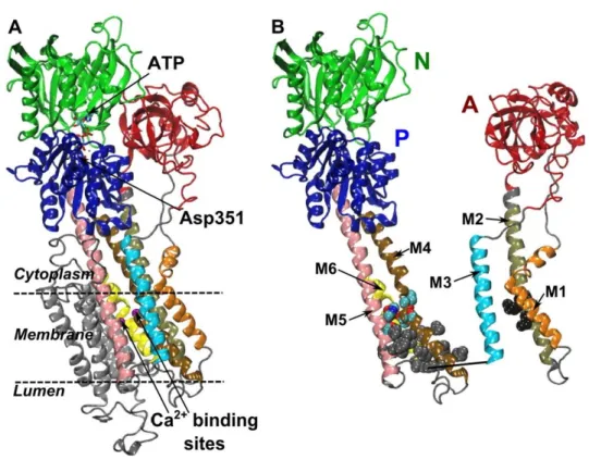
SERCA2, along with SERCA1 and SERCA3, belong to P-type superfamily of Ca2+ pumps that can be detected in the majority of eukaryotic cells’ sarcoplasmic/endoplasmic reticulum. These pumps share a highly conserved general structure, which consists of three cytosolic domains – actuator domain (A), nucleotide-binding domain (N), and phosphorylation domain (P) – and a transmembrane domain (TM), where Ca2+ binding sites are located (Figure 2) [15]. These pumps also share the same reaction cycle which starts with the binding of two calcium ions to the Ca2+ binding sites that are facing the cytoplasm, followed by the binding of one ATP molecule to the N domain and posterior hydrolysis by P domain, leading to the phosphorylation of a highly conserved aspartic acid residue (Asp351) (Figure 3) [15, 21]. This auto-phosphorylation allows the pump to switch conformation so its Ca2+ binding sites can face the luminal space and release the calcium ions into the ER. The
pump is regenerated to its initial state through dephosphorylation of the P domain, catalyzed by H2O and the conserved glutamate in the A domain [15, 21].
Figure 2 | Overall topology of the P-type ATPase calcium pump SERCA. (A) The cytoplasmic headpiece comprises the nucleotide binding (N), phosphorylation (P) and actuator (A) domains. The ATP binds to the N domain. The conserved aspartic acid that gets phosphorylated is in the P domain. The calcium binding sites are located in the transmembrane (TM) domain. The TM domain is made of ten TM helices (labeled M1 to M10 or 11). (B) The A domain is covalently linked to the helices M1, M2 and M3, and the P domain is covalently linked to the helices M4 and M5. Adapted from [22].
Furthermore, several studies have demonstrated, through radiation inactivation analysis and optical diffractometry, that these ATPases appear to be organized as dimers in the sarcoplasmic/endoplasmic membrane [23–26]. Nevertheless, it has not been proved whether monomers are not sufficient for normal function of the pump.
6
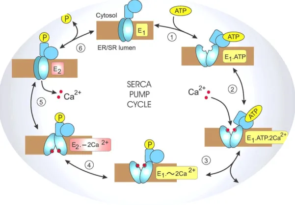
Figure 3 | Reaction cycle for the Sarco/Endoplasmic Reticulum Ca2+-ATPase (SERCA) pumps. During this
cycle, a series of biochemical reactions lead to two major conformational states of the Ca2+ pump: an E 1 state
when two high-affinity Ca2+ -binding sites that are facing the cytoplasm become saturated, and an E
2 state where
the Ca2+ -binding sites have a low-affinity and therefore switch to the opposite side and Ca2+ is released to the
lumen. During each cycle, two Ca2+ ions are pumped for each ATP hydrolyzed. (Adapted from
http://www.cellsignallingbiology.org/csb/005/csb005fig5_SERCA_pump_cycle.htm?resolution=HIGH).
Despite their similarities, these pumps do not share a common expression pattern, and neither do SERCA2 splice variants. SERCA2a (997 amino acids) has been found to be restrained to cardiomyocytes, cardiac slow-twitch muscle, skeletal and smooth muscle, pancreatic, and cerebellar Purkinje neurons (http://www.uniprot.org/uniprot/P16615) [13]. It is expressed at lower levels in the epidermis and has a two-fold lower affinity for Ca2+ and a two-fold higher catalytic turnover rate, when compared to SERCA2b [27]. The reduced expression of isoform SERCA2a, found in animal models of heart failure and in humans with heart failure, confirms its essential role in platelet and cardiac function [28]. In fact, in healthy individuals, SERCA2a is usually expressed at elevated levels in the heart and represents a key player in cardiac myocyte Ca2+ regulation required for excitation contraction coupling [28].
Mutations in the ATP2A2 gene affecting SERCA2a levels should therefore provoke an augment in platelet aggregation and cardiac dysfunction. However, there is a clear paradox since DD patients have been shown to have a normal cardiac function and platelet activity [28]. Researchers suggest that SERCA3, which is also present in platelets, may compensate for the platelet SERCA2a deficit in DD patients [29].
SERCA2b (1042 amino acids) is the ubiquitous housekeeping isoform, expressed in non-muscle cells and highly expressed in epidermal cells [1, 29]. Comparing to SERCA2a, isoform b contains an additional transmembrane domain, encoded by exon 21, and a tail known as 2b tail, which extends to the lumen of the ER. Interestingly, SERCA2b has the highest affinity for Ca2+ among all splice variants [27]. Moreover, a mutation in exon 21, affecting only SERCA2b isoform’s expression appears to be enough to cause DD, suggesting the crucial role of this pump in the skin and the inability of other splice variants to compensate the loss of SERCA2b normal function [13]. Nevertheless, it has been proposed the existence of putative compensatory mechanisms in DD keratinocytes, such as secretory pathway Ca2+-ATPase pump type 1 (SPCA1), that may help overcoming the intracellular [Ca2+] imbalance caused by mutations in the ATP2A2 gene [30].
The latest isoform described, SERCA2c (999 amino acids), is expressed in epithelial, mesenchymal and hematopoietic cell lines and monocytes [24, 28]. This isoform shows a Ca2+ affinity and catalytic turnover rate similar to that of SERCA2a and SERCA2b, respectively [20].
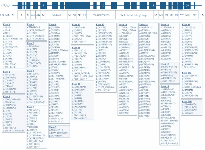
According to Leiden Open Variation Database v3.0 (http://www.lovd.nl/3.0/home), 420 variants have been reported in the ATP2A2 gene. DD mutations are mostly private ones (Figure 4). The majority of these private variants are missense mutations, which have been reported to be associated with a more severe phenotype [13, 31]. Mutations yielding a premature stop codon, nonsense, and splice-site mutations, and in-frame deletions and insertions have also been reported in DD patients [13]. Depending on the protein domain affected by these mutations, one may suggest a series of functional disruptions, including loss of Ca2+ affinity, obstruction of Ca2+-binding sites, decreased ATP affinity, effects in phosphorylation by ATP and Pi, effects on conformational shifts, inhibition of dephosphorylation, or disassembling of Ca2+ transport from ATP hydrolysis [32].
8
Figure 4 | Schematic representation of ATP2A2 gene and encoded protein SERCA2 isoform b and spectrum of ATP2A2 variants responsible for DD. All 21 exons and respective protein domains are represented. S- Stalk domain, M – Transmembrane domain. Exons 5, 10, 15, and 20 are numbered in the
ATP2A2 gene representation. The spectrum of ATP2A2 variants below the gene and protein schemes refer to
1.4 Clinical Manifestations
DD runs a chronic and relapsing course after its onset and may aggravate with age [5, 13]. The onset of the phenotypic abnormalities is usually during the first or second decade of life; however, delayed onset may happen as it has been reported [4, 6]. The disease is characterized by nail abnormalities and focal skin lesions such as greasy, skin-colored (brown or yellow) keratotic papules and eruptions (Figure 5) [1, 34]. The majority of skin lesions are distributed in seborrheic areas of the upper back, folds, neck, scalp, chest, and face. These lesions become easily infected, leading to major discomfort and exacerbation of the condition. Once infected, there is a high prevalence of Staphylococcus aureus colonization in these lesions [35]. Furthermore, external factors such as UVB radiation, heat, friction, sweat, and humidity play a key role in the worsening and relapse of skin lesions [1, 13, 34]. The nail phenotype consists of longitudinal splitting with V-shaped notches at the free margin of the nail, and subungual hyperkeratosis [1]. Palmoplantar pits are quite common, and hyperkeratosis of palms and soles may also occur. Moreover, the oral mucosa and gingiva may also suffer from lesions [1]. However, the above-mentioned clinical manifestations vary in severity from patient to patient and even within affected members of the same family.
1.5 Histopathology
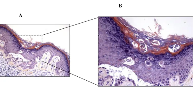
Although genetic testing is the most accurate method to diagnose a genetic disorder, the diagnosis of DD is usually done by histological examination of a skin biopsy [36]. Histologically, two hallmarks of DD can be identified in focal epidermal lesions: (1) acantholysis, which consists of loss of cell-to-cell adhesion and suprabasal clefting as a result of desmosomal disintegration; (2) dyskeratosis, characterized by abnormal/premature keratinocyte differentiation and apoptotic keratinocytes called “corps ronds” with a central basophilic and pyknotic nucleus surrounded by a clear halo (Figure 6) [1, 42]. Acantholysis can be observed in more detail by electron microscopy analysis, which demonstrates an impaired number of desmosomes and desmosomal cadherins [5, 43]. Desmosomes are intercellular junctions, composed by adhesion molecules such as cadherins and desmosomal plaque proteins, responsible for connecting adjacent cells. These cellular structures are
10
essential in mammals’ cardiac and epithelial tissues since these are subject to mechanical stress [39]. Therefore, disruption in the stability and cell surface expression of desmosomal proteins contributes to acantholysis since it impairs cell-to-cell adhesion in lesional keratinocytes. Additionally, cadherins are known for their important role in neuronal functions including the formation of specific neural circuits [40].
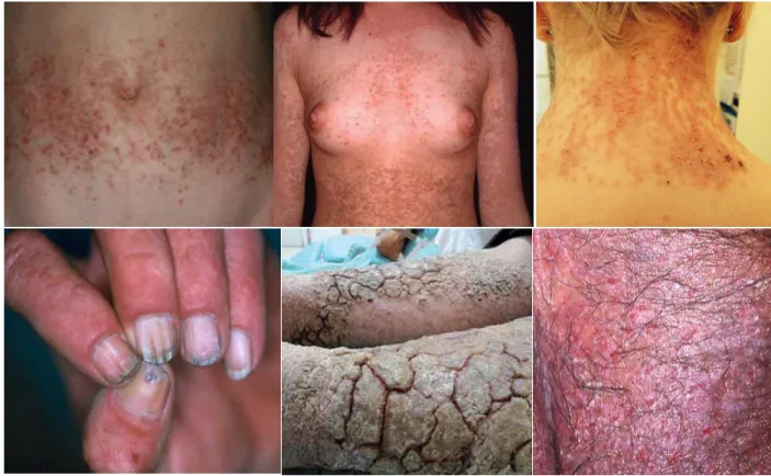
Figure 5 | Clinical manifestations of Darier disease patients. A Reddish, greasy, crusted papules on the abdomen. Adapted from [41]. B Confluent reddish-brownish papules in the seborrheic areas of the chest, submammary areas, and arms. Adapted from [42]. C Papular lesions cover skin in the occipital area of female patient with Darier’s disease. Adapted from [43]. D Nail involvement in Darier’s disease. Adapted from [1]. E Involvement of lower limbs with marked keratosis and fissures, which may lead to secondary infection in a patient with extremely severe condition. Adapted from [44]. F Papillomatous and macerated lesion in the groin. Adapted from [45].
Studies, where authors used thapsigargin as a specific inhibitor of SERCA or small interfering RNA directed to SERCA2, have provided evidence for the intrinsic role of SERCA2 in the disruption of intracellular Ca2+ signaling and consequent mislocalization of desmosomal cadherins [19, 34, 46, 47]. Nevertheless, the intracellular Ca2+ role in the regulation of adhesion molecules needs further investigation, as well as the role of cadherins in acantholysis and dyskeratosis in lesional keratinocytes.
Figure 6 | Histological section of Darier disease skin. Skin biopsy from Darier disease where a suprabasal cleft of the epidermis formed by acantholytic cells can be observed, as well rounded dyskeratotic cells with a dark nucleus, so called “corps ronds”, and hyperkeratosis. B is an enlargement of A. Adapted from [13].
1.6 Associated Manifestations
Although skin lesions represent the major pathophysiological outcome of mutations in the ATP2A2 gene, additional clinical manifestations appear to be linked to DD, being neuropsychiatric features the most common co-occurrences.
B A
12
1.6.1 Neuropsychiatric manifestations
Interestingly, the impairment of Ca2+ homeostasis and connectivity defects due to cadherin disruption are involved in cognitive and psychiatric disorders [45, 48, 49]. In fact, a range of neuropsychiatric features such as depression, suicidal behavior, bipolar disorder, schizophrenia, intellectual disability, epilepsy, cognitive deficit, mental retardation, and learning disability have been reported in DD patients [48–51]. A study involving unrelated DD patients revealed that 31% of these have a lifetime history of suicidal thoughts and 13% have attempted suicide, which did not have a relationship with DD or worsening of the disease [52]. Gordon-Smith et al. have found higher lifetime prevalence rates of mood disorders (50%), especially major depression (30%) and epilepsy (3%), to be present within a sample of one hundred unrelated DD patients compared with that in the general population [52]. In agreement with these findings, Celik et al. highlighted that the overall prevalence of epilepsy is 30–42.9 in 1000 DD patients, while the reported lifetime prevalence rates of epilepsy in the general population are variable, with a percentage of 1.3% [53].
Mutational analysis of a number of DD patients with neuropsychiatric phenotypes has shown predominance of missense mutations affecting the ATP-binding domain, between exons 13 and 19 of the ATP2A2 gene, whereas patients with DD alone have demonstrated a more diverse series of mutations predominantly spread between exon/intron 1 and 19 [32]. Therefore, it is suggestible that missense mutations in the ATP-binding domain turn out to be crucial contributors for abnormalities in neuron signaling. Nevertheless, the reason why psychiatric phenotypes are not co-segregated among every family member with the same DD mutation remains to be unraveled.
Summing up, there are four reasons why these co-occurrences might be present in DD patients: (1) a predisposition of ATP2A2 gene mutation towards the neuropsychiatric illness; (2) ATP2A2 gene location near a susceptibility gene for the neuropsychiatric illness; (3) pleiotropic effects of SERCA2 since it is expressed in both epidermal and neuronal cells; or (4) by pure chance, given that the mentioned psychoses show a high prevalence worldwide [50].
1.6.2 Other manifestations
Besides neuropsychiatric phenotypes, there are other potential associated manifestations in DD that should keep dermatologists alert, such as renal impairment, testicular agenesis, cataracts, skin cancers, bone cysts, and corneal and ocular sequels [54– 57]. Some researchers even point out for the genetic nature of DD as a model of skin carcinogenesis since the disruption of Ca2+ homeostasis, among other disturbances, is capable of inducing several genetic alterations, as it has been observed in cutaneous tumorigenesis models [58]. Opposite to the major mechanism of cancer, in ATP2A2+/- mice, neither RAS nor P53 proto-oncogenes were mutated, suggesting a novel form of cancer susceptibility through mutated SERCA2 [59]. This evidence is in agreement with a case report about a DD patient who had no history of smoking, drugs, or familial history of cancers, and still developed a squamous cell carcinoma (SCC) in the same area of a skin lesion, suggesting that SCC and DD may share common molecular disruption mechanisms [58].
Increasing literature gives more and more evidence that these secondary manifestations are unlikely to occur by mere chance in DD patients. However, further research is needed to clarify the mechanisms underneath these co-phenotypes and their genetic predisposition.
1.7 Intrafamilial phenotypic variability
The incidence and severity of skin lesions are not equally observed from parent to child, or sibling to sibling, who carry the very same pathogenic mutation in the ATP2A2 gene. Furthermore, despite the linkage between neuropsychiatric phenotypes and DD, the formers are not co-segregated in every affected member of the family. Researchers highlight for the presence of nucleotide alterations in the ATP2A2 gene, apart from the pathogenic mutation, as a potential cause for such clinical variability between relatives [30]. Although phenotypic variability can be observed in several families that segregate the disease, linkage studies embracing this matter are scarce and well-defined genotype-phenotype correlations have not yet been achieved.
14
1.8 Mechanism of Action
Throughout literature, haploinsufficiency or dominant-negative effect have been the postulated pathogenic mechanisms of DD, which is characterized by a heterozygous mutation in the ATP2A2 gene in most patients. According to haploinsufficiency, a single wild-type allele is not able to provide enough amount of protein and thus does not allow normal cell viability or function. However, if haploinsufficiency ought to be enough to cause DD, then why is the onset during the first or second decade of life rather than at birth? Why does DD lead to focal lesions rather than generalized dermatoses? How can SERCA2 haploinsufficiency explain the variable clinical severity among family members who carry the same mutation? All these observations have led scientists to further investigate the effect of DD mutations and/or SERCA2 inhibition.
Increasing in vitro studies involving epidermal and other cell types treated with SERCA-specific inhibitors, siRNA directed to SERCA2 mRNA, or site-directed mutations in ATP2A2 gene have demonstrated that SERCA2 haploinsufficiency may not be enough to cause DD [19, 60, 61]. SERCA2 mutants studied in neuronal and epidermal cell lines revealed to be insoluble or partially soluble due to their misfolded state, intrinsic to the mutation itself [60]. Instead of being degraded by proteasomes, the insoluble SERCA2 mutants have shown to gather around the nucleus, originating aggregates that contribute to the rounding up of cells seen in DD epidermis [60]. On the other hand, the perinuclear aggregation of these proteins could rely on the ability of SERCA2 mutant monomers to interact with the wild-type encoded SERCA2 in order to form dimers, as it has been demonstrated by co-immunoprecipitation of SERCA2 mutants and wild-type proteins from human embryonic kidney cells co-transfected with both SERCA2b plasmids [61]. Ahn et al. suggest that different ATP2A2 mutations may lead to different SERCA2 mutants, which may be responsible for the different phenotypes in DD patients according to the mutation’s impact on the activity of the wild-type encoded pump. This study reveals that most mutants not only interact with wild-type SERCA2 to form a dysfunctional dimer but also inhibit the rate of Ca2+ uptake by wild-type SERCA2, causing a dominant-negative effect [61].
Interestingly, it has been demonstrated that both lesional and non-lesional DD keratinocytes suffer ER Ca2+ depletion, but only lesional keratinocytes exhibit mislocalization and altered trafficking of desmosomal cadherins (DCs) [19, 34]. This aspect of DD’s pathology was enlightened by Li et al. who revealed that inhibition of SERCA2 by
thapsigargin in human epidermal keratinocytes leads to the reduction of ER Ca2+ levels below a threshold level, which consequently causes ER retention of nascent DCs [19]. Furthermore, it has been shown that the maturation (ER-to-Golgi transport) of nascent DCs is not affected when SERCA2 activity was inhibited by siRNA, suggesting that haploinsufficiency does not cause enough ER Ca2+ depletion to disrupt adhesion in DD pathology [19]. Presenting similar results, Celli et al. have reported that treatment of normal cells cultured with thapsigargin can disrupt the assembly of desmoplakin and E-cadherin to cell-to-cell boarders in a dose-dependent manner [34]. However, since thapsigargin not only blocks SERCA2 but all other SERCA pumps’ activity, it would be worth to further investigate the assembly of cadherin molecules in SERCA2 mutant cells.
Another hallmark of DD is the abnormal differentiation that occurs in lesional keratinocytes, including hyperproliferation and apoptosis of these cells. It is not surprising that once Ca2+ profile along the epidermis is disrupted, Ca2+ dependent activities such as differentiation, proliferation and apoptosis become deregulated. Calcium signaling may as well be crucial not only to the differentiation markers in each epidermal stratum, but for the BCL2 family which is linked to apoptosis. Bongiorno et. al have found decreased levels of BCL2 and BCL-xL, and deregulated levels of BAX in histological specimens of DD [62]. These results may explain the presence of “corps ronds” in histological sections in lesional epidermis. In addition, BCL-2 was shown to upregulate and possibly interact with SERCA pumps in order to preserve and stabilize ER Ca2+ pools, which is one of the ways that BCL-2 protects cells from apoptosis [62]. These findings suggest that (1) once there is not enough SERCA2 localized in the membrane of the ER, BCL-2 is unable to interact with SERCA2 in order to maintain ER Ca2+ stores, which consequently subjects keratinocytes to apoptosis; (2) with the presence of deregulated BAX, BCL-2 is also deregulated; (3) a decreased level in BCL-xL may also contribute to apoptosis in keratinocytes and to abnormal mitotic process; and finally (4) the imbalance of these three apoptosis-regulator proteins may underline the apoptotic pathway responsible for dyskeratosis seen in DD [62]. Nonetheless, DD hallmarks remain to be fully elucidated.
16
1.9 Treatment
Heretofore there has not been found a treatment whose efficacy does not vary among DD population. Personal hygiene, the use of sunscreen and avoidance from solar exposure, heat, high humidity, and tasks that could lead to excessive sweat are some of the indispensable measures in the day life management of the disease [44]. Currently, oral or systemic retinoids are the most frequently prescribed drugs to manage DD. In some cases, these drugs are able to reduce hyperkeratosis and flatten the papular component of the skin eruptions effectively. However, in most cases, retinoids are associated with a large number of noxious side effects, including mucosal dryness, itching, and severe or worsening of psychiatric symptoms [39, 63, 64]. In addition, most retinoids cannot be prescribed to women of child-bearing age [41, 65].
To date, the conventional treatments (retinoids, emollients and corticosteroids) used to manage DD’s cutaneous manifestations have yield disappointing results in general. Efficacy, safety, and long-term absence of relapses are crucial factors for a successful treatment. Therefore, other alternative drugs and therapies have recently been tested in DD patients.
A promising retinoid is the vitamin A derivate drug, alitretinoin (9-cis retinoic acid), which has shown cutaneous improvement due to its anti-proliferative and immune-modulatory effects on keratinocytes, and absence of relapse or relevant side effects within 15 months [66]. As opposed to other retinoids, alitretinoin’s short half-life suggests that it could be used to treat females of childbearing age as well [66]. Abe et al. reported a great clinical response and safety of vitamin D3 analog, Tacalcitol, in a DD patient [63].
Furthermore, therapies used in cancer patients, such as photodynamic therapy and 5-fluorouracil (5-FU), are also being tested in DD patients with benign or malignant skin lesions. Promising reports on treatment with topical 5-FU reveal that this compound is well tolerated by the majority of patients, who revealed a continuous decrease in cutaneous inflammation, and relief of burning and itching sensations caused by the condition of the disease [67].
In a limited number of patients, photodynamic therapy (PDT) has been reported to be tissue selective and responsible for successful treatment of DD [65, 68]. PDT consists on the combination of a photosensitizer, usually topical 5-aminolaevulinic acid (5-ALA), which accumulates in the target cells, an activating light source, and oxygen which cause a photo-oxidative reaction within the lesional tissue [65]. As a result, reactive oxygen species are
generated, inducing apoptosis and necrosis of target cells. Unlike dermabrasion and laser therapies, PDT prevents the risk of scarring and relapse of the condition due to tissue selectiveness [65].
The compounds and therapies above-mentioned are excellent candidates to manage DD. The increasing promising reports involving single patient or small number sample trials have opened doors to possible wider trials in the future. Randomized clinical trials would be the best way to confirm the effectiveness and safety of these recently tested drugs [67]. Additionally, there have been reported promising targets for DD, including sphingosine phosphate lyase (SGPL1), Transient Receptor Potential Canonical 1 (TRPC1), and cyclooxygenase-2 (COX-2), whose modulation could be a potential treatment for managing abnormal differentiation and loss of intercellular adhesion, keratinocyte proliferation, or for the enhancement of SERCA2 expression, respectively [34, 69, 70].
Despite the progress in gene therapies for heritable skin disorders, there is a considerable need to target DD by correcting the underlying mutation in somatic cells, either by using interfering RNAs, adeno-associated virus, or even CRISPR [71, 72].
2. Aims of the study
This project consists in the molecular characterization of a two-generation Portuguese family with DD history, including co-associated phenotypes and intrafamilial phenotypic variability. Accordingly, there are four aims that must be considered:
Aim no. 1: Identify and describe the pathogenic mutation that is causing the pathology in the family.
Aim no. 2: Examine whether the unaffected individual is a carrier but due to possible incomplete penetrance or later onset of the disease does not yet show clinical manifestations.
Aim no. 3: Analyze the pathogenic mutation’s effect at the transcriptional and translational levels.
Aim no. 4: Investigate genotype-phenotype correlations in an attempt to explain clinical heterogeneity of the affected family members by coupling the results from mutational analysis to their clinical history.
The results from this mutation analysis will determine whether the patients and possible carrier of the mutation should benefit from genetic counseling. Questions such as “Is this disease going to affect my children one day?”, “What should be monitored in my future children?” can eventually be brought up to surface and genetic counseling will help by inform them tightly about the risk of their progeny developing DD and co-occurrences, when planning or expecting to have a baby.
3. Participants and Methods
3.1 Case Report
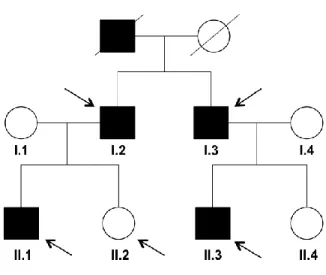
Five individuals from the two-generation family agreed to enroll in this study: four affected relatives (I.2, I.3, II.1 and II.3) and one unaffected relative II.2 (Figure 7). Relatives I.1 and I.4 were not considered to integrate the following study since they do not show any clinical history of the disease.
Figure 7 | Pedigree of the two-generation family. The affected individuals are denoted by solid black symbols whereas unaffected individuals are denoted by open symbols. Arrows point to the individuals who participate in this study.
All affected family members were diagnosed by dermopathology during their first decade of life. These individuals share a monomorphic dermatosis, notably symmetrical, containing multiple non-follicular hyperkeratotic papules with 2-4 mm in diameter, brownish, rounded and occasionally crusted, on skin usually healthy or erythematous skin during periods of inflammatory exacerbation. The lesions are distributed along the entire lateral
22
also in the scalp, and predominant in retroauricular region. Palms of the hands present multiple millimetric depressions filled by keratin (palmar pits). Nails of the hands and feet present discrete longitudinal leukonychia, fragility, fissure and dyskeratosis with V-shaped notches on the distal end of the nail plate. Eventual relapses of greater clinical severity usually lead to skin eruptions or infections by bacterial agents. Patients clinical course is in agreement with the worsening of the disease over the years. Moreover, patients agree that skin lesions are exacerbated by sweat, friction, and sunlight, particularly during the summer.
However, the clinical severity of the disease varies from patient to patient. At the skin level, patient I.1 has more relapses than his brother (I.3) while patient II.1’s relapses are more frequently and more severe than those of his father (I.1). In addition to their skin phenotype, viral, immunologic, and neuropsychiatric features occur in most patients (Table 1).
To manage DD, the patients take medicines prescribed by dermatologists such emollients, topical antibiotics, and retinoids such as acitretin, a derivative of vitamin A. They also avoid being exposed to the sun during the summer in order to prevent focal lesions to emerge. Patients with other occurrences, referred in Table 1, take additional medicines to manage these.
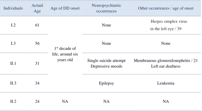
Table 1 | Additional information about participants’ actual age, age of onset, and other phenotypic manifestations.
Individuals Actual
Age Age of DD onset
Neuropsychiatric
occurrences Other occurrences / age of onset
I.2 61
1st decade of
life, around six years old
None Herpes simplex virus
in the left eye / 39
I.3 56 None None
II.1 31 Single suicide attempt Depressive moods
Membranous glomerulonephritis / 21 Left ear deafness
II.3 34 Epilepsy Leukemia
II.2 24 NA NA NA
3.2 Sample collection
Peripheral blood was collected by venipuncture from the enrolled individuals and written informed consent was obtained from each one. In the case of the affected individuals that are followed by a dermatologist, respective doctors were informed about the study. Blood samples were used for the extraction of nucleic acids and preparation of protein homogenates.
3.3 DNA and RNA extraction
Lymphocytes from a total peripheral blood volume of 14 mL from each subject were freshly isolated in a Ficoll density gradient and each pellet was stored at -80ºC before further use. Genomic DNA and total RNA were extracted from peripheral blood lymphocytes using Citogene ® DNA Blood Kit (Citomed) and Isol-RNA Lysis Reagent (5PRIME), respectively. The extraction of genomic DNA was performed based on a standard protocol involving cellular lysis, followed by protein precipitation, and DNA hydration. Total RNA extraction was performed according to the TRIzol method using chloroform to separate RNA from DNA and contaminants, followed by isopropanol precipitation, and pellet solubilization in RNase-free water. DNA and RNA concentrations and A260:A280 ratio from each sample were assessed using the NanoDrop ND-1000 spectrophotometer (Thermo Fisher Scientific). Quality and integrity of nucleic acids were also examined by 1% agarose gel electrophoresis. Extracted DNA samples were preserved at 4ºC before further use, while total RNA samples were stored at -80°C.
3.4 PCR and RT-PCR amplification
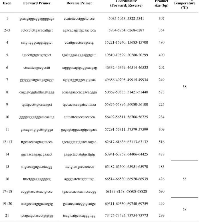
Polymerase chain reaction (PCR) amplification of all 21 exons and flanking intronic boundaries of the ATP2A2 gene was performed using 17 sets of primers, specifically designed with Primer3 program (http://primer3.ut.ee) (Table 2).
24
Table 2 | PCR primers and related parameters for amplification of ATP2A2 from genomic DNA.
a According to NCBI reference sequence NG_007097.2
PCR conditions consisted in an initial denaturation at 98ºC for 30 s, followed by 35 cycles of 98ºC for 10 s, an annealing temperature at either 55ºC or 58ºC for 10 s, and extension at 72ºC for 20 s; a final extension step for 5 minutes was performed. Each PCR reaction was carried out in a total volume of 25 μL containing nuclease-free water, PCR
Exon Forward Primer Reverse Primer Coordinates
a (Forward; Reverse) Product size (bp) Annealing Temperature (ºC) 1 gcaagaggaggaggggaga ccatcttccctggctctccc 5035-5053; 5322-5341 307 58 2+3 cctccctcttgacacattgct agacacagcttgcaactcca 5934-5954; 6268-6287 354 4 catgttgggcaggttggtct ccattgcactccagcctg 15221-15240; 15683-15700 480 5 tgtccttgtgtctgttgcct tgacaggaagggaggtgcta 19810-19829; 20280-20299 490 6 ctcatttcagccgccttt aagggacagtgaggcaagag 46332-46349; 46514-46533 202 7 ggtgggcatgaatgagaggt agtgatggttggcagtgaaa 49686-49705; 49915-49934 249 8 cagcgtcggtatttaagttggg acaaagaaccacgacacgga 50862-50883; 51421-51440 573 9 tgtttgcctttgtcctaagct tgccacaccagatcctttaaa 55876-55896; 56080-56100 225 10 ggggcgggaggaatcaatag ctttcattccacccacccca 56492-56511; 56706-56725 234 11 gacagattgtgcttttgtgga gagagtaggacagtgcagaca 57291-57311; 57579-57599 309 12+13 ttgccacccagtagtatcca tgcagggtgtggacaaagaa 62617-61636; 63113-63132 516 14 ggcaacaagagcgaaact gaggctactatgtgcttgtg 63941-63958; 64406-64425 478 15 tttgccaagagacctacgg tttctgtcttgcccactccc 65482-65500; 65951-65970 483 55 16 ttttctggaggagggcg agggcatctctgtcttttgc 66514-66530; 66920-66939 426 17+18 ccggttaccatcactgtccc tgactacacacaattccccgg 68139-8158; 68808-68828 690 19+20 tactgccactgtgacacgtg gaaatcccatcggtgcatgc 69311-69330; 69740-69759 449 58 21 tctagatgctaccctgtgtgg tcagtcatgcacagggttgg 73475-73495; 73754-73773 299
buffer (5x), 3.0 mM MgCl2, 3% dimethyl sulfoxide, 200 μM of deoxynucleotide triphosphate, 0.5 μM of each primer, 1.0 unit of Phusion® High-Fidelity DNA Polymerase (New England Biolabs), and approximately 200 ng of genomic DNA. To assure PCR products were amplified with the right dimension, an aliquot of each sample was run on a 2% agarose gel.
Total RNA isolated from lymphocytes was reverse transcribed using the Maxima First Strand cDNA Synthesis Kit (Thermo Fisher Scientific). Each RT-PCR reaction was prepared using a Reaction Mix containing reaction buffer, deoxynucleotide triphosphates,
single-stranded 18-mer oligonucleotides and random hexamer primers, Maxima Enzyme Mix, with
both reverse transcriptase (RT) and ribonuclease inhibitor, RiboLock, and finally, 1 μg of RNA, forming a total volume of 20 μL. The conditions for cDNA synthesis consisted in a first step at 25ºC for 10 minutes to allow primers to bind to the transcripts, followed by a second step at 50ºC for 15 minutes so that the RT can synthesize the cDNA strands, and a final step at 85ºC for 5 minutes to denature and degrade the remaining RNA. The total cDNA yield was quantified using NanoDrop ND-1000 spectrophotometer and stored at -20ºC until further use. PCR amplification was performed from SERCA2 cDNA previously synthesized, using six sets of primers, also specifically designed with Primer3 program, which span the whole sequence of SERCA2b that is transcribed (Table 3).
For cDNA amplification, each PCR was performed according to PCR protocol for Phusion® High-Fidelity DNA Polymerase, as previously described, with annealing temperatures of 60 or 61ºC, a final magnesium chloride concentration of 1.5 mM or 3.0 mM, and a total volume of 25 μL.
Table 3 | PCR primers and related parameters for amplification of SERCA2 from cDNA.
a According to NCBI reference sequence NM_170665.4
Exon Forward Primer Reverse Primer Coordinates
a (Forward; Reverse) Product size (bp) Annealing Temperature (ºC) 1-6 aagaggaggaggggagac ttcacctgtgagaattgactgg 417-435; 1071-1092 676 61 6-8 tggtgacaaagttcctgctg tgcagactgacatctggtttg 1008-1027; 1616-1636 629 60 8-13 gctcttggaactcgcagaat accccactctcgaatgacag 1501-1520; 2180-2199 699 13-15 acccacattcgagttggaag gccacaatggtggagaagtt 2116-2135; 2755-2774 659 15-18 gtgaacgatgctcctgctct aaagtccgggttgtcctctt 2653-2672; 3170-3189 537 17-20 ctgctgcatggtggttcat ttcagcagaaatagacagttgctt 3092-3110; 3731-3754 663
26
3.5. Sequence analysis
PCR and RT-PCR products, generated from genomic and coding sequences of ATP2A2 respectively, were purified either from solution or TBE agarose gel slices, using Isolate II PCR and Gel Kit (Bioline). The clean-up protocol was based on a silica-membrane method which encompasses DNA-membrane fixation, membrane washing and DNA elution. PCR forward and/or reverse primers were added to the purified DNA fragments from each individual sample and submitted to GATC Biotech for bi-directional Sanger sequencing. This method uses terminator fluorescent dideoxynucleotides that allow the detection of each distinct nucleotide by laser and automatic storage of data in a computer. As a result, a series of fluorescence intensity peaks correspondent to each nucleotide can be observed in a chromatogram [56]. All chromatograms corresponding to PCR and RT-PCR fragments were carefully analyzed and the pathogenic mutation was found.
3.6 Quantitative PCR
The expression of ATP2A2 gene was quantified by real-time PCR using the Maxima SYBR Green qPCR Master Mix (Thermo Fisher Scientific) and the CFX96 Touch™ Real-Time System (Bio-Rad). Two sets of primers were designed to amplify both mutant and wild-type transcripts (GGTGACAAGTTCCTGCTGA forward and AGTGTGCTTGATGAC AGAGACA reverse), or the wild-type transcripts only (TGTGCTCTTTGTAATGACTCTGC forward and AGAAAGACCCTTCAATTCGGTATCA reverse).
The set of primers designed strictly for wild-type transcripts allows the amplification of exon 10, which is missing in mutant mRNA while the set of primers for both wild-type and mutant anneals in the region of exons 6 and 7. The objective of this experiment was to assess relative levels of mutant and wild-type transcripts and look for a link between these expressions and the variable phenotype between the affected relatives. ATP2A2 mRNA expression was normalized by co-amplification of the housekeeping gene ACTB, coding for beta actin, which worked as an internal control. Primers used for evaluation of ACTB
(CTGGGACGACATGGAGAAAATCTG forward and AGCCTGGATAGCAACGTACATG reverse).
3.7 Western blotting
Lymphocytes isolated from 3 mL of whole blood were homogenized with RIPA lysis and extraction buffer (Pierce, Thermo Fisher Scientific) supplemented with HaltTM Protease and Phosphatase Inhibitor Cocktail (100x) (Thermo Fisher Scientific), followed by sonication. Lymphocyte homogenates were centrifuged for 10 minutes at 8000 rpm and the soluble fraction was collected for Western blot analysis to determine the expression level of the mutant protein in comparison with the wild-type SERCA2. Different procedures for protein extraction were performed in order to find the best strategy to assess SERCA2 expression levels.
A quantity of 25 μg of total protein from each individual (evaluated by the Bicinchoninic Acid method) was mixed with Laemli loading buffer. Two sets of samples were studied as heated and unheated samples. The heated samples were denaturated at 95ºC for 5 minutes. All samples were briefly centrifuged and soluble fraction of cell lysates were resolved in 7.5% sodium dodecyl sulfate-polyacrylamide gel electrophoresis (SDS-PAGE) and transferred onto polyvinylidene difluoride membranes (GE Healthcare Life Sciences, Amersham Hybond) at 300 mA for 100 minutes at 4ºC. Blots were blocked with a solution containing 5% bovine serum albumin (BSA), TBS (1x), and 0.1% Tween® 20 for one hour at room temperature. Membranes were immediately incubated with rabbit polyclonal anti-SERCA2 (dilution 1:10000; #4388 Cell Signaling) overnight at 4ºC, with gentle shaking.
Membranes were probed with anti-rabbit IgG horseradish peroxidase-linked secondary antibody (dilution 1:2000; #7074, Cell Signaling Technologies, Inc.), followed by enhanced chemiluminescent blotting using SuperSignal West Femto Maximum Sensitivity Substrate (Thermo Fisher Scientific). Only after SERCA2 revelation the blots were incubated with primary antibody anti-β-actin (dilution 1:4000; Sigma-Aldrich), followed by secondary antibody incubation and revelation.
28
3.8 Genotype-Phenotype correlation
Since the family under study presents phenotypic variability among the affected individuals, it would be interesting to look for a genotype-phenotype correlation. Accordingly, all chromatograms from genomic DNA PCR products were analyzed closely in search for additional polymorphisms or insertions/deletions within exonic and intronic regions amplified by PCR, Additionally, ATP2A2 and SERCA2 expression levels would also be taken into account for the search of genotype-phenotype correlation.
4. Results and Discussion
4.1 Identification of the pathogenic mutation
Bi-directional sequence analysis of the genomic DNA revealed a transversion in the first nucleotide of intron 10 (c.1287+1G>T or IVS10+1G>T), which corresponds to the G of the wild-type splice donor site dinucleotide (Figure 8A1). Therefore, the pathogenic mutation is considered a splice-site mutation, as proven by 2% gel electrophoresis (Figure 8A2) and bi-directional sequencing of RT-PCR products spanning exons 8 to 13, which confirmed that this variant affects the splicing process, leading to a shorter transcript where the skipping of full exon 10 is observed (Figure 8B). This mutant transcript contains a deletion of 103 base pairs (c.1185_1287del), originating a frameshift which putatively introduces a downstream premature stop codon (p.V395=fs*19). It was also observed that all four patients carry the same mutation in heterozigosity, whereas the unaffected individual carries the wild-type sequence in both alleles. For this reason, the unaffected individual was used as a negative control for the following experiments.
To understand what the splice site mutation does, it is important to know that splicing requires remarkable precision, which is granted by highly conserved intronic and exonic sequences and regulatory elements that are recognized by the spliceosome, a complex of small ribonucleoproteins (U1, U2, U5, U6/U4) and associated proteins [73]. Highly conserved intronic and/or exonic sequences include (1) the splice donor site GU and acceptor AG dinucleotides, recognized by the major spliceosome, at the 5’ and 3’ ends of the pre-mRNA, respectively; (2) the 5’ donor and 3’ acceptor splice site consensus sequences immediately upstream and downstream the GU and AG dinucleotides; and (3) the branch site, approximately 18 to 40 bp upstream the 3’ acceptor splice site [73]. Other relevant exonic and intronic sequences play a regulatory role either by promoting (splicing enhancer sequences) or inhibiting (splicing silencer sequences) the splicing process [73]. For instance, exon splicing enhancers are thought to compensate for the presence of splice sites with lower consensus values since it has been revealed that these sequences are in higher abundance in
30
Mutant allele
c.1287+1G>T in heterozygosity Wild-type allele
1) Exon 10 + intronic boarders
2) RT-PCR products (exon 8 – exon 13)
M- Molecular marker (pBR322 x MspI, New England Biolabs)
Figure 8A | Identification and expression of ATP2A2 pathogenic mutation. (1) Sequence analysis of the genomic sequence of ATP2A2 gene, corresponding to exon 10. The transversion IVS10+1G>T resulted in a splice-site mutation in intron 10. (2) RT-PCR products from every patient revealed that the splice site mutation yielded one wild-type allele transcript and a shorter transcript, while RT-PCR product from the unaffected member revealed two wild-type allele transcripts.
699 bp 596 bp
1) Fragment of cDNA sequence originating from the wild-type allele
2) Fragment of cDNA sequence originating from the mutant allele
Figure 8B | Sequence analysis of the coding sequence of ATP2A2 gene. Chromatograms correspondent to the wild-type sequence (1) and to the mutant sequence (2) which confirm the skipping of the entire region of exon 10 from every smaller transcript.
Mutations in these highly conserved sequences can either result in two consequences: the most frequent one, exon skipping, when an exon is no longer recognized as such, or cryptic splice site use, when the resulting mature RNA lacks a portion of an exon or contains an additional sequence from an intron. In the reported case, the reason why c.1287+1G>T led to exon skipping rather than cryptic splice site may rely on potential spliceosome’s functional
![Figure 1 | Intracellular and extracellular calcium concentration profile and differentiation markers along the epidermal strata (X100 Hematoxylin and Eosin staining) (https://basicmedicalkey.com/skin-6/#ch18lev9) [11, 12]](https://thumb-eu.123doks.com/thumbv2/123dok_br/19177058.943658/23.892.118.782.137.439/intracellular-extracellular-concentration-differentiation-epidermal-hematoxylin-staining-basicmedicalkey.webp)