F
ACULDADE DE
F
ARMÁCIA
D
ESIGN AND
S
YNTHESIS OF
S
MALL
M
OLECULE
M
ODULATORS OF P
53
C
ARLOSJ
ORGEA
ZEVEDOC
OSTAR
IBEIROOrientadores: Doutora Maria Manuel Duque Vieira Marques dos Santos Prof. Doutor Rui Ferreira Alves Moreira
Prof.ª Doutora Cecília Maria Pereira Rodrigues
Tese especialmente elaborada para obtenção do grau de Doutor em Farmácia, especialidade de Química Farmacêutica e Terapêutica
F
ACULDADE DE
F
ARMÁCIA
D
ESIGN AND
S
YNTHESIS OF
S
MALL
M
OLECULE
M
ODULATORS OF P
53
C
ARLOSJ
ORGEA
ZEVEDOC
OSTAR
IBEIROOrientadores: Doutora Maria Manuel Duque Vieira Marques dos Santos Prof. Doutor Rui Ferreira Alves Moreira
Prof.ª Doutora Cecília Maria Pereira Rodrigues
Tese especialmente elaborada para obtenção do grau de Doutor em Farmácia, especialidade de Química Farmacêutica e Terapêutica
JÚRI:
PRESIDENTE:Doutora Matilde da Luz dos Santos Duque da Fonseca e Castro VOGAIS:
-Doutor Jorge António Ribeiro Salvador
- Doutora Maria Emília da Silva Pereira de Sousa
- Doutora Maria Alexandra Núncio de Carvalho Ramos Fernandes - Doutora Maria Matilde Soares Duarte Marques
- Doutora Rita Alexandra do Nascimento Cardoso Guedes - Doutora Joana São José Dias Amaral
- Doutora Maria Manuel Duque Vieira Marques dos Santos
Este trabalho foi financiado pela Fundação para a Ciência e a Tecnologia através da bolsa de doutoramento SFRH/BD/69258/2010 e dos projectos PTDC/QUI-QUI/111664/2009, PTDC/SAU-FAR/110848/2009, PTDC/SAU-ORG/119842/2010 e Pest-OE/SAU/UI4013/2014.
- In the Scope of the PhD Thesis:
1. C. J. A. Ribeiro, R. Moreira, M. M. M. Santos, Synthesis of a Spiroisoxazoline Oxindole by 1,3-Dipolar Cycloaddition. In: Comprehensive Organic Chemistry Experiments for the Laboratory Classroom Book, Royal Society of Chemistry (accepted).
2. J. J. Badillo, C. J. A. Ribeiro, M. M. Olmstead, A. K. Franz, Titanium(IV)-Catalyzed Stereoselective Synthesis of Spirooxindole-1-Pyrrolines. Org. Lett. 2014, 16: p. 6270−6273
3. C. J. A. Ribeiro, J. D. Amaral, C. M. P. Rodrigues, R. Moreira, M. M. M. Santos, Synthesis and Evaluation of Spiroisoxazoline Oxindoles as Anticancer Agents. Bioorg. Med. Chem. 2014, 22(1): p. 77−84
4. C. J. A. Ribeiro, S. P. Kumar, R. Moreira, M. M. M. Santos, Efficient Synthesis of Spiroisoxazoline Oxindoles. Tetrahedron Lett. 2012, 53(3): p. 281−284.
- Other publications:
1. C. J. A. Ribeiro, S. P. Kumar, J. Gut, L. M. Gonçalves, P. J. Rosenthal, R. Moreira, M. M. M. Santos, Squaric Acid/4-Aminoquinoline Conjugates: Novel Potent Antiplasmodial Agents. Eur. J. Med. Chem. 2013, 69: p. 365−72.
Oral Communications in Scientific Conferences
1. C. J. A. Ribeiro, J. D. Amaral, C. M. P. Rodrigues, R. Moreira, M. M. M. Santos. “Synthesis and Biological Evaluation of Spiroisoxazoline and Spirooxadiazoline Oxindoles as Anticancer Agents”. Flash Poster Presentation. EFMC-YMCS 2014. 1st EFMC Young Medicinal Chemist Symposium. Lisbon, Portugal. 12/09/2014.
2. C. J. A. Ribeiro, J. D. Amaral, C. M. P. Rodrigues, R. Moreira, M. M. M. Santos. “Synthesis and Biological Evaluation of Spiroisoxazoline Oxindoles as Anticancer Agents”. 4rd Portuguese Young Chemists Meeting. Coimbra, Portugal. 29-30/04-01/05/2014.
3. C. J. A. Ribeiro, J. D. Amaral, C. M. P. Rodrigues, R. Moreira, M. M. M. Santos. “Spiroisoxazoline Oxindoles: a New Class of p53-MDM2 Interaction Inhibitors”. 4th iMed.UL Post-Graduate Students Meeting. Lisbon, Portugal. 20/12/2012.
4. C. J. A. Ribeiro. “Synthesis and Biological Evaluation of New Spiroisoxazoline Oxindoles as Potential Anticancer Agents”. Poster Selected for Oral Presentation. 32nd
7/07/2012.
5. C. J. A. Ribeiro, R. Moreira, M. M. M. Santos. “Efficient Synthesis of New Spiroisoxazoline Oxindoles”. 3rd Portuguese Young Chemists Meeting. Porto, Portugal. 9-11/05/2012.
6. C. J. A. Ribeiro, R. Moreira, M. M. M. Santos. "Structural Characterization of a Novel Spiroisoxazoline Oxindole". Flash Presentation. Modern Methods of Structure Elucidation (MMSE). Lisbon, Portugal. 14-18/11/2011.
Poster Communications in Scientific Conferences
1. C. J. A. Ribeiro, J. D. Amaral, C. M. P. Rodrigues, R. Moreira, M. M. M. Santos. “Synthesis and Biological Evaluation of Spiroisoxazoline and Spirooxadiazoline Oxindoles as Anticancer Agents”. EFMC-YMCS 2014. 1st EFMC Young Medicinal Chemist Symposium. Lisbon, Portugal. 12/09/2014.
2. C. J. A. Ribeiro, J. D. Amaral, C. M. P. Rodrigues, R. Moreira, M. M. M. Santos. “Spiroisoxazoline and Spirooxadiazoline Oxindoles as Anticancer Agents”. EFMC-ISMC 2014, 23nd International Symposium on Medicinal Chemistry. Lisbon, Portugal. 07-11/09/2014.
3. C. J. A. Ribeiro, J. D. Amaral, C. M. P. Rodrigues, R. Moreira, M. M. M. Santos. “Spiroisoxazoline Oxindoles: Novel Small-Molecule Inhibitors of the p53-MDM2 Interaction”. COST Action CM1106: Chemical Approaches to Targeting Drug Resistance in Cancer Stem Cells. 2nd Working Group Meeting. Warsaw, Poland. 19-20/09/2013.
4. C. J. A. Ribeiro, S. P. Kumar, J. Gut, L. M. Gonçalves, P. J. Rosenthal, R. Moreira, M. M. M. Santos. “Synthesis of novel antiplasmodial agents containing squaramide and 4-amino-7-chloroquinoline moieties”. 10th Portuguese National Meeting of Organic Chemistry/1st Portuguese-Brazilian Organic Chemistry Symposium. Lisbon, Portugal. 4-6/09/2013.
5. C. J. A. Ribeiro, J. D. Amaral, C. M. P. Rodrigues, R. Moreira, M. M. M. Santos. “Spiroisoxazoline Oxindoles: New p53-MDM2 Interaction Inhibitors”. 4th Frontiers in Medicinal Chemistry. San Francisco, USA. 23-26/06/2013.
6. C. J. A. Ribeiro, J. D. Amaral, C. M. P. Rodrigues, R. Moreira, M. M. M. Santos. “Evaluation of New Spiro-Oxindoles as Anticancer Agents”. 3rd Portuguese Meeting on Medicinal Chemistry. Aveiro, Portugal. 28-30/11/2012.
7. C. J. A. Ribeiro, J. D. Amaral, C. M. P. Rodrigues, R. Moreira, M. M. M. Santos. “Synthesis and Biological Evaluation of New Spiroisoxazoline Oxindoles as Potential
Chemistry. Berlin, Germany. 2-6/09/2012.
8. C. J. A. Ribeiro. “Synthesis and Biological Evaluation of New Spiroisoxazoline Oxindoles as Potential Anticancer Agents”. 32nd Edition of the European School of Medicinal Chemistry (ESMEC). Urbino, Italy. 2-7/07/2012.
9. C. J. A. Ribeiro, R. Moreira, M. M. M. Santos, “Simple and Efficient Synthesis of Spiroisoxazoline Oxindoles, Using Zinc as Dehydrochlorinating Agent”. 3rd iMed.UL Post-Graduate Students Meeting. Lisbon, Portugal. 11/12/2011.
10. C. J. A. Ribeiro, R. Moreira, M. M. M. Santos. “Synthesis of Spiroisoxazoline oxindoles from Methyleneindolinones and Nitrile oxides”. XXII National Meeting of SPQ - 100 years of Chemistry in Portugal, Braga, Portugal. 03-06/07/2011.
Table of Contents
Chapter 1. State of the Art 1
1.1. p53 activation in Health and Disease ... 3
1.2. Reactivation of p53 as a Therapeutic Strategy ... 4
1.2.1. Targeting p53-MDM2 Interaction ... 5
1.2.1.1. Cis-imidazolines (Nutlins). Road to RG7112 ... 8
1.2.1.2. Spirooxindoles ... 10
1.2.1.2a. Spiropyrrolidine Oxindoles. Road to MI-77301 ... 10
1.2.1.2b. Spirothiazolidine Oxindoles ... 13
1.2.1.3. Pyrrolidine-2-carboxamides. Road to RG7388. ... 15
1.2.1.4. Piperidinones and Morpholinones. Road to AMG232. ... 16
1.2.1.5. Other MDM2 Inhibitors in Clinical Trials ... 19
1.2.1.6. Benzodiazepinediones ... 19
1.2.1.7. Isoindolinones ... 21
1.2.1.8. Chromenotriazolopyrimidines ... 23
1.2.1.9. 3-Imidazoyl Indoles and Other Indolyl Derivatives ... 25
1.2.1.10. Other Compounds ... 26
1.2.2. Inhibition of E3 Ligase Activity of MDM2 ... 28
1.2.3. MDMX and Dual MDM2/MDMX Inhibitors ... 29
1.2.4. Targeting Upstream Regulators ... 31
1.2.5. Sirtuins Inhibitors ... 33
1.2.6. S100B Inhibitors ... 34
1.2.7. p53-Targeting Compounds ... 35
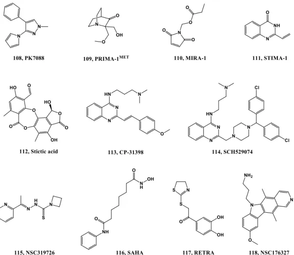
1.2.8. Mutant p53 Reactivation ... 35
1.3. Concluding Remarks ... 38
Chapter 2. Scope and General Goals 39
2.1. Synthesis of Spirooxindole Derivatives ... 41
2.2. Biological Studies ... 42
2.3. Stability Assessment ... 43
2.4. Thesis Layout ... 43
Chapter 3. Spiroisoxazoline Oxindoles: Synthetic Methodology Optimization 45
3.1. Introduction ... 47
3.2. Synthesis of Intermediates ... 48
3.3. Synthesis of Spiroisoxazoline Oxindoles and Optimization ... 49
3.4. Regio and Stereoselectivity Considerations ... 52
Chapter 4. Spiroisoxazoline Oxindoles:
Increasing Synthetic Scope and Biological Evaluation 57
4.1. Introduction ... 59
4.2. Synthesis of Intermediates ... 61
4.3. Synthesis of Spiroisoxazoline Oxindole Derivatives ... 62
4.4. Regio and Stereoselectivity Considerations ... 64
4.5. Biological Studies ... 64
4.5.1. Assessment of Cell Viability ... 64
4.5.2. Evaluation of Compounds Ability to Block the Intracellular p53-MDM2 Interaction ... 66
4.5.3. Evaluation of Apoptosis ... 68
4.6. Stability ... 68
4.7. Concluding Remarks ... 69
Chapter 5. Spiroisoxazoline Oxindoles: Enantioselective Approach 71
5.1. Enatioselective Approaches towards Spiroisoxazoline Oxindoles ... 73
5.1.1. Introduction ... 73
5.1.2. Synthesis of intermediate 121n. ... 74
5.1.3. Attempts to Enanteoselectively Synthesize Compound 119ac ... 75
5.2. Enatioselective Approaches towards Spiropyrroline Oxindoles ... 81
5.2.1. Introduction ... 81
5.2.2. Synthesis of Intermediate 121p. ... 81
5.2.3. Attempts to Enantioselectively Synthesize compound 163 ... 82
5.3. Concluding Remarks ... 84
Chapter 6. Spirooxadiazoline Oxindoles: Synthesis and Biological Evaluation 85
6.1. Introduction ... 87
6.2. Synthesis of Intermediates ... 88
6.3. Synthesis of Spiro[indoline-3,5’-[1,2,4]oxadiazoline]-2-ones ... 89
6.4. Biological Studies ... 90
6.4.1. Assessment of Cell Viability ... 90
6.4.2. Evaluation of Compounds Ability to Block the Intracellular p53-MDM2 Interaction ... 93
6.4.3. Evaluation of Apoptosis ... 94
6.5. Stability ... 95
Chapter 7. Spirotriazoline Oxindoles:
Synthesis and Biological Evaluation 97
7.1. Introduction ... 99
7.2. Synthesis of Intermediates ... 101
7.3. Synthesis of 2’,4’-Dihydrospiro[indoline-3,3’-[1,2,4]triazol]-2-ones ... 103
7.4. Synthesis of Spiropyrazoline Oxindole 167f ... 103
7.5. Biological Studies ... 104
7.5.1. Assessment of Cell Viability and SAR Study ... 104
7.5.2. Evaluation of Compounds Ability to Block the Intracellular p53-MDM2 Interaction .... 108
7.5.3. Evaluation of Apoptosis ... 109
7.6. Stability ... 110
7.7. Concluding Remarks ... 111
Chapter 8. General Conclusions and Future Perspectives 113
Chapter 9. Experimental Section 117
9.1. Experimental Section: Chemistry ... 119
9.1.2. General Procedure for the Synthesis of 3-Methylene indoline-2-ones. ... 121
9.1.2.1. Method A. Wittig Reaction. ... 121
9.1.2.2. Method B. Aldolic Condensation. ... 121
9.1.3. Synthesis of Indolin-2,3-diones and 5-Bromoindolin-2-one. ... 126
9.1.4. General Procedure for the Synthesis of 3-Imino-indoline-2-ones. ... 128
9.1.5. General Procedure for the Synthesis of Aldoximes. ... 134
9.1.6. General Procedure for the Synthesis of Chlorooximes. ... 135
9.1.6.1. Aromatic Derivatives ... 135
9.1.6.2. Ester Derivatives ... 137
9.1.7. General Procedure for the Synthesis of Hydrazones ... 138
9.1.8. General procedure for the synthesis of Hydrazonoyl Chlorides. ... 141
9.1.9. General Procedure for the Synthesis of 4'H-Spiro[indoline-3,5'-isoxazol]-2-ones. ... 144
9.1.9.1. Method A (Et3N). ... 144
9.1.9.2. Method B (Zn). ... 146
9.1.9.3. Method C (synthesis directly from aldoxime). ... 151
9.1.10. Synthesis of 4'-ethyl 5'-methyl (3S,4’R,5’S)-N-acetyl-5-fluoro-2'-(4-methoxyphenyl)-2-oxo-4',5'-dihydrospiro[indoline-3,3'-pyrrole]-4',5'-dicarboxylate (epi-163). ... 160
9.1.11. General Procedure for the Synthesis of Spiro[indoline-3,5'-[1,2,4]oxadiazoline]-2-ones. ... 161
9.1.12. General Procedure for the Synthesis of 2',4'-Dihydrospiro[indoline-3,3'-[1,2,4]triazol]-2-ones. ... 176
9.2. Experimental Section: Biology. ... 192
9.2.2. Western Blot Analysis. ... 193
9.2.3. Evaluation of Caspase-3/7 Activity ... 194
9.2.4. Bimolecular Fluorescence Complementation (BiFC) Assay. ... 194
9.3. Experimental section: Stability. ... 195
9.3.1. HPLC Analysis. ... 195
9.3.2. Stability in pH 7.4 Phosphate Buffer. ... 195
9.3.3. Stability in Human Plasma. ... 195
9.3.4. Stability in Rat Microsomes. ... 195
9.4. Experimental Section: Docking Studies ... 196
Figure Index
Figure 1.1. Simplified p53 activation and response upon acute DNA damage. ... 4
Figure 1.2. Cellular regulation of p53 by MDM2. ... 5
Figure 1.3. The p53-MDM2 interaction representation (PDB 1YCR) ... 6
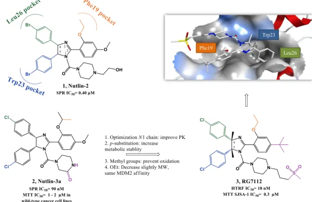
Figure 1.4. Nutlins optimization to RG7112. Right upper quadrant: crystal structure of compound 3 bound to MDM2 (PDB 4IPF).. ... 9
Figure 1.5. Nutlin and derivatives. ... 9
Figure 1.6. Spiropyrrolidines optimization to MI-106. Docking pose of compound 13 in MDM2 (PDB 3LBL). ... 12
Figure 1.7. Spiropyrrolidines and others spiro-heterocyclic-oxindole derivatives. ... 13
Figure 1.8. Spiropyrrolidines and spirothiazolidines derivatives. ... 14
Figure 1.9. Pyrrolidine-2-carboxamide optimization to RG7388. Right upper quadrant: crystal structure of compound 26 bound to MDM2 (PDB 4JRG) ... 15
Figure 1.10. Piperidinone optimization to AMG232. ... 18
Figure 1.11. Piperidinone and morpholinone derivatives. Right lower quadrant: crystal structure of compound 34 bound to MDM2 (PDB 4OAS) ... 19
Figure 1.12. Benzodiazepinediones scaffold optimization. Right upper quadrant: crystal structure of compound 40 bound to MDM2 (PDB 1T4E) ... 21
Figure 1.13. Examples of benzodiazepinediones derivatizations. ... 21
Figure 1.14. Isoindolinone scaffold optimization... 23
Figure 1.15. Oxazoloisoindolinone derivative 52. ... 23
Figure 1.16. Chromenotriazolopyridines scaffold optimization. Right upper quadrant: crystal structure of compound 53 bound to MDM2 (PDB 3JZK) ... 24
Figure 1.17. Indolyl derivatives. Right upper quadrant: structure of compound 56 bound to MDM2 (PDB 1YCR). ... 25
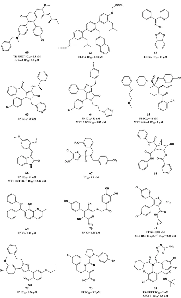
Figure 1.18. p53-MDM2 interaction inhibitors ... 27
Figure 1.19. MDM2 E3 Ligase activity inhibitors ... 29
Figure 1.20. MDMX and dual MDM2/MDMX inhibitors ... 30
Figure 1.21. Compounds that target upstream regulators of p53 activating pathway. ... 32
Figure 1.22. SIRT1and/or SIRT2 inhibitors. ... 34
Figure 1.23. p53-S100B interaction inhibitors. ... 34
Figure 1.24. RITA. ... 35
Figure 1.25. Compounds targeting mutant p53. ... 37
Figure 1.26. Different strategies for targeting wild-type and mutant p53 by small molecules. ... 38
Figure 3.1. Spiroisoxazoline oxindoles obtained by 1,3-dipolar cycloaddition described in literature. ... 54
Figure 4.1. Examples of spirooxindoles with biological activity ... 59
Figure 4.2. Number of publications per year in the last 15 years for the topic: (A) spirooxindole(s) and (B) p53-MDM2 interaction inhibitor(s) ... 60
Figure 4.3. Examples of spirooxindoles with anticancerl activity ... 60
Figure 4.4. p53 and MDM2 are linked to different non-fluorescent fragments of a fluorescent reporter protein ... 67
Figure 4.5. Compound 119z decreases p53-MDM2 interaction by BiFC. ... 67
Figure 4.6. Compound 119aa and 119ac induces caspase-3 activation and PARP cleavage ... 68
Figure 5.1. Examples of catalysts, ligands and counterions employed. ... 78
Figure 5.2. X-ray crystallography of 163 and epi-163 [339] ... 83
Figure 6.1. Compound 165ab decreases p53-MDM2 interaction by BiFC. ... 94
Figure 6.2. Compound 165ab and 165ad induce PARP cleavage and compound 165ad induce caspase 3/7 activity. ... 95
Figure 7.1. A. Best docking pose for 119ac (depicted in stick model and colored in green) and MI-77301 (13, depicted in stick model and colored in white) in the p53 binding pocket (grey surface) of MDM2 (4WT2). B. Schematic representation of the moieties that mimic (13) or potentially mimic (119ac) p53 Phe19, Trp23, Leu26 ... 100
Figure 7.2. A. Best docking pose for 168h (depicted in stick model and colored in green) and MI-77301 (13, depicted in stick model and colored in white) in the p53 binding pocket (grey surface) of MDM2 (4WT2). B. Schematic representation of the moieties that mimic (13) or potentially mimic (168h) p53 Phe19, Trp23, Leu26. ... 108
Figure 7.3. Compound 168h decreases p53-MDM2 interaction by BiFC ... 109
Table Index
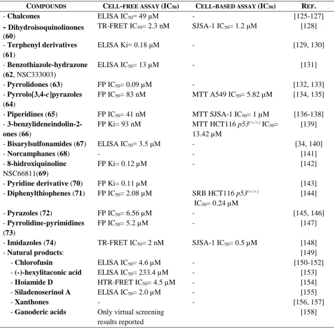
Table 1.1. Cell-free and cell-based in vitro assays. ... 7
Table 1.2. Other inhibitors or potential inhibitors of p53-MDM2 interaction... 26
Table 1.3. Therapy strategies when p53 is mutated in cancers. ... 36
Table 3.1. Cycloadditions attempts, using primary nitro compound 123a as precursors of nitrile oxides. ... 48
Table 3.2. Intermediates synthesized. ... 49
Table 3.3. Optimization attempts of the 1,3-dipolar cycloaddition reaction using clorooxime 121a ... 50
Table 3.4. Optimization of the 1,3-dipolar cycloaddition reaction using chlorooxime 123b. ... 51
Table 3.5. Cycloaddition reaction scope: Using different dipolarophiles with chlorooxime 123b. 51 Table 3.6. Cycloaddition reaction scope: Using three equivalents of chlorooxime and zinc. ... 52
Table 3.7. NMR chemical shifts (ppm) for representative spirooxindoles derivatives ... 54
Table 4.1. Synthesis of 3-methylene indolin-2-ones. ... 61
Table 4.2. Synthesis of aldoximes ... 62
Table 4.3. New spiroisoxazoline oxindole synthesized. ... 63
Table 4.4. Synthesis of compound 119q. ... 64
Table 4.5. In vitro antiproliferative activities in HepG2 cell line ... 65
Table 4.6. In vitro antiproliferative activities ... 66
Table 5.1. Cycloaddition reaction between 121n and 123e... 76
Table 5.2. Cycloaddition reaction between 121k and 123e... 77
Table 5.3. Cycloaddition reaction between 121k and 123e... 78
Table 5.4. Cycloaddition reaction between 121k and 123e... 79
Table 5.5. In vitro antiproliferative activities ... 80
Table 5.6. Synthesis of enantioenriched spiropyrroline epi-163. ... 82
Table 6.1. Synthesis of 3-imino-indolin-2-ones. ... 88
Table 6.2. Synthesis of hydroximoyl chlorides. ... 89
Table 6.3. In vitro antiproliferative activities in HCT116 p53(+/+) cell line. ... 91
Table 6.4. In vitro antiproliferative activities in HCT116 p53(–/–), HepG2 and SW620 cell lines ... 92
Table 7.1. Synthesis of hydrazones. ... 101
Table 7.2. Synthesis of hydrazonyl chlorides. ... 102
Table 7.3. Spiropyrazoline oxindoles reported in literature ... 105
Table 7.4. In vitro antiproliferative activities. ... 106
Scheme Index
Scheme 2.1. Different spirooxindole scaffolds synthesized during the PhD. ... 42
Scheme 3.1. Retrosynthesis of spiroisoxazoline oxindoles derivatives highlighting the final step between 3-methylene indolin-2-ones (121) and chlorooximes (123). ... 47
Scheme 3.2. Synthesis of ester 3-methylene indolin-2-ones. ... 49
Scheme 3.3. Synthesis of chlorooximes. ... 49
Scheme 3.4. Possible regioisomers formed as a result of 1,3-dipolar cycloaddition between 121a and 120a. ... 53
Scheme 4.1. Synthesis of 3-methylene indolin-2-one. ... 61
Scheme 4.2. Synthesis of aldoximes ... 62
Scheme 4.3. Synthesis of spiroisoxazoline oxindoles ... 63
Scheme 5.1. Example of chiral Lewis acid-catalysed cycloaddion reaction with nitrile oxides (Sibi et al). ... 74
Scheme 5.2. Synthesis of alkylidene oxindole 121n. ... 74
Scheme 5.3. Asymmetric Synthesis of Spiroisoxazoline oxindoles by Liang et al. ... 80
Scheme 5.4. Titanium-catalysed stereoselective synthesis of spirooxazoline oxindole 162 ... 81
Scheme 5.5. Titanium-catalysed stereoselective synthesis of spiropyrazole oxindole 163 ... 81
Scheme 5.6. Synthesis of 3-methylene indolin-2-one 121p ... 81
Scheme 5.7. Proposed mechanism for Lewis acid catalyzed formation of 163... 83
Scheme 6.1. Optimization Strategy ... 87
Scheme 6.2. Retrosynthesis of spirooxadiazoline oxindoles derivatives, highlighting the final step between 3-imino indolin-2-ones (165) and chlorooximes (123) ... 87
Scheme 6.3. Synthesis of 3-imino-indolin-2-ones ... 88
Scheme 6.4. Synthesis of hydroximoyl chlorides ... 89
Scheme 6.5. Synthesis of spirooxadiazoline oxindoles. ... 89
Scheme 6.6. Possible regioisomers formed as a result of 1,3-dipolar cycloaddition between 166 and 120. ... 90
Scheme 7.1. Optimization strategy first from spiroisoxazoline to spiropyrazoline (A) and then to spirotriazoline (B) oxindoles. ... 99
Scheme 7.2. Retrosynthesis of spirotriazoline oxindoles derivatives, highlighting the final between imino indolin-2-ones (166) and nitrile imines (169). ... 100
Scheme 7.3. Synthesis of 3-imino-indoline-2-one ... 101
Scheme 7.4. Synthesis of hydrazones ... 101
Scheme 7.5. Synthesis of hydrazonyl chlorides ... 102
Scheme 7.6. Synthesis of spirotriazoline oxindoles ... 102
Scheme 7.7. Possible regioisomers formed as a result of 1,3-dipolar cycloaddition between 166 and 169. ... 103
Acknowledgments
First of all I would like to thank Fundação para a Ciência e a Tecnologia for financial support (SFRH/BD/69258/2010) and the three supervisors that embarked in this journey with me: Dr. Maria M. M. Santos, Professor Rui Moreira and Professor Cecília M. P. Rodrigues.
I would like to thank the Liquid Chromatography and Mass Spectrometry Laboratory of the Faculty of Pharmacy of Lisbon University, especially João Fereira for the elemental analyses and LRMS; and the FT-ICR and Advanced Proteomics Laboratory from Faculty of Sciences of Lisbon University for HRMS analyses.
I also acknowledge Sara Silva (iMed.ULisboa) and Dr. Rita Guedes (iMed.ULisboa) for the docking studies.
I would like to thank Lídia Gonçalves (iMed.ULisboa) for the MTT antiproliferative assays and for all the know how behind biological assays transmitted.
I also thank Dr Joana Amaral for all the help in the biology department, especially for performing the western blotting assays. I want to extend this acknowledgement to all the colleagues from the Cellular Function and Therapeutic Targeting group (iMed.ULisboa) that help me in all the logistics that biology assays entail.
I would like to thank Dr. Annaliese K. Franz for accepting me in her lab in University of California – Davis for a period of three months. I want to thank all Franz group, and especially Joseph Badillo and Nicholas Ball-Jones, for all the help in the lab, and for open my mind on how to approach science. And also for remind me once again that Portuguese hospitality is overrated.
I would like to thank the entire medicinal chemistry group (from 2011-2015) for all the help, the stories, the camaraderie, and good times. I want to special acknowledge the post docs that are in reality the lab supervisors.
Since the PhD work represents most of the time period that I lived in Lisbon, I want to especially acknowledge the people that disrupted the coworker bubble and arose into friendship domain outside Faculty. Therefore, I want to especially mention two folks that are going to stay with me for a very long time: Catarina Charneira and Marta Figueiras; the former for representing the exception of what I think about people that live bellow Douro River and the latter for corroborating that there is something special about people from the North. In addition I want to thank my tango partner Mariana “Marisol” Reis for all the adventures, Marta Carrasco for turning out to be a very good surprise, the newbie Sara
Silva, Ana Rita “pompetinha” Duarte for all the songs (or not!), and Ana da Ress for all the scientific and life discussions throughout the years.
Outside Lisbon bubble I want to acknowledge my longtime friends (you know who you are!), with a special shout out to Isabel Duarte that represents what I think a scientist should aspired to be.
Finalmente quero agradecer à minha família, em particular ao Guedes e à Rosinha, pelo apoio incondicional, mesmo quando as coisas deixaram de correr como deviam. Um muito obrigado!
A
BBREVIATIONS
1-NMI 1-Methylimidazole
5-HT6 5-hydroxytryptamine receptor 6
A549 Human lung carcinoma cell line
ARF (or p14ARF) Alternate reading frame protein product of the CDKN2A locus
ATM Ataxia-telangiectasia mutated
ATR Ataxia telangiectasia and RAD3-related
BDP 1,4-benzodiazepine-2,5-dione
BiFC Bimolecular fluorescence complementation assay
Boc tert-Butyloxycarbonyl
Box Bisoxazoline
BrdU Bromo-2’-deoxyuridine
casp-3 caspase 3
CCK-8 Cell Counting Kit-8
CDK cyclin-dependent kinase
CHK1 Checkpoint kinase 1
CHK2 Checkpoint kinase 2
CK2 casein kinase 2
Compds Compounds
CRL Cullin-RING ubiquitin E3 ligase
CRM1 Chromosomal maintenance 1 D. A. Dihydrochlorinating agent DABCO 1,4-diazabicyclo[2.2.2]octane DCE Dichloroethane DCM Dichloromehane Del Deleted DIEA N,N-Diisopropylethylamine DMAP 4-Dimethylaminopyridine DMF Dimethylformamide
DMSO Dimethyl sulfoxide
DNA Deoxyribonucleic acid
dr Diasteriomeric ratio
E2F1 E2F transcription factor 1
EdU 5-Ethynyl-2’-deoxyuridine
ee Enantiomeric excess
ELISA Enzyme-linked immunosorbent assay
er Enantiomeric ratio
ESI Electrospray Ionization
Et Ethyl
EtOAc Ethyl Acetate
FACT Facilitates chromatin transcription
FP Fluorescence polarization
Fro Human anaplastic thyroid carcinoma cell line G1 phase Growth 1/Gap 1 phase
GC Guanine-cytosine
GI50 Concentration for 50% of maximal inhibition of cell proliferation
Gln Glutamine
HCT116 p53(-/-) Human colorectal cancer cell line with null p53 HCT116 p53(+/+) Human colorectal cancer cell line with wild-type p53
HDAC Histone deacetylase
HDACi Histone deacetylase inhibitor
HIC-1 Hypermethylated in cancer 1
His Histidine
HPLC High-performance liquid chromatography HRMS High-resolution mass stectrometry
HSP90 Heat shock protein 90
HTRF Homogeneous time resolved fluorescence
HTS High-throughput screening
Hz Hertz
hNav1.7 Voltage-gated sodium channel
IC50 Concentration for 50% of maximal inhibition
Ile Isoleucin
ipr Isopropyl
IR Infra-red
JAR Human choriocarcinoma cell line
Kat-4 Human thyroid tumor cell line
Ki Inhibition constant
LCVA Luminescent cell viability assay
Leu Leucine
LNCaP Human prostatic adenocarcinoma cell line
Lys Lysine
LRMS Low resolution mass spectrometry
M phase Mitotic phase
M. S. Molecular sieves
M.W. Microwave
MCF-7 Human breast adenocarcinoma cell line MDA-MB-231 Human breast adenocarcinoma cell line
MDM2 Murine double minute-2
MDM4 Murine double minute-4
Me Methyl
MHM Human osteosarcoma cell line
mp Melting point
mRNA Messenger ribonucleic acid
MTS (3-(4,5-dimethylthiazol-2-yl)-5-(3-carboxymethoxyphenyl)-2-(4-sulfophenyl)-2H-tetrazolium)
MTT 3-(4,5-dimethylthiazol-2-yl)-2,5-diphenyltetrazolium bromide
Mut Mutated
NaBArF sodium tetrakis[3,5-bis(trifluoromethyl)-phenyl]borate NADPH Nicotinamide adenine dinucleotide phosphate
NAE NEDD8-activating enzyme
NCS N-Chlorosuccinimide
NF-κB Nuclear factor kappa-light-chain-enhancer of activated B cells
NMR Nuclear magnetic resonance
NMR-AIDA NMR-based antagonist induced dissociation assay
NOE Nuclear Overhauser Effect
Otf Triflate
PARP poly ADP ribose polymerase
PDB Protein data bank
Ph Phenyl
Phe Phenylalanine
PLK-4 Serine/threonine-protein kinase 4 also known as polo-like kinase 4
PMP para-methoxyphenyl
PPA Phenyl phosphinic acid
py Pyridine
Pybox bis(oxazolinyl)pyridine
R,R-TUC
3,5-Bis(trifluoromethyl)phenyl]-3-[(1R,2R)-(-)-2-(dimethylamino)cyclohexyl]thiourea
RITA Reactivation of p53 and induction of tumor cell apoptosis
RNA Ribonucleic acid
rRNA Ribosomal ribonucleic acid
r.t. Room temperature
S phase Synthesis phase
S. M. Starting material
S100B S100 calcium-binding protein B SAR Structure-activity relationship
SEM Standard Error of the Mean
Ser Serine
SI Selectivity index
SINE Selective inhibitor of nuclear export
SIRT Sirtuin
SJSA-1 Human osteosarcoma cell line
SPR Surface plasmon resonance
SRB Sulforhodamine B
TDZ 1,4 thienodiazepine-2,5-diones
TEA Triethylamine
temp. Temperature
ThermoFluor Thermal denaturation screening assay
THF Tetrahydrofuran
TLC Thin layer chromatography
TR-FRET Time-resolved fluorescence energy transfer
Trp Tryptophan
U-2OS Human osteosarcoma cell line U937 Human lung lymphoblast cell line Weri1 Retinoblastoma cell line
WST-8 Water soluble tetrazolium-8: 2-(2-methoxy-4-nitrophenyl)-3-(4-nitrophenyl)-5-(2,4-disulfophenyl)-2H-tetrazolium
Wt Wild-type
A
BSTRACT
Among the tumor suppressor genes, p53 is one of the most studied. It is widely regarded as the “guardian of the genome”, playing a pivotal part in the preservation of genomic integrity by regulating cell cycle, apoptosis, DNA repair, senescence and angiogenesis, and consequently has a major role in carcinogenesis. The function played by p53 in tumor suppression is further highlighted by the fact that direct inactivation of this gene occurs in more than 50% of malignancies. In addition, in tumors that retain wild type p53 status, its function is usually inactivated by overexpression of negative regulators, primarily murine double minute-2 (MDM2), mainly through MDM2 gene amplification or by activity loss of MDM2 inhibitor ARF. Hence, restoring p53 function in cancer cells represents a valuable anticancer approach. Several strategies are being developed, and in particular targeting p53-MDM2 interaction has emerged as a promising viable approach when dealing with cancers that retain wild type p53 function. These two proteins regulate each other through an autoregulatory feedback loop: activation of p53 stimulates the transcription of MDM2, which in turn binds to the N-terminal transactivation domain of p53, disabling its transcriptional function. p53-MDM2 interaction inhibitors share common structural features: a rigid heterocyclic scaffold with three lipophilic groups that mimic the three pivotal p53 Phe19, Trp23 and Leu26 that interact with MDM2. Seven compounds have already entered clinical trials.
The main goal of this PhD thesis was to develop new anticancer agents containing a spirooxindole scaffold with different spiro five-membered rings: isoxazoline, oxadiazoline, and triazolines. The spirocycle can potentially function as the rigid heterocyclic scaffold, from which three lipophilic groups are projected to mimic the three pivotal p53 amino acids.
This work followed three major strategies: synthesis of spirooxindole derivatives by 1,3 dipolar cycloaddition; biological evaluation of the compounds synthesized; and stability assessment.
Overall this PhD thesis contributed with three new families of spirooxindoles with in vitro anti-cancer activity. The most active derivative possessed a GI50 of 1.72 µM in HCT116 p53(+/+) cell line. Furthermore their ability to disrupt the interaction between p53 and MDM2 was confirmed by implementing a cell-base in vitro bimolecular fluorescence complementation assay (BiFC) and the apoptotic outcome verified by immunoblotting analysis and luminescent caspase 3/7 activity assay.
KEYWORDS: spirooxindoles, anticancer drugs, protein-protein interaction, p53,
R
ESUMO
A proteína p53 foi descoberta há mais de 35 anos e é actualmente um dos mais estudados supressores tumorais, sendo designada até por “guardião do genoma”. A p53 desempenha um papel crucial na célula: regulação do ciclo celular, apoptose, reparação do DNA, senescência e angiogénese e, consequentemente, tem um papel decisivo na carcinogénese. Este papel central da p53 como supressor tumoral é incontestável, como observado no aumento da predisposição para o desenvolvimento de tumores em indivíduos com a síndrome Li-Fraumeni, caracterizados por serem detentores de uma mutação no gene da p53 (TP53) e em ratos sem o gene que codifica esta proteína (Trp-null). Actualmente acredita-se que em virtualmente todos os cancros se verifica algum tipo de perda da função supressora tumoral da p53. Em cerca de 50 % dos tumores, esta perda é directa, isto é, é devido à presença de uma mutação no gene Tp53 ou ocorre indirectamente, por inactivação das vias de sinalização celular da p53. Em tumores que retêm o fenótipo selvagem da p53, normalmente ocorre aumento da expressão de reguladores negativos da p53, como por exemplo a MDM2 e MDM4.
Em condições normais, os níveis de expressão da p53 são mantidos em valores baixos. Contudo em resposta a sinais de stress, como por exemplo a danos genotóxicos, a p53 é estabilizada e activada por meio de modificações pós-tradução. Como esta proteína é um factor de transcrição, a sua activação levará a um aumento da expressão de determinados genes-alvo que culminará numa resposta apropriada, incluindo prevenção da tumorigénese. Em contraste a inactivação da função celular da p53 pode levar à proliferação de células danificadas, podendo resultar no aparecimento e desenvolvimento de tumores.
Devido à importância da inactivação da via da p53 para o desenvolvimento tumoral, conceber estratégias que visam a reactivação desta proteína é de grande interesse, especialmente se permitirem um efeito apoptótico selectivo em células cancerígenas. Estas estratégias podem ser agrupadas em duas categorias: direccionadas para cancros detentores de uma versão mutada da p53 ou para cancros que conservam a sua forma nativa. No primeiro grupo, o objectivo será focado em estratégias que permitam o correcto enrolamento da p53 mutada com o intuito de restaurar a sua função. No segundo grupo, o objectivo é inibir a função de reguladores negativos da p53. A segunda categoria compreende diversas estratégias, incluindo: inibidores das sirtuínas, inibidores da S100B, inibidores da actividade E2 ligase da MDM2, inibidores da proteína MDM4, compostos que promovem a disrupção do nucléolo, agentes intercalantes e inibidores da exportação nuclear de proteínas. Contudo, a estratégia que tem suscitado mais interesse nos últimos 10 anos corresponde à inibição da interacção
entre a p53 e a MDM2. Estas duas proteínas auto-regulam-se: a activação da p53 estimula a transcrição da MDM2 e esta proteína, por sua vez, liga-se ao terminal N da p53 desactivando a sua função transcricional. A proteína MDM2 também promove a exportação da p53 do núcleo e favorece a sua degradação mediada pelo proteasoma, através da função de E3 ubiquitina ligase. Estes eventos conduzem a uma diminuição dos níveis de p53 que, por sua vez, promovem uma diminuição da expressão da MDM2, permitindo assim que a proteína p53 possa ser novamente activada. A estrutura cristalográfica da p53 ligada à MDM2 revelou que esta interacção ocorre numa pequena fenda hidrofóbica superficial na proteína MDM2. Os resíduos mais importantes da p53 que contribuem para esta interacção são a Phe19, o Trp23 e a Leu26. Portanto, quando se desenham inibidores da interacção p53-MDM2 é necessário que estes sejam capazes de mimetizar os três aminoácidos da p53 supracitados. Diversas famílias de compostos já foram descritas na literatura capazes de inibir esta interacção, sendo as mais importantes: cis-imidazolinas, spiropirrolidina oxindoles, pirrolina-2-carboxamidas, piperidinonas, benzodiazepinedionas e isoindolinonas. Sete compostos inibidores da interacção p53-MDM2 encontram-se em ensaios clínicos.
Compostos com um núcleo spirooxindole são encontrados em produtos naturais e apresentam diversas actividades biológicas. Em particular, já se encontram descritos na literatura compostos spiropirrolidina oxindoles com diversas actividades cancerígenas, como por exemplo inibidores da polimerização dos microtúbulos e, consequente, paragem da mitose e inibidores da interacção p53-MDM2. Deste segundo grupo, o composto MI-77301 encontra-se em ensaios clínicos.
O objectivo principal desta tese de doutoramento consistiu em desenvolver novos agentes anticancerígenos detentores de um esqueleto spirooxindole fundido com diferentes anéis de cinco membros: isoxazoline, oxadiazolina e triazolina. Diferentes grupos essencialmente hidrofóbicos foram introduzidos no esqueleto central, numa tentativa de encontrar aqueles que melhor mimetizam os três aminoácidos da p53. Para cada uma destas famílias foram descritos os métodos sintéticos envolvidos e a respectiva avaliação biológica in vitro dos derivados sintetizados. Os compostos foram sintetizados por cicloadição 1,3-dipolar entre 3-imino ou 3-metileno indolina-2-onas e dipolos 1,3 gerados in situ por desidrohalogenação de cloretos de hidroximoílo e cloretos de hidrazonoílo. Para o primeiro núcleo sintetizado – spiroisoxazoline oxindoles – são ainda descritas tentativas de síntese enanteosselectiva do composto mais activo. Esta parte do trabalho foi realizada num período de três meses no grupo da Dr. Annaliese K. Franz na Universidade da Califórnia – Davis (EUA). Em paralelo, neste período na Califórnia, foi também desenvolvida uma metodologia de síntese enanteosselectiva de um derivado spiropirrolina oxindole.
Os estudos in vitro permitiram avaliar e demonstrar a potencialidade dos compostos como agentes anticancerígenos, através da realização de ensaios anti-proliferativos em diversas linhas celulares cancerígenas: par isogénico HCT116 p53(+/+) e p53(-/-) de cancro colorrectal humano, carcinoma hepatocelular humano HepG2, adenocarcinoma colorrectal humano SW620, cancros da mama MCF-7 e MDA-MB-231 e uma linha celular epitelial embrionária humana de rim. A capacidade dos compostos interferirem com a interacção entre a p53 e a MDM2 foi também avaliada e confirmada in vitro através da implementação de um método de fluorescência de complementação biomolecular (BiFC) realizado em células. A capacidade dos compostos induzirem a apoptose foi avaliada por ensaios de Western blotting e luminescência, onde se verificou um aumento da actividade das caspases e um aumento da clivagem do seu substrato PARP.
Por último, foram efectuados ensaios de estabilidade química em tampão fosfato pH 7.4, em plasma e em microssomas de rato, para dois compostos de cada família. Estes ensaios servirão para ajudar na escolha dos compostos a seguir para ensaios in vivo numa futura continuação do trabalho.
Foram sintetizados trinta e três compostos da primeira família (spiroisoxazoline oxindoles). O derivado mais activo possui um GI50 de 26.50 µM em HCT116 p53(+/+). Com a segunda família de compostos (spirooxadiazoline oxindoles) conseguiu-se aumentar a actividade para 1.72 µM, na mesma linha celular. Nesta série de compostos foram sintetizados trinta e dois derivados, dos quais nove revelaram possuir um GI50 inferior a 10 µM. Na terceira família sintetizada (spirotriazolina oxindoles) foi possível encontrar cinco derivados com GI50 abaixo dos 10 µM na linha celular MCF-7 (p53 fenótipo selvagem). Curiosamente outros cinco derivados revelaram ser selectivos para a linha celular tumoral MDA-MD-231 (p53 mutada). Desta série foram sintetizados vinte e sete derivados. Estudos preliminares de docking molecular corroboraram a possibilidade destes compostos conseguirem mimetizar os três resíduos da p53.
PALAVRAS-CHAVE: spirooxindoles, compostos anticancerígenos, interacção
Chapter
1
Tumor suppressor p53 was discovered over 35 years ago and since then it emerged as a key protein of scientific interest, and became widely regarded as the “guardian of the genome” [1]. p53 plays a pivotal role in the regulation of cell cycle, apoptosis, DNA repair, senescence and angiogenesis, and consequently has a major function in carcinogenesis. The central role of p53 as tumor suppressor is undeniable, as observed by the increased predisposition to cancer in individuals with Li-Fraumeni syndrome, who inherit a mutant p53 gene TP53, and in Trp-null mice [2]. Additionally, it is now believed that in virtually all cancers, loss of p53 function occurs, either directly due to the presence of a mutated form of TP53, or by inactivation of the p53 signal transduction pathways. In tumors that retain wild type p53 status, corresponding to 50% of all cancers, its function is usually inactivated by overexpression of negative regulators, primarily murine double minute-2 (MDM2) and MDM4 (also known as MDMX) [3].
1.1. P53 ACTIVATION IN HEALTH AND DISEASE
The cellular levels of the p53 protein are tightly regulated. In normal cells, and under physiological conditions, steady-state values of p53 are maintained at very low levels by its negative regulators, mainly MDM2 and MDM4. However, under cellular stress, such as DNA damage, hypoxia or oncogene activation, a range of differential posttranslational modifications of p53 are triggered that lead to p53 stabilization and activation, by promoting its release from repression and by inhibiting its degradation. For instance, upon acute DNA damage, p53 stabilization is mostly achieved by phosphorylation mediated by upstream kinases such as ATM/ATR and/or CHK1/CHK2 (Figure 1.1). Activated p53 binds to DNA and promotes the transcription of several target genes, culminating in a proper cellular response that is dictated by the nature of the stress, cell type and environment milieu. Under low levels of stress, p53 induces a transient G1 cell cycle arrest, while cells attempt to repair their genome. However, if the damage is too severe, activation of the p53 pathway results in cell death by apoptosis or senescence. By contrast, loss of p53 tumor suppressor activity allows the proliferation of cells that are damaged under stress conditions, potentially leading to uncontrolled proliferation that can result in tumor development [4-6].
Canonical p53 responses that lead to cellular functions of cell cycle arrest, senescence and apoptosis are extensively studied specially when triggered upon acute DNA damage. However, recently more attention is given to understanding p53 signaling in a tumor context, since distinct stresses and different responses that can facilitate/trigger tumor suppression have been described. These interesting p53 responses include: inhibition of oncogenic metabolic reprogramming, activation of autophagy, communication endorsement within the tumor microenvironment, inhibition of stem cell self-renewal and
differentiated cells reprogramming into stem cells, and limiting invasion and metastasis [2, 7].
Figure 1.1. Simplified p53 activation and response upon acute DNA damage (adapted from [2]).
1.2.REACTIVATION OF P53 AS A THERAPEUTIC STRATEGY
It is well documented that the loss of p53 can induce tumor formation in mice, whereas its restoration leads universally to a rapid regression of established in situ tumors, showcasing the anticancer therapeutic potential of p53 reactivation. Furthermore, the key question for p53 reactivation strategy is whether or not this event will result in a selective effect on tumor cells as opposed to healthy tissues. It seems that a simple overexpression of p53 in cells is not sufficient to activate the p53 pathway. The restored p53 protein needs to be properly activated and for that the transformed environment of tumor cells appears to be required [8, 9]. For instance, studies using p53-MDM2 interaction inhibitors showed that in fact, in normal cells, the activation of p53 induces preferentially cell cycle arrest and not cell death, revealing therefore a more selective apoptotic effect on tumor cells. The effect of p53 activation by this type of inhibitor in normal tissues has immense interest from a therapeutic perspective due to the possibility of using it in monotherapy, as well as a normal cells protector in combination with more aggressive agents [10, 11].
Throughout the last ten years, great advance was made in devising strategies to modulate p53, giving rise to several review papers on the subject [3, 11-24]. Pharmacological p53 reactivation strategies for cancer therapy can be clustered in two major approaches based on p53 status. In tumors that retain wild-type p53 but have defects in p53 regulatory pathways, the main goal is to inhibit the function of negative regulators of p53 activation outcome. When p53 is mutated in tumors, the most common strategy consists in refolding the protein into a wild-type conformation to restore its function. In
this chapter, it will be given emphasis to small-molecules that use the former strategy and in particular to the interaction between p53 and its inhibitor MDM2. However other strategies are also being pursued to restore p53 function in cancer cells such as using peptides, stapled peptides and other oligomers as inhibitors on p53-MDM2/X interaction [20] and adenovirus-mediated p53 cancer gene therapy [25].
1.2.1.TARGETING P53-MDM2INTERACTION
Increased levels of p53 repressor MDM2 are present in many cancers, mainly through MDM2 gene amplification or by activity loss of MDM2 inhibitor ARF. Therefore, targeting the p53-MDM2 interaction to reactivate p53 has emerged as a promising new cancer therapeutic strategy, in the last fifteen years or so, with already in vitro an in vivo established proof-of-concept [10, 26-45]. These two proteins regulate each other through an autoregulatory feedback loop [46]. Activation of p53 stimulates the transcription of MDM2, which in turn binds to the N-terminal transactivation domain of p53, disabling its transcriptional function. MDM2 also promotes the nuclear export of p53 and p53 proteasome-mediated degradation through its E3 ubiquitin ligase activity by promoting mono and polyubiquitination, respectively, at several lysine residues (Figure 1.2). These events result in decreased levels of p53 that will therefore reduce MDM2 expression, allowing p53 protein to potentially be activated again [44, 47].
Figure 1.2. Cellular regulation of p53 by MDM2 (adapted from [39]).
The crystal structure of the p53 binding domain of MDM2 (109-residue amino-terminal) with a short peptide of the p53 transactivaction domain (15-residues) has been solved and published, providing detailed information about the interaction between these two proteins [48]. The co-crystal revealed that MDM2 has a deep hydrophobic cleft on which the p53 peptide binds as an amphipathic alpha helix. In the bound conformation, the p53 amphipathic α-residues 19-26 of the transactivation domain projects residues Phe19,
Trp23 and Leu26 into the deep hydrophobic cleft of the MDM2 protein, representing the critical residues for binding between this two proteins to occur (Figure 1.3). In the crystal structure, Phe19 and Trp23 align in the deeper portion of the cleft. Phe19, through its backbone amine, forms one hydrogen bond with the backbone carbonyl Gln72 at the entrance of the cleft, while establishing hydrophobic interactions with Gly58 and Ile61 of MDM2. Trp23 occupies the deepest part of the binging pocket, forming a solvent protected hydrogen bond between the NH from its indole side chain and Leu54 of MDM2, and makes hydrophobic interactions with Gly58 and Ile61 of MDM2. Leu26 is the final residue of the alpha helix to be projected into the hydrophobic pocket. Furthermore, the interaction is strengthened by additional van der Waals contacts provided by p53 Leu22 [39, 48].
Figure 1.3. The p53-MDM2 interaction representation (PDB 1YCR). Phe19, Trp23 and Leu26 from a small
amphipathic p53 derived α-helix (blue) are projected into the MDM2 pocket (grey surface).
After publication of the crystal structure of p53 bound to MDM2, several efforts were made to design more potent peptide derivatives. The search for small molecules that could interfere with the protein-protein interaction only flourished after the publication of a co-crystal structure of a small-molecule in MDM2 pocket. To date more than 20 different chemical classes have been described as inhibitors of p53-MDM2 interaction. Recently, several molecules entered, and are still in, clinical trials, revealing the continuous relevance and efforts in finding new and improved derivatives/ scaffolds.
In this chapter, a detailed understanding of the medicinal chemistry and optimization approach is going to be given especially to scaffolds that provided molecules that entered clinical trials [45, 49]: cis-imidazolines, spiropyrrolidine oxindoles, pyrrolidine-2-carboxamides and piperidinones, as well as other relevant families, such as morpholinones, benzodiazepinediones, isoindolinones, chromenotriazolopyridines and imidazole-indoles.
To facilitate the comprehension of this section, it is depicted in table 1.1 all in vitro cell-free and cell-based methods used to determine the IC50s presented, as well as the cell lines employed and their p53 status. In general, the description will be given as follows for cell-based assays: “assay” “cell line” IC50= “value” (e.g. compound 3, MTT SJSA-1 IC50= 0.3 µM, Figure 1.4).
Table 1.1. Cell-free and cell-based in vitro assays.
CELL-FREE BINDING ASSAYS:
SPR Surface plasmon resonance
HTRF Homogeneous time resolved fluorescence FP Fluorescence polarization
NMR-AIDA NMR-based antagonist induced dissociation assay ThermoFluor Thermal denaturation screening assay
TR-FRET Time-resolved fluorescence energy transfer ELISA Enzyme-linked immunosorbent assay
CELL-BASED ASSAYS:
BrdU Bromo-2’-deoxyuridine EdU 5-Ethynyl-2’-deoxyuridine LCVA Luminescent cell viability assay MTT Tetrazolium salt
SRB Sulforhodamine B
WST-8 Water soluble tetrazolium salt
CELL LINES:
A549 Human lung carcinoma – wild-type p53 Fro Human anaplastic thyroid carcinoma – null p53 HCT116 p53(+/+) Human colorectal cancer – wild-type p53
JAR Human choriocarcinoma – wild-type p53 Kat-4 Human thyroid tumor – mutant p53
LNCaP Human prostatic adenocarcinoma – wild-type p53 MCF-7 Human breast adenocarcinoma – wild-type p53 MDA-MB-231 Human breast adenocarcinoma – mutant p53 MHM Human osteosarcoma – wild-type p53 SJSA-1 Human osteosarcoma – wild-type p53 U-2OS Human osteosarcoma – wild-type p53 U937 Human lung lymphoblast – wild-type p53
1.2.1.1.CIS-IMIDAZOLINES (NUTLINS).ROAD TO RG7112
The Nutlin scaffold, consisting of a tetrasubstituted imidazoline unit, was first discovered by high-throughput screening (HTS) of a diverse library of synthetic compounds, using a surface plasmon resonance (SPR) assay, followed by structure-based optimization (Hoffman-La Roche). Three compounds arose from this study in 2004. It also provided the first crystallographic structure published of a small-molecule (Nutlin-2: 1, Figure 1.4) in complex with MDM2 [50]. The para-bromophenyl ring at position 4 occupies Leu26(p53) pocket while the other para-bromophenyl substituent at position 5 inserts deeply into the Trp23(p53) pocket with the bromo atom enhancing the binding by filling a small cavity not normally occupied by the indole ring of p53 Trp23. The Phe19(p53) pocket is occupied by the ethyl ether side chain of the third aromatic ring while its para-methoxy group mimics the p53 Leu22. The N1 chain functions mainly as a “solubility-tag” but also contributes to activity by possibly establishing polar interactions between the hydroxyl group and Gln72 side chain [50, 51]. The most potent of these three compounds is the enantiopure Nutlin-3a (2, SPR IC50 of 0.09 µM, MTT IC50= 1 - 2 µM in wild-type p53 cancer cell lines). Nutlin-3a usage in monotherapy and in combination with other anti-cancer drugs and radiation has already been extensively published, serving as proof-of-concept for Nutlins and establishing p53-MDM2 interaction as a promising and valuable target [52-57].
Since biological and pharmacokinetic (PK) properties achieved by Nutlin-3a were still suboptimal for clinical development, further research was performed. The strategy mainly focused on probing different N1 side chains in an attempt to enhance PK properties and MDM2 binding and on removing stability liabilities found in the previous compounds: oxidation of the main core to imidazole, and metabolization of the para-methoxyphenyl group to phenol. They were amended by adding methyl groups to position 4 and 5 of the imidazoline ring, and by replacing the methoxy with tert-butyl group, respectively [58]. One of the best compounds, RG7112 (3, HTRF IC50= 18 nM, MTT IC50= 0.18 - 2.2 µM in wild-type p53 cancer cell lines) entered clinical trials [59]. RG7112 shows good selectivity over mutated p53 cancer cells (MTT IC50= 5.7 - 20.3 µM), and it is able to activate the p53 signaling pathway in wild-type p53 cells, leading to cell cycle arrest and apoptosis. Furthermore a daily dose of 100 mg/kg is capable of promoting partly regression of SJSA-1 and MHM tumor xenograft mice models [45, 60].
Hu et al published in 2011 [61] and 2012 [62] novel derivatives based on the imidazoline scaffold, mainly by varying the N1 side chain of Nutlin-3. Compound 4 (FP IC50= 0.59 µM, MTT HCT116 p53+/+ IC50= 3.73 µM, Figure 1.5) was one of the most potent compounds obtained, however not representing an improvement of potency when
compared with Nutlin-3a. Nevertheless, these publications helped once again to establish that changing N1 side chain interferes mainly with PK properties but also with potency.
Several analogs are disclosed in patents from Hoffman-La Roche, possessing the same imidazoline core and others structure variations such as imidazopyridinones (5) [29, 38, 63, 64]. Miyazaki et al also published a new series of dihydroimidazothiazole derivatives based on Nutlin-3a structure, such as DS-5272 (6, HTRF IC50= 2.4 µM, LCVA SJSA-1 IC50= 0.2 µM) [65, 66].
Figure 1.4. Nutlins optimization to RG7112. Right upper quadrant: crystal structure of compound 3 bound to
MDM2 (PDB 4IPF). MDM2 surface is colored in blue for hydrophilic areas and grey for hydrophobic areas. Compound 3 is depicted in stick model and is colored according to element type: white for carbon atoms, blue for nitrogen atoms, red for oxygen atoms, yellow for the sulfur atom, and green for chlorine atoms.
1.2.1.2.SPIROOXINDOLES
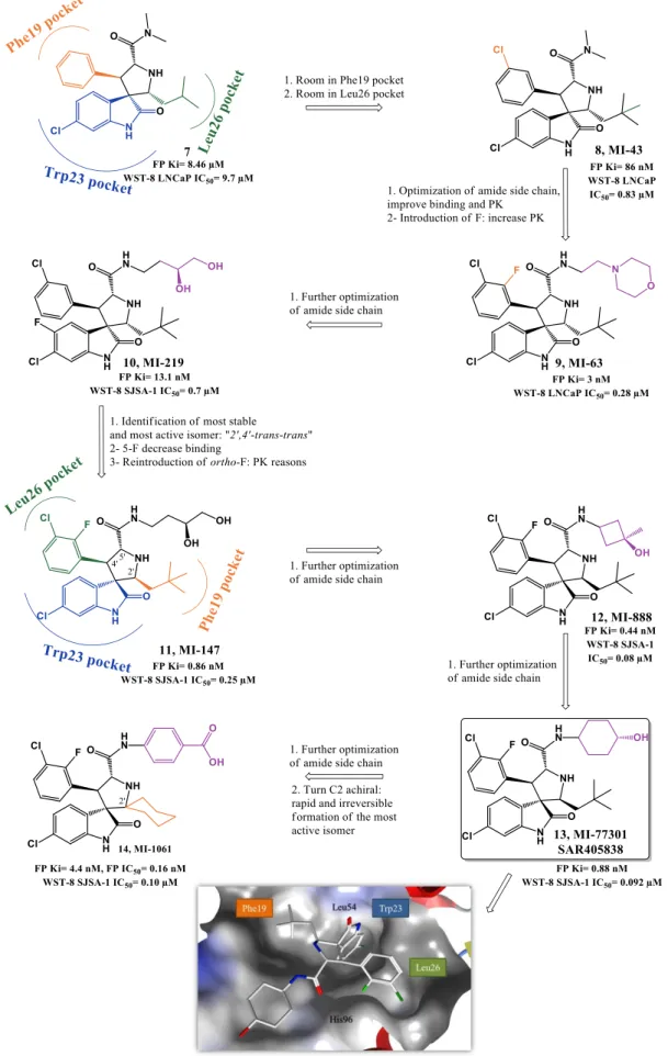
1.2.1.2a. Spiropyrrolidine oxindoles. Road to MI-77301
Taking in consideration that p53 Trp23 side chain (indole group) seems to be critical for p53-MDM2 interaction, by burring deeply inside p53 hydrophobic pocket and by establishing a hydrogen bond (NH) to MDM2 backbone (carbonyl), the oxindole moiety was believed to perfectly mimic this residue. Moreover since many natural anticancer products, such spirotryprostatin A and alstonisine, have a spirooxindole main core, it was rationalized and later corroborated by in silico studies that the spiropyrrolidine ring could provide the necessary rigidity to the spirooxindole scaffold, from which two extra hydrophobic groups could be projected in the required orientation to mimic the other two residues of p53. From this structure-based design an initial lead compound arose in 2005 (7, Figure 1.6) with a FP Ki of 8.46 µM (Wang group, University of Michigan). Computational modeling suggested that interaction optimization could be achieved by varying the isobutyl substituent and by introducing small substituents in the meta-position of the phenyl ring (room in Leu26(p53) and Phe19(p53) pocket respectively still available). Therefore, structure-activity relationship (SAR) studies led to MI-43 (8, FP Ki= 86 nM, WST-8 LNCaP IC50= 0.83 µM). This compound also showed good selectivity over normal cells and cancer cells with deleted p53 [67].
Further virtual investigation into the interaction of compound 8 and p53 with MDM2, suggested that a possible fourth residue (Leu22) could be mimicked. Leu22(p53) pocket is partially exposed to solvent and therefore mimicking this residue could result not only in an increase of potency but also in PK improvement, since it could allow the presence of polar groups. A second round of SAR studies was devised, having mainly this observation in consideration, leading to MI-63 (9) with a 2-morpholin-4-yl-ethylamine group. Docking studies revealed that not only this side chain could mimic Leu22 but the morpholine oxygen could possibly interact by H bond with Lys90 in MDM2 (mimicking p53 Glu17). Furthermore the introduction of a fluor atom in the phenyl group, a frequently effective strategy to increase metabolic stability, augmented the potency (FP Ki of 3 nM, WST-8 LNCaP IC50= 0.28 µM) [68, 69]. However, due to the fact that compound 9 had only a modest oral bioavailability, additional investigations, especially on the polar morpholinyl substituent were performed. It was found that changing to a butyl-1,2-diol significantly improved PK (MI-219: 10, FP Ki of 13.1 nM, WST-8 SJSA-1 IC50= 0.7 µM and MI-147: 11, FP Ki of 0.6 nM, WST-8 SJSA-1 IC50= 0.2 µM) [70]. Nevertheless these new derivatives still required high oral doses (200-300 mg/kg) to achieve tumor growth inhibition in mice xenograft models, and most important a complete tumor regression was still elusive [71]. More recently this last goal was attained with MI-888 (12, FP Ki of 0.44 nM, WST-8 SJSA-1 IC50= 0.08 µM) [72] and MI-77301/SAR405838 (13, FP Ki of 0.88
nM, WST-8 SJSA-1 IC50= 0.092 µM) [73]. These compounds were synthesized in a new series of optimizations that continued to focus on the 5’ pyrrolidine tail chain, especially by introducing conformational constrain, while attempting to increase metabolic stability [72].
Recently it was discovered that some of these spiropyrrolidine oxindoles can suffer reversible ring-opening and cyclization of the pyrrolidine ring in protic solution, giving rise to different stereoisomers with different binding affinities to MDM2 [74]. Therefore a second generation of spirooxindoles emerged in 2015 that possess symmetrical substituents at C2’ position of the pyrrolidine ring that allow a rapid and irreversible conversion to the most active diastereoisomer (MI-1061: 14, FP Ki of 0.16 nM, WST-8 SJSA-1 IC50= 0.10 µM) [75]. Compounds 12 and 13 from the first generation were already synthesized having in consideration the desired stereochemistry. Interestingly the best diastereomer revealed a different and better binding to MDM2 with the neopentyl and phenyl ring occupying now Phe19(p53) and Leu26(p53) pockets respectively (Figure 1.6, represented for compound 11). Furthermore their side chain carbonyl is capable of stablish an H bond with the imidazole side chain of His96 and terminal hydroxyl group with Lys94 side chain [73].
Compound 13 advanced into clinical trials in 2012 sponsored by Sanofi. It displays more than 100-fold selectivity over cell lines with mutated or deleted p53, activating a p53-dependent pathway leading to cell-cycle arrest and/or apoptosis in cancer cells in vitro and in vivo xenograft tumor models. A complete tumor regression was achieved at 100 mg/kg with a daily dose for 9 days and at 200 mg/kg with a single oral dose in SJSA-1 mice xenograft [73].
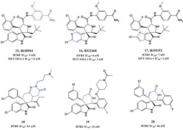
In 2014, Hoffmann-La Roche published two other papers describing further optimizations of spiro[oxindole-3,3’-pyrrolidines], having in consideration the beneficial PK and potency improvement obtained when a phenyl derivative group is attached to the amide side chain as in their p53-MDM2 inhibitor RG7388 (see section 1.2.1.3). RO8994 (15, HTRF IC50 of 5 nM, SJSA-1 IC50= 13 nM, Figure 1.7) emerged in a SAR study focused especially in additional modifications to this side chain [76, 77]. Compounds RO2468 (16, HTRF IC50 of 6 nM, MTT SJSA-1 IC50= 3 nM), and RO5353 (17, HTRF IC50 of 7 nM, MTT SJSA-1 IC50= 2 nM) were based on RO8994, and represent a bioisosteric substitution study of the 6-chlorooxindole moiety. Although with in vitro and in vivo activity comparable with RO8994, these compounds showed improve selectivity between wt p53 cell lines and mut p53 cell lines [78].
Several patents emerged from Hoffmann-La Roche throughout the last 10 years covering spiro[oxindole-3,3’-pyrrolidines] and others spiro-heterocyclic-oxindole based compounds (e.g. 18, 19 and 20) [38, 41, 64].
Figure 1.6. Spiropyrrolidines optimization to MI-106. Docking pose of compound 13 in MDM2 (PDB
3LBL). MDM2 surface is colored in blue for hydrophilic areas and grey for hydrophobic areas. Compound
13 is depicted in stick model and is colored according to element type: white for carbon atoms, blue for
Figure 1.7. Spiropyrrolidines and others spiro-heterocyclic-oxindole derivatives.
Kumar et al published this year a new family of spiro[oxindole-3,2’-pyrrolidines] [79]. They focused in breast cancer, providing good evidence that the best compound modulates p53 in vitro and in vivo. However, the compounds did not show selectivity between breast cancer cell lines with wt p53 (MCF-7) and mut p53 (MDA-MB-231), and although it was observed an increase in MDM2 levels, no studies were focused in the p53-MDM2 interaction (21, MTT MCF-7 IC50= 6.5 µM, Figure 1.8). Also this year, Ivanenkov et al reported dispiros with spiropyrrolidine oxindole moiety that can potentially interfere with p53-MDM2 interaction by in silico comparison with known MDM2 antagonists (22, MTT MCF-7 IC50= 4.88 µM) [80].
1.2.1.2b. Spirothiazolidine oxindoles
In 2010, Gomez-Monterrey et al synthesized a series of spiro[oxindole-3,3’-thiazolidines], with ISA27 emerging as the most potent derivative in cell lines (23, MTT U937 IC50= 0.87 µM, Figure 1.8). Destabilization of p53-MDM2 interaction by compound
23 was established first by NMR analysis (AIDA method) [81], and later complemented by
an in depth in vitro validation in human glioblastoma multiforme [82]. A second round of SAR studies was focused on opening ISA27 imidazole ring in an attempt to increase structural diversity and introduce extra potential binding points. Therefore, it could allow a better fitting to p53 pocket, since ISA27 most likely only mimic two of the three pivotal p53 amino acids [83]. SM13 (24, MTT MCF-7 IC50= 0.04 µM) emerged from this study.
Docking studies suggested that 3-cyclohexylcarboxy moiety occupies the Trp23(p53) pocket, and ethyl ester side chain the Phe19(p53) pocket. Leu26(p53) pocket is only slightly occupied by the oxindole ring, but the authors suggested that this drawback seems to be somewhat compensated by extra hydrophobic interactions gained through the N1-methyl group. In vitro inhibition of p53-MDM2 interaction was evaluated for both compounds using an ELISA binding assay. At 5 µM ISA27 (23) and SM13 (24) inhibited 25% and 30% of the interaction respectively (nutlin-3 inhibited 19%). However, both compounds were also effective in cancer cell lines with mutated p53. A detailed study to clarify the mechanism of action of SM13 suggested that besides inhibiting p53-MDM2 interaction, this compound acts in a manner strictly dependent on p53. No apoptosis induction was observed in FRO cells (null p53) and only activation of p53-dependent mitochondrial apoptotic pathway was observed in Kat-4 (mut p53) due to its lack of p53 transcriptional activity [84].
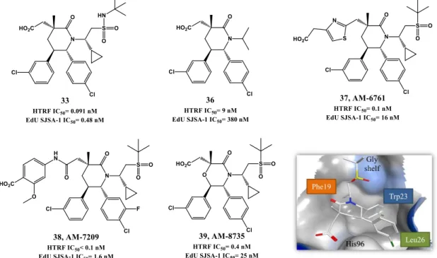
1.2.1.3. PYRROLIDINE-2-CARBOXAMIDES.ROAD TO RG7388.
RG7388 from Hoffman-La Roche (25, Figure 1.9) was design based on the spiropyrrolidine oxindole MI-219 structure (10) and on the notion that an aromatic moiety would be better to mimic Leu26 of p53. They reported that the presence of a nitrile group favored the necessary “trans-trans” configuration (between rings A and B, and ring A and neopentyl group, Figure 1.9) to obtain the proper configuration and the better interaction with MDM2. As referred previously, Wang group later also described that a different configuration for MI-219 was preferred to obtain more potent spiropyrrolidine oxindoles. Optimization to RG7388 was mainly focused on the amide side chain of compound 26 (HTRF IC50= 74 nM, MTT SJSA-1 IC50= 2.1 µM), with best PK properties and potency obtained with a methoxy para-benzoic acid moiety (25, HTRF IC50= 6 nM, MTT SJSA-1 IC50= 10 nM). Furthermore, the addition of fluor to both phenyl rings also contributed to increase binding to MDM2. RG7388 exhibits more than 100-fold selectivity over cell lines with mutated p53, and activates the p53 pathway. It promoted tumor regression at 25 mg/kg with daily doses in SJSA-1 mice xenograft [85, 86] and is currently in clinical trials.
Figure 1.9. Pyrrolidine-2-carboxamide optimization to RG7388. Right upper quadrant: crystal structure of
compound 26 bound to MDM2 (PDB 4JRG). MDM2 surface is colored in blue for hydrophilic areas and grey for hydrophobic areas. Compound 26 is depicted in stick model and is colored according to element type: white for carbon atoms, blue for nitrogen atoms, red for oxygen atoms, and green for chlorine atoms.
![Figure 1.2. Cellular regulation of p53 by MDM2 (adapted from [39]).](https://thumb-eu.123doks.com/thumbv2/123dok_br/19184165.946785/34.892.235.685.644.920/figure-cellular-regulation-p-mdm-adapted.webp)
![Figure 1.14. Isoindolinone scaffold optimization. Representations of binding modes were determined from chemical shift data and molecular docking [115, 116]](https://thumb-eu.123doks.com/thumbv2/123dok_br/19184165.946785/52.892.164.744.84.607/figure-isoindolinone-scaffold-optimization-representations-determined-chemical-molecular.webp)