Contents lists available atScienceDirect
Journal of Inorganic Biochemistry
journal homepage:www.elsevier.com/locate/jinorgbioInhibition of Na
+
/K
+
- and Ca
2+
-ATPase activities by
phosphotetradecavanadate
Gil Fraqueza
a,b,1, Juan Fuentes
b,1, Lukáš Krivosudský
c,d, Saikat Dutta
e, Sib Sankar Mal
e,⁎,
Alexander Roller
f, Gerald Giester
g, Annette Rompel
c,⁎, Manuel Aureliano
b,h,⁎⁎aISE, University of Algarve, 8005-139 Faro, Portugal bCCMar, University of Algarve, 8005-139 Faro, Portugal
cUniversität Wien, Fakultät für Chemie, Institut für Biophysikalische Chemie, Althanstr. 14, 1090 Wien, Austria
dComenius University, Faculty of Natural Sciences, Department of Inorganic Chemistry, Mlynská dolina, Ilkovičova 6, 842 15 Bratislava, Slovakia eDepartment of Chemistry, National Institute of Technology Karnataka, Mangalore 575025, Karnataka, India
fUniversität Wien, Fakultät für Chemie, Zentrum für Röntgenstrukturanalyse, 1090 Wien, Austria
gUniversität Wien, Fakultät für Geowissenschaften, Geographie und Astronomie, Institut für Mineralogie und Kristallographie, 1090 Wien, Austria hFCT, University of Algarve, 8005-139 Faro, Portugal
A R T I C L E I N F O Keywords: Polyoxometalates Phosphotetradecavanadate Decavanadate P-type ATPases
Epithelial chloride secretion
A B S T R A C T
Polyoxometalates (POMs) are promising inorganic inhibitors for P-type ATPases. The experimental models used to study the effects of POMs on these ATPases are usually in vitro models using vesicles from several membrane sources. Very recently, some polyoxotungstates, such as the Dawson anion [P2W18O62]6−, were shown to be potent P-type ATPase inhibitors; being active in vitro as well as in ex-vivo. In the present study we broaden the spectrum of highly active inhibitors of Na+/K+-ATPase from basal membrane of epithelial skin to the bi-capped Keggin-type anion phosphotetradecavanadate Cs5.6H3.4PV14O42(PV14) and we confront the data with activity of other commonly encountered polyoxovanadates, decavanadate (V10) and monovanadate (V1). The X-ray crystal structure of PV14was solved and contains two trans-bicapped α-Keggin anions HxPV14O42(9-x)-. The anion is built up from the classical Keggin structure [(PO4)@(V12O36)] capped by two [VO] units. PV14(10 μM) exhibited higher ex-vivo inhibitory effect on Na+/K+-ATPase (78%) than was observed at the same concentrations of V
10 (66%) or V1(33%). Moreover, PV14is also a potent in vitro inhibitor of the Ca2+-ATPase activity (IC505 μM) exhibiting stronger inhibition than the previously reported activities for V10(15 μM) and V1(80 μM). Putting it all together, when compared both P-typye ATPases it is suggested that PV14exibited a high potential to act as an
in vivo inhibitor of the Na+/K+-ATPase associated with chloride secretion.
1. Introduction
Polyoxometalates (POMs) are inorganic anionic metal oxide clusters exhibiting a broad diversity of structures and outstanding properties leading to their application in many fields [1–5]. The usage of biolo-gically active POMs for medical purposes is continuously increasing and thus attracting more and more attention from scientist coming from medical- and biology-related research areas [5–8]. P-type ATPases constitute a large family of ion pumps, which are found in all kingdoms of life and are responsible for many biologically essential processes assigning them important roles in health and diseases [9–12]. There-fore, P-type ATPases represent important pharmacological targets being
reflected by the substantial number of drugs targeting these ion pumps [11].
Forty years have passed since the discovery that the muscle in-hibitor factor (MIF) and the Na+/K+-ATPase inhibitor (present in commercial ATP) contained vanadium in the +V oxidation state (as vanadate VO43−) [13]. Actually, it is well known that vanadate ions or vanadium complexes inhibit or stimulate the activity of many enzymes [14]. In fact, the serendipitous discovery of vanadate as a Na+/K+ -ATPase inhibitor [13] leads us, 40 years after, to the discussion on POMs as putative drugs in the treatment of several diseases in which the molecular targets are established to be precisely the ion pumps such as Na+/K+-ATPase and Ca2+-ATPase [12,14–16]. Furthermore, POMs are
https://doi.org/10.1016/j.jinorgbio.2019.110700
Received 15 January 2019; Received in revised form 16 April 2019; Accepted 25 April 2019 ⁎Corresponding authors.
⁎⁎Correspondence to: M. Aureliano, CCMar, University of Algarve, 8005-139 Faro, Portugal.
E-mail addresses:malss@nitk.edu.in(S.S. Mal),annette.rompel@univie.ac.at(A. Rompel),maalves@ualg.pt(M. Aureliano). 1Both authors contributed equally for this manuscript.
Journal of Inorganic Biochemistry 197 (2019) 110700
Available online 29 April 2019
0162-0134/ © 2019 The Authors. Published by Elsevier Inc. This is an open access article under the CC BY-NC-ND license (http://creativecommons.org/licenses/BY-NC-ND/4.0/).
particularly known as inhibitors of others enzymes such as phospha-tases, ecto-ATPases or cholinesterases [6–8,14,17–21]. For the majority of common drugs used in therapy as P-type ATPase inhibitors, the main target is the Na+/K+-ATPase [11]. These drugs are being investigated for several disease treatments such as heart failure, antipsychotic, anti-malaria and also used as anesthetics, tumor promoter, antibiotic and insulin mimetic agents, and, very importantly, they present inhibitory capacity not so different from POMs [11,12]. Previously published data regarding the Ca2+-ATPase inhibition are available for decaniobate (abbreviated as Nb10) [Nb10O28]6− (IC50= 35 μM) [16] and for Keggin-based polyoxotungstates (POTs) such as mono-substituted Keggin structure [TiW11CoO40]8− (4 μM), tri-lacunary [A-α-SiW9O34]10−(16 μM) and [B-α-AsW9O33]9−(20 μM), lacunary Dawson type [α-H2P2W12O48]12−(11 μM), and also for [As2W19O67(H2O)]14− (28 μM) [22].
Herein, we report and compare for the first time the effects of three polyoxovanadates (POVs), namely phosphotetradecavanadate, HxPV14O42(9-x)-(abbreviated asPV14), monovanadate HxVO4(3-x)-(V1) and decavanadate HxV10O28(6-x)-(V10) on the activity of P-type ATPases using two different experimental models: the in vitro Ca2+-ATPase ac-tivity of sarcoplasmic reticulum (SR) vesicles and the ex-vivo activity of Na+/K+-ATPase using a model obtained from basal membrane of epithelial skin. To our knowledge those represent the first ex-vivo stu-dies describing the effects of PV14and V10in the processes of epithelial chloride secretion energized by basolateral Na+/K+-ATPase activity. Furthermore, we also provide full characterization of the PV14prepared as Cs5.6H3.4PV14O42including X-ray crystal structure analysis.
2. Experimental section
2.1. Materials and characterization methods
Vanadium and cesium contents were determined by ICP-MS using Perkin-Elmer ELAN 6000 instrument. FT-IR spectra were collected on a Bruker Vertex 70 instrument by ATR method. 51V nuclear magnetic resonance spectroscopy measurement of 1 mM PV14aqueous solution (with 10% of D2O, 20 °C) was taken on a Bruker AV II+ 500 MHz in-strument operating at 131.60 MHz for51V nucleus (2500 scans, accu-mulation time 0.05 s, relaxation delay 0.01 s). Chemical shift values are given with reference to VOCl3(0 ppm) as a standard.
The X-ray intensity data were measured on a Bruker X8 Apex2 diffractometer equipped with multilayer monochromators, Mo K/α (λ = 0.71073 Å) INCOATEC micro focus sealed tubes and Oxford cooling system. The structure was solved by Patterson methods and refined by full-matrix least-squares techniques. Non‑hydrogen atoms were refined with anisotropic displacement parameters. The following software was used: Bruker SAINT software package [23] using a narrow-frame algorithm for frame integration, SADABS [24] for ab-sorption correction, OLEX2 [25] for structure solution, refinement, molecular diagrams and graphical user-interface, Shelxl [26] for re-finement and graphical user-interface, SHELXS-2016 [27] for structure solution, SHELXL-2016 [28] for refinement, Platon [29] for symmetry search and check. The graphic forFig. 1was obtained with Diamond 4.5.3 [30].
Common analytical solutions, reagents and materials used for the preparation of the calcium pump vesicles and for the kinetic studies described below were prepared from reagents obtained from Sigma-Aldrich (Portugal, Austria). The stock solutions of monovanadate (V1, 50 mM, pH 10.5) and decavanadate (V10, 5 mM, pH 4.0) were prepared similarly as in our previous studies on V10interactions with proteins and in vivo studies [14,16,17]. Briefly, NH4VO3was dissolved in milliQ water with heating, the pH was adjusted to 10.5 with NaOH and the solution was heated until colorless solution was obtained. After cooling, the solution was splitted into two parts. The pH of one of them was adjusted to 4.0 with HCl to obtain an orange solution of V10. The other solution is named monomeric vanadate solution V1.
2.2. Preparation and characterization of phosphotetradecavanadate The bicapped Keggin–type phosphotetradecavanadate Cs5.6H3.4PV14O42·12H2O (PV14) was synthesized by modification of published procedures [31–33]: NaVO3(4.5 g, 37 mmol) was dissolved in 40 mL of hot distilled water. H3PO4(0.636 mL, 9.3 mmol) was added and the pH was adjusted to 2.5 with conc. HNO3. The solution was heated to 50 °C, CsCl (2 g, 12 mmol) was gradually added into it and the solution was maintained at 50 °C several hours to provide red cubic crystals of the title compound. Elemental analysis for Cs5.6PV14H27.4O54 (calc.): V 31.2 (31.3), Cs 29.7 (30.0). Stock solutions (1 mM aqueous solution, pH 5.5) of PV14 for inhibition studies were prepared daily wherever adequate by dissolution of the solid compound in water and kept on ice during the utilization.
2.3. Ca2+-ATPase (in vitro) and Na+/K+-ATPase (ex-vivo) inhibition
studies
Detailed description of experimental procedures necessary for in-hibition studies (preparation of sarcoplasmatic reticulum Ca2+-ATPase vesicles and epithelial short circuit current in Ussing chambers ex-vivo) can be found in [16,22]. In short: Isolated sarcoplasmic reticulum (SR) vesicles prepared from rabbit skeletal muscles as described elsewhere [16] were suspended in 0.1 M KCl, 10 mM HEPES (pH 7.0), diluted 1:1 with 2.0 M sucrose and frozen in liquid nitrogen prior to storage at −80 °C. The ATPase activity and the inhibition of POVs solutions was measured taken into consideration the decrease of the OD (Optical density) per minute in the absence (100% activity) and in the presence of several PV14concentrations, according to described elsewhere [16]. All experiments were performed at least in triplicate. The inhibitory power of the investigated PV14was evaluated determining IC50values meaning the POM concentration inducing 50% of Ca2+-ATPase in-hibition of the enzyme activity. Aqueous stock solutions of these PV14 were prepared in concentrations up to 1 or 0.5 mM stock solution in water considering a Mrof 2380.01 g/mol. Solutions of PV14were pre-pared daily wherever adequate by dissolution of the solid compounds in water and kept on ice during the utilization to avoid putative POM decomposition.
For the ex-vivo study killifish (F. heteroclitus, 4–8 g) was collected with fish traps from the saltmarshes of Ria Formosa (Faro) and main-tained in Ramalhete Marine Station (CCMar, University of Algarve, Faro, Portugal). All animal protocols were performed under a “Group C” license from the Direcção-Geral de Veterinária, Ministério da Agricultura, do Desenvolvimento Rural e das Pescas, Portugal.
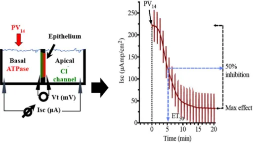
The experimental setup for the ex-vivo study is illustrated inFig. 1A. ET50is the effective time (in minutes) necessary to reach 50% of the maximum effects for each POM concentration. The maximum in-hibitory effect (%) of the ATPase activity by the POVs solutions and the effective time (ET50) necessary to reach 50% of the maximum effects (in minutes) were measured, taken into consideration the % decrease of short circuit current (Isc, μA/cm2) in the absence (100% activity) and upon addition of PV14(Fig. 1B). In the opercular epithelium of killifish used for our studies, Isc is a direct measure of apical chloride secretion mediated by chloride channels, which relies on an intact basolateral Na+/K+-ATPase to function [34,35]. Methodology for ex-vivo oper-cular epithelia preparation followed our current methods [22,36]. Fish were anaesthetized with 2-phenoxyethanol (1:2000 v/v), sacrificed by decapitation and the cranium was cut longitudinally. The gills and other tissue remains were removed carefully and the epithelial skin covering the opercular bone were dissected out and transferred to fresh-gassed saline (99.7:0.3 O2/CO2) with the following composition (all values in mM): NaCl, 160; MgSO4, 0.93; NaH2PO4, 3; CaCl2 1.5, NaHCO317.85, KCl 3; Glucose 5.5 HEPES 5; pH 7.8. Measurement of short circuit current (Isc, μA/cm2) was performed in symmetric con-ditions under voltage clamp to 0 mV. Open circuit potential (Vt, mV) and Isc were monitored by means of Ag/AgCl electrodes connected to
the chambers by 3 mm bore agar bridges (1 M KCl in 3% agar). Clamping of epithelia to 0 mV and recording of Isc was performed by means of VCC600 voltage clamp amplifiers (Physiologic Instruments, San Diego, USA). Bioelectrical data was continuously digitized trough a Lab-Trax-4 (WPI, Sarasota, US) onto a Macbook laptop using Labscribe3 Software (Iworks systems, Dover, US). All experiments were performed at least in triplicate. Calculations of ET50and Maximum effect were performed using GraphPad Prism version 6.00 for Macintosh (GraphPad Software, La Jolla California USA).
3. Results and discussion
3.1. Characterization of phosphotetradecavanadate
The non-stochiometric composition found for Cs5.6H3.4PV14O42·12H2O (PV14) is in good agreement with the pre-viously reported analogical salts K5.72H3.28[PV14O42] [31] and Rb5.89H3.11[PV14O42] [37]. The characteristic bands in IR spectra re-present PeO (1053 m cm−1) and V]O stretching vibrations (934 vs + 862 s cm−1), as well as vibrations of various V–O–V bridges (two groups of bands at 800, 741, 709 cm−1+ 590, 557, 476, 428 cm−1) (Fig. S2).
51V NMR spectrum of a 1 mM solution of PV
14 (Fig. 2) revealed three characteristic peaks at −592 ppm, −574 ppm, −524 ppm in the ratio 4:8:2. The chemical shift at −524 ppm corresponds to vanadium atoms of the two capping V]O units that are pentacoordinated, the remaining peaks corresponds to octacoordinated vanadium atoms of the Keggin cage connected to the capping V atoms through oxido bridges (−574 ppm) or not connected (−592 ppm). Based on published speciation studies in the ternary H+/H
2VO4−/H2PO4− system with found chemical shifts −589 ppm, −572 ppm, −521 ppm for
H3[PV14O42]6− species and −598 ppm, −580 ppm, −530 ppm for H4[PV14O42]5−species [38], the NMR shows that the found chemical shifts lie in the expected region.
The X-ray crystal structure of PV14(Fig. S1; Table S2 and Table S3) was solved in P-4n2 space group and the unit cell contains two trans-bicapped α-Keggin anions HxPV14O42(9-x)-(Fig. 3). The anion is built up from the classical Keggin structure [(PO4)@(V12O36)] capped by two [VO] units. The V]O groups are located in surface moieties formed by four adjacent oxygen atoms at VeO distances in the range 1.838–2.238 Å (Fig. S1). The two [VO] units are in opposition and occupy three different pairs of positions on the surface of the Keggin anion with various occupancies giving the overall sum formula as de-termined by ICP-MS analysis. The high degree of disorder in the area of counter ions and water molecules forced the use of squeeze. According to the result of elemental analysis (5.6 Cs), the excluded volume (1208.2 Å3) and number of electrons (1112.0), the following maximum content is possible: 12 Cs and up to ~45 H2O molecules. This leads to ~1112 electrons and ~1200 Å3by the model. This is in very good ac-cordance to approximately twelve charges of the two POMs in the unit cell needed. The analogous compounds K5.72H3.28[PV14O42] [32] and Rb5.89H3.11[PV14O42] [37] also exhibited the same disorder that was treated similarly.
3.2. Inhibition of Ca2+-ATPase by phosphotetradecavanadate: in vitro
studies
The effect of phosphotetradecavanadate (PV14) on the activity of sarcoplasmic reticulum Ca2+-ATPase from skeletal muscle (in vitro
Fig. 1. A) Schematic representation of the
experi-mental setup of the opercular epithelium of killifish used for our ex vivo studies. Isc was measured in voltage clamp, and in this model represents chloride secretion. The process is energized by basolateral Na+/K+-ATPase and chloride is secreted apically
via a chloride channel. B) Original trace of the effect
of short circuit current (Isc, μA/cm2) in the oper-cular epithelium of killifish mounted in Ussing chambers and kept under voltage clamp (Vt = 0 mV) after PV14addition. Effective time 50 (ET50), time necessary to reach 50% of the max-imum effects is shown in minutes; and maxmax-imum inhibitory effects are calculated as the % of basal values.
Fig. 2.51V NMR spectrum of 1 mM aqueous PV
14solution at autogenous pH (5.5). Legend: a′, a″, a‴ HxPV14O42(9-x)-; b H3VPO7−.
Fig. 3. Polyhedral representation of the structure of HxPV14O42(9-x)-anion in
PV14as revealed by X-ray structure analysis. Polyhedron legend: pink {PO4}, blue {VO6}, orange {VO} capping unit.
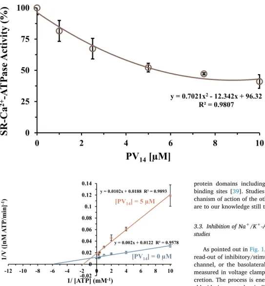
model) was investigated. It was observed that PV14inhibits the Ca2+ -ATPase activity, expressed as a percentage of the control obtained without inhibitor, in a concentration dependent manner (Fig. 4). The inhibitory power of PV14was evaluated using IC50values (POM con-centration at which it induces 50% of Ca2+-ATPase inhibition of the enzyme activity) and the IC50 value of 5.4 ± 0.5 μM was obtained. Ca2+-ATPase IC
50 values of inhibition below 1 μM were previously determined for polyoxotungstates (POTs) such as [α-P2W18O62]6− (0.6 μM) and [H10Se2W29O103]14− (0.3 μM), whereas the lowest po-tency was observed for [TeW6O24]6− (IC50= 200 μM) [22]. It was previously determined that decavanadate V10(IC50= 15 μM) is more potent Ca2+-ATPase inhibitors than V
1 (IC50= 80 μM). Herein, we show that PV14 is the strongest inhibitor of the calcium pump (IC50= 5 μM) among so far investigated vanadates. For both Nb10and V10, it was previously observed that they showed to be Ca2+-ATPase non-competitive inhibitors regarding the natural ligand Mg-ATP [16]. PV14presented a mixed type inhibition (Fig. 5), as was previously ob-served for [α-P2W18O62]6−and [TeW6O24]6−[22] suggesting that it can interact with the Ca2+-ATPase whether or not the enzyme has al-ready bound substrate and pointing out to the existence of two distinct protein binding sites for these types of POMs. Only for V10a binding site in the Ca2+-ATPase was previously described, involving at least three
protein domains including the phosphorylation and the nucleotide binding sites [39]. Studies about the type of inhibition and the me-chanism of action of the other POMs interactions with P-type ATPases are to our knowledge still to be determined [16,17,22].
3.3. Inhibition of Na+/K+-ATPase by phosphotetradecavanadate: ex-vivo
studies
As pointed out inFig. 1, modification of Isc provides an immediate read-out of inhibitory/stimulatory effects on either the apical chloride channel, or the basolateral Na+/K+-ATPase induced by PV
14. Isc is measured in voltage clamp, and in this model represents chloride se-cretion. The process is energized by basolateral Na+/K+-ATPase and chloride is secreted apically via a chloride channel. In this polarized epithelium, both mechanisms are required to be intact to sustain the process of secretion. Therefore, inhibitory effects of POMs on the ba-solateral side (where Na+/K+-ATPase is located) result in inhibitory effects on Isc. Herein, it was observed that when PV14was added to the basal side (Fig. 1), it does inhibit the Na+/K+-ATPase activity (ex-pressed as the % of maximum inhibition, in a concentration dependent manner,Fig. 6A). Thus, for 10 μM a maximum inhibition of 78% of the basal current was observed whereas a 50% inhibition is obtained for even less than 1 μM PV14. On the other hand, the addition of PV14to the apical side was not accompanied by an effect on Isc, ruling out chloride channels as putative targets of PV14, at least in a range of concentra-tions up to 10 μM. From the data of Fig. 6A, we calculated an IC50 values of 1.4 ± 0.1 μM for the ex-vivo Na+/K+-ATPase inhibition. In addition, it has to be noted that besides the maximum inhibitory effect (providing information about inhibitor efficacy) also ET50 values (providing information about inhibition velocity) can be appreciated to characterize the biological effects of the inhibitor. It was observed that the value of ET50decreases from 21 to 7 min upon increasing PV14 concentration (Fig. 6B). When comparing different POMs, this negative correlation does necessary mean that the lowest ET50value implies a higher inhibition. In fact, it was observed that decavanadate at the same concentration (10 μM) exhibits the maximum inhibition of 66% and an ET50of 14 min; whereas for the monomeric vanadate (at 10 μM) only 33% of maximum inhibition was observed, but a lower ET50was de-termined (6 min).
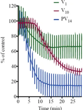
A simultaneous comparison of these three vanadate species, PV14, V10and V1at the same concentration of 10 μM (Fig. 7) clearly illus-trates that PV14(blue line) is the most potent ex-vivo inhibitor (78%
y = 0.7021x
2- 12.342x + 96.32
R² = 0.9807
0
25
50
75
100
0
2
4
6
8
10
SR
-C
a
2+
-)
%(
yt
i
vi
t
c
A
es
a
P
T
A
PV
14
[µM]
Fig. 4. Inhibition of Ca2+-ATPase activity by
PV14. Ca2+-ATPase was measured spectro-photometrically at 340 nm and 22 °C, using the coupled enzyme pyruvate kinase/lactate dehydrogenase assay. The experiments were initiated after the addition of 10 μg/mL cal-cium ATPase, in the presence or absence of 4% (w/w) of calcium ionophore A23187. Data are plotted as means ± SD. The results shown are the average of triplicate experi-ments. y = 0.002x + 0.0122 R² = 0.9578 y = 0.0102x + 0.0188 R² = 0.9893 -0.02 0 0.02 0.04 0.06 0.08 0.1 0.12 0.14 -12 -10 -8 -6 -4 -2 0 2 4 6 8 10
]
ni
m/
P
T
A
M
n[
(
V/
1
-1)
1/ [ATP] (mM
-1)
[PV
14] = 0 µM
[PV
14] = 5 µM
Fig. 5. Lineweaver-Burk plots of Ca2+-ATPase activity in the absence (blue)
and in the presence (orange) of 5 μM of the polyoxometalate PV14.The plots were used for determining the type of enzyme inhibition. The POM PV14 pre-sented a mixed type of inhibition. Data are plotted as means ± SD. The results shown are the average of triplicate experiments.
after 30 min upon addition), whereas for decavanadate (red) and va-nadate (green) only minor effects were observed. It can be also ob-served, a constant height of the current deflections used to calculate tissue resistance implying that the ex-vivo preparation maintained its integrity and selectivity before and after POM exposure (Fig. 7). For these studies a positive control experiment was performed with the conventional Na+/K+-ATPase inhibitor ouabain. Ouabain (at 10 μM) showed a maximum inhibition value of 100% and an ET50of 3.2 min [22]. By inhibiting the basolateral Na+/K+-ATPase activity, ouabain concomitantly prevents apical chloride secretion. Cs+, in contrast with rubidium, is known not to affect the activity of this type of enzyme [40].
Let us briefly comment on the mechanism of P-type ATPases in-hibition by POMs. POMs clearly exhibit different types of interaction with different P-type ATPases [12,14]. For example, the orthotungstate (HWO42−) presents a very low inhibition capacity (IC50= 1.5 mM) for the Na+/K+-ATPase [15], while for another ion pump, SR Ca2+ -AT-Pase is the IC50= 400 μM [16]. Herein, taking into account the Ca2+ -ATPase IC50 values of inhibition for PV14 (IC50= 5 μM) and V10
(IC50= 15 μM) apparently the lower negative charge (−6) for V10,at physiological pH and according to its pKas values [14,16], in compar-ison to PV14(−9) is not favoring enzyme inhibition. Very recently, for high affinity POTs, exhibiting IC50 values lower than 16 μM, it was described a correlation between their activity (IC50 value) and their charge density, as well as by volume of POM anion [22]. Also recently, a combination of NMR studies, ab initio calculations and crystal-lographic analysis point out to specific molecular interactions between H3PW12O40or H4SiW12O40and the Na+/K+-ATPase [41]. POMs-pro-tein interactions observed previously with myosin, actin and calcium ATPase [12,14,16,17,22,42,43] were described to be mostly of elec-trostatic nature including hydrogen bonds, but could also involve direct interaction with cysteins, as described for the P-type ATPase and actin [17,43]. Thus, besides electrostatic interactions, specific bonding modes underpinning their relative affinities and site recognition, and the structure and accessibility of the ATPase site for each crystal-lographically defined enzyme/pump relative to the inhibitory effec-tiveness of each POM are still to be determined [44–46]. In conclusion, the study of protein-POMs interactions is of utmost importance to ex-plain and improve inhibition of certain enzymes for potential drug development [47–49].
As referred above, only few studies described POMs as inhibitors of the hydrolytic activity of several P-type ATPases [15,16]. In these P-type ATPases studies, normally vesicles from cellular membranes or organelles are used and constitute in vitro models for the study of POMs toxicity effects. However, in the majority of these studies, the stability, as well others factors, of the compounds and/or the putative decom-position into others species are not taken in consideration. Thus, this possibility must be considered and several papers were published over the last years [49–56]. Therefore, if the polyoxometalates species re-sponsible for the observed effects are not always possible to be de-termined, participation of other species than the ones initially added to the biological system should be taken into account [49–57].
In fact, regarding the in vitro studies of POMs for the P-type ATPases
Fig. 6. A) Maximum inhibitory effects are calculated as the % of basal values
and B) Effective time 50 (ET50), time necessary to reach 50% of the maximum effects (shown in minutes); obtained for 1, 2.5 and 10 μM PV14concentrations. Data are plotted as means ± SD. The results shown are the average of triplicate experiments at 22 °C.
Fig. 7. PV14, V10and V1inhibition (%) of the Na+/K+-ATPase activity from
basal membrane of the skin epithelia. PV14(10 μM) inhibited 78% with an ET50 of 6.4 min (blue) whereas lower effects were observed for V10(red) (66% in-hibition, ET5013.7) and for V1(green) (33% and ET506.4 min). Perpendicular lines were used to calculate tissue resistance. As it can be observed, the ex vivo epithelia preparations retained integrity and selectivity after compounds ex-posure.
[15,16] it is possible to find, for instance, different experimental con-ditions such as temperature (25 and 37 °C), different times upon in-cubation depending on the kinetic studies used (2, 15 or 30 min) and of course different concentration range due to different IC50 values of ATPase activity inhibition. After incubation, both higher temperature and time would favor the decomposition of the added POM and therefore the appearance of different species that might also have a contribution for the observed inhibitory effects.
In this paper, the temperature was maintained for both in vitro and ex-vivo studies but the time after addition of the PV14 were clearly different, 1 min for the Ca2+-ATPase assays, and 5 to 10 min incubation for the Na+/K+-ATPase assays. The determination of the inhibitory capacity needs also different times, 2–3 min for the Ca2+-ATPase and 30 min for the Na+/K+-ATPase. Moreover, the mediums are different and also with slight different pH values. Therefore, the experimental conditions are not always favoring the putative comparison with dif-ferent experimental conditions and methodologies.
In previous studies with V10 and Ca2+-ATPase and other muscle proteins (such as myosin and actin) the putative reduction of vanadate was always taken in consideration [14,43,49]. In fact, reduction of V10 to oxidovanadium(IV) (VO2+), was previously observed upon actin interaction, but only after 90 min incubation and with huge amounts of protein. Moreover, in the presence of the natural ligand (ATP) the va-nadate reduction was not observed, suggesting that decavava-nadate in-teraction at the actin ATP binding is needed for the reduction of va-nadate [43,49]. Herein, at the experimental in vitro and in vivo conditions the redox stability of PV14during the biological measure-ments would be hard or impossible to be determined not only because of the concentrations used, in the order of microM, but also because of the time upon exposition, that is in the scale of a few minutes. Still, it is well known that intracellular V(V) can be reduced to oxidovanadium (IV) [58–61].
Putting it all together, and taking into account the IC50values de-termined, the Keggin type polyoxovanadate PV14exibited a high po-tential to act as an in vivo inhibitor of the Na+/K+-ATPase than V
10or V1.However, as referred before for V10[49], and also due to the va-nadium complex chemistry and biochemistry we cannot exclude that besides PV14the observed effects might be due to other vanadate or even to vanadyl species. Therefore, strong efforts are needed to confirm the biologically active POMs species.
4. Conclusions
The X-ray crystal structure of PV14 was solved and contains two trans-bicapped α-Keggin anions HxPV14O42(9-x)-. The anion is built up from the classical Keggin structure [(PO4)@(V12O36)] capped by two [VO] units. Phosphotetradecavanadate (Cs5.6H3.4PV14O42·12H2O) syn-thesized and characterized in this work is the best so far investigated member of the alkali metals family of phosphotetradecavanadates, therefore it was reasonable to employ it in the inhibition studies of P-type ATPases. Ca2+-ATPase activity from sarcoplasmic reticulum is inhibited by PV14with the IC50= 5 μM. This is about ten times higher than IC50 values reported for highly active POMs such as [α-P2W18O62]6−(0.6 μM), but on the other hand PV14is 3 times more potent than V10(15 μM) and much more potent than Nb10(35 μM) and [TeW6O24]6− (≈200 μM). Similarly as described before for [α-P2W18O62]6−and [TeW6O24]6− a mixed type of inhibition was ob-served for PV14. Therefore, a different mode of interaction with the Ca2+-ATPase than the one observed for V
10and Nb10(shown to be non-competitive inhibitors) must be involved. However, PV14was shown to be the most potent Na+/K+-ATPase inhibitor when using an ex-vivo model obtained from basal membrane of the skin epithelia. Thus, for the ex-vivo Na+/K+-ATPase activity an IC
50value of 1.4 μM was ob-served. This ex-vivo model seems to be a very specific model to study the effects of POMs in the processes of epithelial chloride secretion energized by basolateral Na+/K+-ATPase activity.
Abbreviations
IC50 concentration that induces 50% of Ca2+-ATPase inhibition of the enzyme activity
ET50 the effective time (in minutes) necessary to reach 50% of the maximum effects.
Nb10 decaniobate, niobate oligomer containing 10 niobate units POMs Polyoxometalates
POTs Polyoxotunsgtates POVs Polyoxovanadates PV14 phosphotetradecavanadate SR sarcoplasmic reticulum
V1 vanadate, monomeric vanadate containing 1 vanadate units V10 decavanadate, vanadate oligomer containing 10 vanadate
units
Acknowledgments
MA thanks to national funds through FCT, Foundation for Science and Technology (UID/Multi/04326/2013; SFRH/BSAB/129821/2017). This research was funded by the Austrian Science Fund (FWF): P27534 (AR) and M2200 (LK). SSM thanks to Council of Scientific & Industrial Research (CSIR) for providing financial support (01(2906)/17/EMR-II). This work was supported by the Grant Agency of the Ministry of Education of the Slovak Republic and Slovak Academy of Sciences VEGA Project No. 1/0507/17. The authors are grateful to Professor Markus Galanski (Universität Wien, Wien) for NMR measurements and Dr. Marek Bujdoš (Comenius University, Bratislava) for ICP-MS ana-lysis.
Appendix A. Supplementary data
Supplementary data CCDC 1886317 contains the supplementary crystallographic data for PV14. These data can be obtained free of charge viahttp://www.ccdc.cam.ac.uk/conts/retrieving.html, or from the Cambridge Crystallographic Data Centre, 12 Union Road, Cambridge CB2 1EZ, UK; fax: +44 1223 336 033; or e-mail:deposit@ ccdc.cam.ac.uk. These data include experimental details on inhibition studies, crystal data and structure refinement details. MOL files and InChiKeys of the most important compounds described in this article. Supplementary data to this article can be found online at doi:https:// doi.org/10.1016/j.jinorgbio.2019.110700.
References
[1] X. Chen, S. Yan, H. Wang, Z. Hu, X. Wang, M. Huo, Carbohydr. Polym. 117 (2015) 673–680.
[2] S.S. Wang, G.Y. Yang, Chem. Rev. 115 (2015) 4893–4962.
[3] L. Mohapatra, K.M. Parida, Phys. Chem. Chem. Phys. 16 (2014) 16985–16996. [4] M.A. Moussawi, N. Leclerc-Laronze, S. Floquet, P.A. Abramov, M.N. Sokolov,
S. Cordier, A. Ponchel, E. Monflier, H. Bricout, D. Landy, M. Haouas, J. Marrot, E. Cadot, J. Am. Chem. Soc. 139 (2017) 12793–12803.
[5] A. Bijelic, A. Rompel, Acc. Chem. Res. 50 (2017) 1441–1448.
[6] T. Sun, W. Cui, M. Yan, G. Qin, W. Guo, H. Gu, S. Liu, Q. Wu, Adv. Mater. 28 (2016) 7397–7404.
[7] A. Galani, V. Tsitsias, D. Stellas, V. Psycharis, C.P. Raptopoulou, A. Karaliota, J. Inorg. Biochem. 142 (2015) 109–117.
[8] S. Treviño, D. Velázquez-Vázquez, E. Sánchez-Lara, A. Diaz-Fonseca, J.A. Flores-Hernandez, A. Pérez-Benítez, E. Brambila-Colombres, E. González-Vergara, Oxidative Med. Cell. Longev. 2016 (2016) 605870514 pages.
[9] P.L. Pedersen, E. Carafoli, Trends Biochem. Sci. 12 (1987) 146–150.
[10] C. Toyoshima, M. Nakasako, H. Nomura, H. Ogawa, Nature 405 (2000) 647–655. [11] L. Yatime, M.J. Buch-Pedersen, M. Musgaard, J.P. Morth, A.-M.L. Winther,
B.P. Pedersen, C. Olesen, J.P. Andersen, B. Vilsen, B. Schiøtt, M.G. Palmgre, J.V. Møller, P. Nissen, N. Fedosova, Biochim. Biophys. Acta 1787 (2009) 207–220. [12] M. Aureliano, G. Fraqueza, C.A. Ohlin, Dalton Trans. 42 (2013) 11770–11777. [13] L.C. Cantley Jr., L. Josephson, R. Warner, M. Yanagisawa, C. Lechene, G. Guidotti,
J. Biol. Chem. 252 (1977) 7421–7423.
[14] M. Aureliano, D.C. Crans, J. Inorg. Biochem. 103 (2009) 536–546.
[15] M.B. Čolović, D.V. Bajuk-Bogdanović, N.S. Avramović, I.D. Holclajtner-Antunović, N.S. Bošnjaković-Pavlović, V.M. Vasić, D.Z. Krstić, Bioorg. Med. Chem. 19 (2011) 7063–7069.
[16] G. Fraqueza, C.A. Ohlin, W.H. Casey, M. Aureliano, J. Inorg. Biochem. 107 (2012) 82–89.
[17] G. Fraqueza, L.A.E. Batista de Carvalho, M. Paula, M. Marques, L. Maia, C. André Ohlin, W.H. Casey, M. Aureliano, Dalton Trans. 41 (2012) 12749–12758. [18] H. Stephan, M. Kubeil, F. Emmerling, C.E. Müller, Eur. J. Inorg. Chem. 10–11
(2013) 1585–1594.
[19] S.H. Saeed, R. Al-Oweini, A. Haider, U. Kortz, J. Iqbal, Toxicol. Rep. 1 (2014) 341–352.
[20] S.Y. Lee, A. Fiene, W. Li, T. Hanck, K.A. Brylev, V.E. Fedorov, J. Lecka, A. Haider, H.J. Pietzsch, H. Zimmermann, J. Sévigny, U. Kortz, H. Stephan, C.E. Müller, Biochem. Pharmacol. 93 (2015) 171–181.
[21] J. Iqbal, M. Barsukova-Stuckart, M. Ibrahim, S.U. Ali, A.A. Khan, U. Kortz, Med. Chem. Res. 22 (2013) 1224–1228.
[22] N.I. Gumerova, L. Krivosudský, G. Fraqueza, J. Breibeck, E. Al-Sayed, E. Tanuhadi, A. Bijelic, J. Fuentes, M. Aureliano, A. Rompel, Metallomics 10 (2018) 287–295. [23] Bruker SAINT v8.37A & V7.68A Copyright © 2005–2018 Bruker AXS. [24] G.M. Sheldrick, SADABS, University of Göttingen, Germany, 1996. [25] O.V. Dolomanov, L.J. Bourhis, R.J. Gildea, J.A.K. Howard, H. Puschmann, J.:
OLEX2: a complete structure solution, refinement and analysis program, J. Appl. Crystallogr. 42 (2009) 339–341.
[26] C.B. Huebschle, G.M. Sheldrick, B. Dittrich, ShelXle: a Qt graphical user interface for SHELXL, J. Appl. Crystallogr. 44 (2011) 1281–1284.
[27] G.M. Sheldrick, SHELXS v 2016/4, University of Göttingen, Germany, 2015. [28] G.M. Sheldrick, SHELXL v 2016/4, University of Göttingen, Germany, 2015. [29] A.L. Spek, Structure validation in chemical crystallography, Acta Cryst D65 (2009)
148–155.
[30] Diamond - Crystal and Molecular Structure Visualization. Crystal Impact - Dr. H. Putz & Dr. K. Brandenburg GbR, Kreuzherrenstr. 102, 53227 Bonn, Germany. http://www.crystalimpact.com/diamond
[31] R. Kato, A. Kobayashi, Y. Sasaki, Inorg. Chem. 21 (1982) 240–246. [32] K. Nomiya, K. Kato, M. Miwa, Polyhedron 5 (1986) 811–813.
[33] S. Uematsu, Z. Quan, Y. Suganuma, N. Sonoyama, J. Power Sources 217 (2012) 13–20.
[34] K.J. Karnaky Jr., K.J. Degnan, J.A. Zadunaisky, Science 195 (1977) 203–205. [35] J.A. Zadunaisky, The chloride cell: the active transport of chloride and the
para-cellular pathways, in: W.S. Hoar, D.J. Randall (Eds.), Fish Physiology, XB, Academic Press, New York, 1984, pp. 129–176.
[36] J.A. Martos-Sitcha, G. Martínez-Rodríguez, J.M. Mancera, J. Fuentes, Comp. Biochem. Physiol. A Physiol. 182 (2015) 93–101.
[37] E.V. Murashova, A.B. Iluikhin, N.N. Chudinova, Russ. J. Inorg. Chem. 46 (2001) 1292–1295.
[38] I. Andersson, A. Gorzsás, C. Kerezsi, I. Tóth, L. Pettersson, Dalton Trans. (2005) 3658–3666.
[39] S. Hua, G. Inesi, C. Toyoshima, J. Biol. Chem. 275 (2000) 30546–30550. [40] R. Krulík, I. Farská, J. Prokeš, Neuropsychobiology 3 (1977) 129–134. [41] N. Bošnjaković-Pavlović, D. Bajuk-Bogdanović, J. Zakrzewska, Z. Yan,
I. Holclajtner-Antunović, J.-M. Gillet, A. Spasojević-de Biré, J. Inorg. Biochem. 176 (2017) 90–99.
[42] T. Tiago, P. Martel, C. Gutiérrez-Merino, M. Aureliano, Biochim. Biophys. Acta 1774 (2007) 474–480.
[43] M.P.M. Marques, D. Gianolio, S. Ramos, L.A.E. Batista de Carvalho, M. Aureliano, Inorg. Chem. 56 (2017) 10893–10903.
[44] A. Bijelic, A. Rompel, Coord. Chem. Rev. 299 (2015) 22–38.
[45] A. Solé-Daura, V. Goovaerts, K. Stroobants, G. Absillis, P. Jiménez-Lozano, J.M. Poblet, J.D. Hirst, T.N. Parac-Vogt, J.J. Carbó, Chem. Eur. J. 22 (2016) 15280–15289.
[46] M. Arefian, M. Mirzaei, H. Eshtiagh-Hosseini, A. Frontera, Dalton Trans. 46 (2017) 6812–6829.
[47] A. Bijelic, M. Aureliano, A. Rompel, Chem. Commun. 54 (2018) 1153–1169. [48] A. Bijelic, M. Aureliano, A. Rompel, Angew. Chem. Int. Ed. 58 (2019) 2980–2999. [49] M. Aureliano, Oxidative Med. Cell. Longev. 2016 (2016) 61034578 pages. [50] A. Levina, D.C. Crans, P.A. Lay, Coord. Chem. Rev. 352 (2017) 473–498. [51] A. Levina, P.A. Lay, Chem. Asian J. 12 (2017) 1692–1699.
[52] M. Le, O. Rathje, A. Levina, P.A. Lay, J. Biol. Inorg. Chem. 22 (2017) 663–672. [53] K.A. Doucette, K.N. Hassell, D.C. Crans, K.A. Doucette, J. Inorg. Biochem. 165
(2016) 56–70.
[54] T. Jakusch, T. Kiss, Coord. Chem. Rev. 351 (2017) 118–126.
[55] D. Sanna, V. Ugone, G. Micera, P. Bugly,ó, L. Bír,ó, E. Garribba, Dalton Trans. 46 (2017) 8950–8967.
[56] D. Sanna, J. Palomba, G. Lubinu, P. Buglyó, S. Nagy, F. Perdih, J. Med. Chem. 62 (2019) 654–664.
[57] R. Prudent, V. Moucadel, B. Laudet, C. Barette, L. Lafanechère, B. Hasenknopf, J. Li, S. Bareyt, E. Lacôte, S. Thorimbert, M. Malacria, P. Gouzerh, C. Cochet, Chem. Biol. 15 (2008) 683–692.
[58] M. Garner, J. Reglinski, W.E.J. Mcmurray, I. Abdullah, R. Wilson, J. Biol. Inorg. Chem. 2 (1997) 235–241.
[59] T. C. Delgado, A. I. Tomaz, I. Correia, J. C. Pessoa, J.G. Jones, C.F. Geraldes et al., J. Inorg. Biochem. 99 (2005) 2328–2339.
[60] T. Jakusch, É.A. Enyedy, K. Kozma, Z. Paár, A. Bényei, T. Kiss, Inorg. Chim. Acta 420 (2014) 92–102.