Stage-specific Activity of Potential Antimalarial Compounds
Measured in Vitro by Flow Cytometry in Comparison to Optical
Microscopy and Hypoxanthine Uptake
Carmen E Contreras/
+, María A Rivas, José Domínguez*, Jaime Charris*, Mario Palacios,
Nicolás E Bianco, Isaac Blanca
Instituto de Inmunología, Facultad de Medicina *Laboratorio de Síntesis Orgánica, Facultad de Farmacia, Universidad Central, Apartado Postal 50109, Caracas 1050 A, Venezuela
The evaluation of new antimalarial agents using older methods of monitoring sensitivity to antimalarial drugs are laborious and poorly suited to discriminate stage-specific activity. We used flow cytometry to study the effect of established antimalarial compounds, cysteine protease inhibitors, and a quinolone against asexual stages of Plas-modium falciparum. Cultured P. falciparum parasites were treated for 48 h with different drug concentrations and the parasitemia was determined by flow cytometry methods after DNA staining with propidium iodide. P. falciparum
erythrocytic life cycle stages were readily distinguished by flow cytometry. Activities of established and new antima-larial compounds measured by flow cytometry were equivalent to results obtained with microscopy and metabolite uptake assays. The antimalarial activity of all compounds was higher against P. falciparum trophozoite stages. Advantages of flow cytometry analysis over traditional assays included higher throughput for data collection, insight into the stage-specificity of antimalarial activity avoiding use of radioactive isotopes.
Key words: Plasmodium falciparum - antimalarial compounds - flow cytometry - hypoxanthine uptake - chloroquine - quinolone
Drug resistance of Plasmodium falciparum, the most deadly human malaria parasite, is a major factor in the widespread persistence of malaria (Ouellette & Kunding 1997, Macreadi et al. 2000). Current efforts focus on re-search into novel compounds and on measures to pre-vent or delay resistance once new drugs are introduced. However, malaria therapy has generally not taken into consideration the stage-specificity of action of different drugs. This is an important consideration, since inappro-priate timing of administration of antimalarial drugs might limit drug efficacy and favor the selection of drug-resis-tant parasites.
Few studies have focused on the in vitro stage-spe-cific efficacy of antimalarial compounds (Chimanuka et al. 2001). Most in vitro studies monitoring resistance and susceptibility to antimalarial compounds have been per-formed by microscopy and by uptake of a radiolabelled nucleic acid precursor 3[H]-hypoxanthine (Desjardins et al. 1979). They are poorly suited to discriminate stage-specific activity. More recently, flow cytometry analysis (FCA) using different fluorescent dyes has been reported (van Vianen et al. 1990, Pattanapanyasat et al. 1996, 1997). We used propidium iodide (PI) whose incorporation as fluorescent molecule has been shown to be propor-tional to the DNA parasite content. In the present study, results of in vitro effect of antimalarial compounds on P. falciparum parasitized erythrocytes obtained from FCA
Financial support: Conicit, grant S1-97001305
+Corresponding author. Fax: +58-212-693.2815. E-mail:
[email protected] Received 14 July 2003 Accepted 18 February 2004
were compared with other assays. FCA results were com-parable to optical microscopy observation and to [3 H]-hypoxanthine uptake assay. Moreover, FCA offers advan-tages of high throughput, ready automation, simplified data processing, and easy determination of the stage-spe-cific effectiveness of potential antimalarial compounds.
MATERIALS AND METHODS
Parasites - Chloroquine sensitive NF54 (kindly pro-vided by Dr T Shapiro, Johns Hopkins University, Balti-more, MD) and FCB2 strains, anda chloroquine resistant FCBstrain(MR4 Resource Center, ATCC), as well as a Venezuelan (VEN, unknown chloroquine sensitivity) P. falciparum strains were used in this study. All strains were maintained in continuous culture as previously de-scribed (Trager & Jensen 1976) at 2.4% hematocrit using type 0+ human erythrocytes in RPMI 1640 medium supple-mented with 2 mM L-glutamine (Gibco, Grand Island, NY), 10% heat inactivated pooled AB+ human serum (Univer-sity Hospital Blood Bank, Caracas, Venezuela), 25 mM HEPES (Calbiochem, San Diego, CA), and 5% NaHCO3 under a 3% O2, 4% CO2, and 93% N2 gas mixture. Parasite synchronization was performed by sorbitol treatment (D-Sorbitol, Sigma) as previously described (Lambros & Vanderberg 1979). Viability and parasitemia of cultured parasites were calculated by light microscopy analysis of blood smear stained with Giemsa (5000 erythrocytes counted per blood smear).
et al. 1996). Quinolone 3 (purity assessed by HPLC was > 99%) previously showed antimalarial activity (Dominguez et al. 1996). Vinyl sulfone (Mu-Leu-Hph-VSPh), and fluoromethyl ketone (Mu-Phe-Hph-CH2F) protease inhibi-tors (Rosenthal et al. 1996) were kindly provided by Dr P Rosenthal, San Francisco General Hospital, University of California, CA; chloroquine (Sigma, St. Louis, MO) or artemisinin (Aldrich Chemical CO, Milwaukee, WI) were also included in this study as controls.
Drug susceptibility assays - Different techniques were used to measure antimalarial activity of compounds in-cluded in this study, FCA of PI incorporation into parasite DNA, [3H]-hypoxanthine uptake, and parasitemia by light microscopy.Antimalarial activity was performed in three parallel microtiter plate wells (Costar, Corning, NY, 96 flat-bottom plates). Triplicate wells containing 100 µl of drug were mixed with 100 µl of P. falciparum parasitized eryth-rocytes (0.25% parasitemia, and 2.4% hematocrit). Qua-druplicates of three different controls were included in each microplate: nonparasitized human erythrocytes (nega-tive control), P. falciparum parasitized human erythro-cytes without drug, and P. falciparum parasitized human erythrocytes without drug in the presence of 0.2% DMSO (positive control).Stock solutions of HPLC-purified or recrystallized compound were dissolved in either DMSO (quinolone 3, protease inhibitors, and artemisinin) or dis-tilled water (chloroquine). DMSO soluble drugs were later diluted 500 folds in medium, and finally serially diluted in 0.2% DMSO-medium (to maintain constant solvent con-centration). Dose-response curves of each drug included 10 concentrations of 1.8 fold dilutions yielding final con-centrations between 0.16-2500 ng/ml. Final concon-centrations were expressed in nM.
Calculation of IC50 -IC50 was calculated from each drug dose-response curve after logarithmic transforma-tion and fit to a generalized sigmoidal functransforma-tion by means of the Marquardt algorithm (Bard 1974), and analyzed for goodness of fit (r2 value).
DNA staining and flow cytometry analysis - Drug activity against malarial parasites was determined by mea-suring incorporation of PI (Sigma) as previously described (Pattanapanyasat et al. 1993, 1997) with some modifica-tions. After 48 h incubation, drug-treated and control para-sitized erythrocytes, were washed twice with PBS by cen-trifugation each time at 200 x g, and fixed overnight with 0.25% v/v glutaraldehyde (Sigma) in PBS. After washing with PBS, fixed cells were mixed with a recently prepared and filtered PI/H2O solution (final concentration 50 µg/ ml) in Tris 1 M, pH 8 containing RNAse, Nonidet P-40 and NaCl as described by Vindelov (1977), and incubated for 4 h in dark at 37ºC. Cells were vigorously mixed and intrac-ellular parasite DNA content was determined by PI incor-poration using a flow cytometer (Epics-Elite, Beckman-Coulter, Miami, FL) equipped with a Enterprise Argon-ion laser tuned at 488 nm. PI red fluorescence intensity was collected through a 575 nm band pass filter. List mode data from 100,000 erythrocytes was stored and processed for each well in an Elite-ESP Software (Coulter). Results were reported as DNA stained cells (%).
Optimal FCA conditions were initially established us-ing chicken erythrocytes since they are nucleated cells
able to incorporate PI in their DNA and localized differ-ently than the normal human erythrocytes in flow cytometry histograms. Suspensions of uninfected human erythrocytes/chicken erythrocytes (2.5 x 10⁶ normal hu-man erythrocytes/250-2.5 x 105 chicken erythrocytes) yielded histograms which clearly differentiated both popu-lations from contaminating human peripheral blood leu-kocytes, platelets, or reticulocytes (data not shown). Such contaminating cells were subsequently eliminated by ex-tensive washing of uninfected human erythrocyte sus-pensions. In initial trials, a PI concentration of 50 µg/ml and 4 h incubation at 37ºC in the dark was optimal to discriminate among parasitized and non parasitized eryth-rocytes and other nucleated cells.
Tritiated hypoxanthine uptake - Incorporation of 3 [H]-hypoxanthine was performed as previously described (Desjardins et al. 1979) with modifications (Posner et al. 1997). Briefly, nonparasitized and parasitized erythrocytes (with or without drug) were incubated for 24 h prior to addition of 2 µCi/well of [3H]-hypoxanthine [1 mCi/ml ethanol-water (1:1) solution, Amersham, Pharmacia, En-gland] diluted with medium. After an additional 18 h incu-bation, cells were harvested (Skatron Instruments) onto glass fiber filters (Wallac, TurKu, Finland) which were exhaustively washed. Radioactivity from dry filters was determined with a scintillation counter (LKB, Stockholm, Sweden).
Microscopic observation - Thin smears were made from nonparasitized and parasitized erythrocytes (with or without drug) after 24-48 h incubation at 37ºC. Parasitemias were calculated by counting the number of parasites per 5000 Giemsa-stained human erythrocytes using light microscopy.
RESULTS
inhibition compared to that of the untreated parasites. Inhibition of parasitemia on ring and schizont stages was 33 and 46% respectively. To confirm this observa-tion, we set up further experiments using synchronized parasites at different stages of their life cycle. Table I il-lustrates results obtained when P. falciparum parasitized erythrocytes were incubated (young rings: 16 h, young trophozoites: 20 h) with different concentrations of quinolone 3 and analyzed by FCA and optical micros-copy. Similar results were observed with both techniques (Table I). A slight inhibition of ring-stage parasitemia (20%) was observed only by FCA, at the highest quinolone 3 concentration (1.4 µM). In contrast, again by FCA a sig-nificant inhibition of trophozoite-stage parasitemia of 80% was observed at 1.4 µM, and of 40% at 0.8 µM. A similar response was observed by optical microscopy where 88 and 50% inhibition were obtained at 1.4 and 0.8 µM of quinolone 3 concentrations respectively.
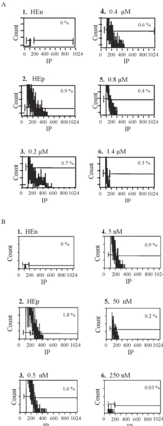
Based on these results a dose-response curve was obtained for quinolone 3 using P. falciparum NF54 tro-phozoites. Fluorescent histograms were acquired for non-parasitized and non-parasitized erythrocytes incubated with increasing quinolone concentrations. As shown in Fig. 2A progressive drug effects were demonstrated as de-creasing fluorescence. The antimalarial effective range of quinolone 3 was between 0.2 and 1.4 µM. We further tested the effect of other antimalarial compounds, two cysteine protease inhibitors, and artemisinin and chloroquine used as positive controls. As shown in Fig. 2B increasing drug-effects of fluoromethyl ketone (Mu-Phe-Hph-CH2F) cys-teine protease inhibitor was demonstrated as decreasing fluorescence. The antimalarial effect of this drug was be-tween 0.5 and 250 nM. Similar results were obtained using another cysteine protease inhibitor such as vinyl sulfone (Mu-Leu-Hph-VSPh), as well as traditional drugs as chlo-roquine and artemisinin (data not shown).
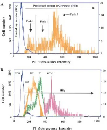
Antimalarial IC50- Dose-response curves of some of the compounds studied by FCA indicate that tropho-zoite parasitemia inhibition is proportional to drug con-centration. Fig. 3A shows representative dose-response curves of quinolone 3 and of two antimalarial drugs (chlo-roquine and artemisinin). An excellent fit of the data to the regression equation was obtained with each compound (r2: 0.999, data not shown). Antimalarials IC50 calculated from dose-response curves were consistent between tech-Fig. 1: flow cytometry assay of Plasmodium falciparum
parasit-ized erythrocytes (FCB2 strain) stained with propidium iodide (PI). A: overlap of typical histograms of HEn and HEp from asynchro-nous P. falciparum cultures gated on red fluorescent light. The scale of HEp fluorescent histogram was fixed 100 time lower to visualize the parasitized erythrocyte population. B: overlap of fluo-rescent histograms from synchronous P. falciparum cultures with ER (early rings), ET (early trophozoites), LT (late trophozoites), and SCH (schizonts).
TABLE I
Stage-specific antimalarial activity of quinolone 3 on synchronized Plasmodium falciparum parasites Optical microscopy a,b Flow cytometry a,c
Rings % Inhib % Troph % Inhib % Rings % Inhib % Troph % Inhib %
HEn 0.1 ± 0.1 0.1 ± 0.1
HEp 0.2 ± 0.03 0.8 ± 0.2 0.45 ± 0.1 0.5 ± 0.1
1.4 µM 0.3 ± 0.04 0 0.1 ± 0.02 88 0.4 ± 0.1 20 0.1 ± 0 80 0.8 µM 0.2 ± 0.04 0 0.4 ± 0.03 50 0.5 ± 0.1 0 0.3 ± 0.1 40 0.1 µM 0.3 ± 0.03 0 0.7 ± 0.1 13 0.5 ± 0.1 0 0.5 ± 0.1 0
a:synchronous parasites (NF54 strain) incubated with drug decreasing concentrations; b: parasitemia, mean ± SD, n = 3 (Giemsa smears read by two persons); c:parasitemia, mean ± SD, n = 3; Troph: trophozoites; Inhib: parasitemia inhibition; HEn: non-parasitized human erythrocytes; HEp: non-parasitized human erythrocytes
niques and P. falciparum strains (data not shown). IC50’s of P. falciparum VEN strain, were of 800 nM for quinolone 3, 29 nM for chloroquine, 8.5 nM for artemisinin, 330 nM for vinyl sulfone, and 15.7 nM for fluoromethyl ketone cysteine protease inhibitors. As expected, chloroquine IC50 for the chloroquine-resistant strain FCB, and for the chloroquine-sensitive strain NF54 were of 170 nM and 10 nM respectively.
Comparison between IC50’s of four antimalarial com-pounds measured by FCA, hypoxanthine uptake, and optical microscopy - Antimalarial activities of standard and new compounds measured by FCA were compared with those obtained by the hypoxanthine uptake assay, and optical microscopy, standard methods of assessing in vitro antimalarial drug activity. As depicted in Fig. 3B, a high correlation (r2: 0.999) was observed among IC50 cal-culated by FCA and hypoxanthine uptake. In addition, IC50’s of FCB, VEN, and NF54 strains calculated from chloroquine optical microscopy dose-response curves were of 151 nM, 33 nM, and 9 nM respectively. Such IC50’s were consistent with the ones calculated from FCA dose-response curves which were of 170 nM, 29 nM, and 10 nM respectively.
Correlation of antimalarial activity of quinolone measured by FCA and optical microscopy - Antimalarial activity of quinolone evaluated by FCA and optical mi-croscopy was similar (Table II). Inhibitory growth effect was proportional to drug concentration after 48 h incuba-tion. A significant correlation between both techniques was observed only at 0.8 µM (p < 0.05). Quinolone activ-ity at 2.5 and 0.8 µM was evident since microscopy exami-nation showed parasitemias of 0 and 0.4% respectively. Moreover, parasites look unhealthy with a compact chro-matin and could be easily distinguished as dead para-sites. In contrast, FCA does not detect morphological changes, indeed damaged or dead parasites may incorpo-rate PI at their DNA as was demonstincorpo-rated by the increased parasitemias observed (0.2 and 0.7% at 2.5, and 0.8 µM respectively).
DISCUSSION
FCA have proved to be useful to study diverse fea-tures related with human and experimental malaria (Janse et al. 1987, Pattanapanyasat et al. 1993, 1999, Kumaratilake & Ferrante 2000, Saito-Ito et al. 2001).
TABLE II
Quinolone antimalarial activity correlation between flow cytometry and optical microscopy
Parasitemia % (Mean ± SD)
48 h O M FCA p
HEp 2.3 ± 0.1 1.9 ± 0.2 NS 2.5 µM 0.0 ± 0.0 0.2 ± 0.1 NS 0.8 µM 0.4 ± 0.0 0.7 ± 0.1 < 0.05 0.1 µM 1.9 ± 0.3 1.6 ± 0.4 NS OM: optical microscopy; FCA: flow cytometry assay; HEp: parasitized human erythrocytes (Plasmodium falciparum NF54 strain) without drug; NS: not significant
Fig. 2: effect of quinolone 3 (A), and protease inhibitor Mu-Phe-Hph-CH2F (B) on Plasmodium falciparum synchronized tropho-zoites (VEN and NF54 strains respectively) measured by flow cytometry. Individual histograms show the % of parasitemia (FCA) corresponding to each drug concentration. Non-parasitized human erythrocytes (HEn) and parasitized human erythrocytes (HEp) without drug were included as negative and positive controls respec-tively.
A
In this study, we have described by flow cytometry the anti-malarial activity of four drugs using continuos cultures of P. falciparum geographically different strains. Moreover, we compared the IC50 calculated by this method with those obtained by microscopy and 3 [H]-hy-poxanthine uptake.
Flow cytometry method enabled to discriminate three different populations of infected erythrocytes, corre-sponding to different asexual P. falciparum stages. Such populations can be electronically gated to determine the mean channel fluorescence intensity of PI uptake charac-teristic of each one. By monitoring the P. falciparum de-velopmental stages and drug sensitivity, it was observed that the inhibitory effect of all tested antimalarial com-pounds was primarily directed to trophozoite stages, with little or not effect on ring stages.
These results agree with previous reports, showing that P. falciparum trophozoite stage was the most
sensi-tive to in vitro treatment with chloroquine (Yayon et al. 1983, Cambie et al. 1991). Relevance of chronotherapy in malaria has been recently demonstrated in vivo studies of the rodent malaria parasite P. chabaudi chabaudi. This study indicates that the best timing for treatment with chloroquine was the trophozoite stage (Chimanuka et al. 2001).
In contrast with previous reports (Saito-Ito et al. 2001), our results based on FCA data, showed that IC50’s of each antimalarial compound were consistent with those obtained by microscopy, and 3[H]-hypoxanthine uptake. In summary, despite hypoxanthine uptake is the gold standard method for testing in vitro antimalarial activity, our results confirm FCA as a better method to assess stage-specific antimalarial activity showing high reducibility, automation of analysis, and efficient data pro-cessing. In contrast, hypoxanthine gives limited informa-tion on parasite stages and produces radioactive waste.
ACKNOWLEDGEMENTS
To Drs Juan De Sanctis for advice and support and Diana Ajami for manuscript review, and to Patricia Rodríguez, Eladia Paolini, and Angelyseb Dorta for assistance.
REFERENCES
Bard Y 1974. The Marquardt method. In Non Linear Param-eter Estimation, Academic Press Inc., New York, p. 94-96. Cambie G, Caillard V, Beaute-Laffite A, Ginsburg H, Chabaud A, Landau I 1991. Chronotherapy of malaria: identification of drug-sensitive stage of parasite and timing of drug deliv-ery for improved therapy. Ann Parasitol Hum Comp66: 14-26.
Chimanuka B, Francois G, Timperman G, Heyden YV, Holenz J, Plaizier-Vercammen J, Bringmann G 2001. A comparison of the stage-specific efficacy of chloroquine, artemether and dioncophylline B against the rodent malaria parasite
Plasmodiun chabaudi chabaudi in vivo. Parasitol Res87: 795-803.
Desjardins RE, Canfield CJ, Haynes JD, Chulay JD 1979. Quan-titative assessment of antimalarial activity in vitro by a semiautomated microdilution technique. Antimicrob Agents Chemother 16: 710-718.
Domínguez J, Basante W, Charris J, Riggione F 1996. Synthe-sis and activity of some quinolone derivatives against Plas-modium falciparum in vitro. Il Farmaco51: 407-412. Dominguez JN, Charris J, Mendez B 1998. 13C NMR spectral
characterization of some antimalarial tricyclic quinolone derivatives. Magn Reson Chem36: 454-456.
Hooper DC, Wolfson JS 1985. The fluoroquinolones: pharma-cology, clinical uses, and toxicities in humans. Antimicrob Agents Chemother28: 716-721.
Janse CJ, van Vianen PH, Tanke HJ, Mons B, Ponnudurai T, Overdulve JP 1987. Plasmodium species: flow cytometry and microfluorometry assessments of DNA content and synthesis. Experimental Parasitol64: 88-94.
Kumaratilake LM, Ferrante A 2000. Opsonization and phago-cytosis of Plasmodium falciparum merozoites measured by flow cytometry. Clin Diag Lab Immunol7: 9-13. Lambros C, Vanderberg JP 1979. Synchronization of
Plasmo-dium falciparum erythrocytic stages in culture. J Parasitol 65: 418-420.
Macreadie I, Ginsburg H, Sirawaraporn W, Tilley L 2000. An-timalarial drug development and new targets. Parasitol To-day16: 438-444.
Ouellette M, Kunding C 1997. Microbial multidrug resistance.
Int J Antimicrob Agents8: 179-187.
Pattanapanyasat K, Thaithong S, Kyle DE, Udomsangpetch R, Yongvanitchit K, Hider RC, Webster HK 1997. Flow cytometric assessment of hydroxy piridinone iron chela-tors on in vitro growth of drug-resistant malaria. Cytometry 27: 84-91.
Pattanapanyasat K, Udomsangpecth R, Webster HK 1993. Two-color flow cytometric analysis of intraerythrocytic malaria parasite DNA and surface membrane-associated antigen in erythrocytes infected with Plasmodium falciparum. Cytometry14: 449-454.
Pattanapanyasat K, Yongvanitchit K, Heppner DG, Tongtawe P, Kyle DE, Wepster HK 1996. Culture of malaria para-sites in two different red blood cell populations using bi-otin and flow cytometry. Cytometry25: 287-294. Pattanapanyasat K, Yongvanitchitk K, Tongtawe P, Tachavanich
K, Wanachiwanawin W, Fucharoen S, Walsh DS 1999. Im-pairment of Plasmodium falciparum growth in thalassemic red blood cells: further evidence by using biotin labeling and flow cytometry. Blood 93: 3116-3119.
Posner G, González L, Cumming J, Klinedinst D, Shapiro T 1997. Synthesis and antimalarial activity of heteroatoms containing bicyclic endoperoxides. Tetrahedron53: 37-50 Rosenthal PJ, Olson JE, Lee GK, Plamer JT, Klaus JL, Rasnick
D 1996. Antimalarial effects of vinyl sulfone cysteine pro-teinase inhibitors. Antimicrob Agents Chemother40: 1600-1603.
Saito-Ito A, Akai Y, Shenyi H, Kimura M, Kawabata M 2001. A rapid, simple and sensitive flow cytometric system for de-tection of Plasmodium falciparum. Parasitol Int50: 249-257.
Trager W, Jensen JB 1976. Human malaria parasite in continu-ous culture. Science 193: 674-675.
Tripathi KD, Sharma AK, Valecha N, Biswas S 1993. In vitro activity of fluoroquinolones against chloroquine-sensitive and chloroquine-resistant Plasmodium falciparum. Indian J Malariol30: 67-73.
van Vianen PH, Thaithong S, Reinders PP, van Engen A, van der Keur M, Tanke HJ, van der Kaay HJ, Mons B 1990. Auto-mated flow cytometric analysis of drug susceptibility of malaria parasites. Am J Trop Med Hyg43: 602-607. Vindelov L 1977. Flow microfluorometric analysis of nuclear
DNA in cells from solid tumors and cell suspensions. A new method for rapid isolation and staining of nuclei. Virchows Arch B Cell Pathol24: 227-242