1. rheumatology department, centro Hospitalar são João, porto, portugal; faculty of medicine of porto university
2. Nephrology department, centro Hospitalar são João, porto, portugal; Nephrology and infeciology group, iNeB – National institute of Biomedical engineer, university of porto, portugal; i3s, university of porto
Keywords: Chronic kidney disease–mineral and bone disorder; Renal osteodystrophy; Fragility fracture; Bone histomorphometry.
IntroductIon
Chronic kidney disease (CKD) and fragility fractures are important public health concerns, both increasing as the population ages and co-occurring frequently. Fracture susceptibility and incidence are increased in CKD and this risk gradually rises as renal disease pro-gresses1,2. Of note, morbidity and mortality associated with fractures are also higher in these patients3,4.
The increased fracture risk in CKD is multifactorial. On one hand, CKD patients have impaired bone strength due to deterioration of both bone volume and bone quality (incorporating changes in turnover, mi -neralization, cortical porosity and trabecular bone ar-chitecture) that increase in severity as the disease pro-gresses5. On the other hand, these patients are commonly elderly with multiple comorbidities, such as diabetes mellitus, also associated with increased fractu -re risk, along with sarcopenia and cognitive dysfunction leading to higher risk for falls1,6. In addition, previous fracture is a known risk factor for a subsequent one7.
The management of a CKD patient with one or more fragility fractures is challenging, first due to the need to determine whether the patient has osteoporosis and/or the various forms of metabolic bone disease and secondly to choose the therapeutic approach. Herein the authors review the contribution of bone biopsy with histomor-phometric analysis in the management of these patients.
osteoporosIs And cKd–mInerAl And bone dIsorder (cKd-mbd)
Osteoporosis has been mechanistically defined as a metabolic skeletal disorder characterized by
compro-Bone biopsy: an ally in the management of
fragility fractures in chronic kidney disease
Aguiar F1, Meng C2, Pereira L2, Brito I1, Frazão JM2
acta reumatol port. 2018;43:201-209
AbstrAct
Patients with chronic kidney disease (CKD) have an in-creased susceptibility to fracture and this risk gradual-ly rises as renal disease progresses. Chronic kidney di sease–mineral and bone disorder (CKDMBD) en -compasses the mineral, bone, hormonal and calcific cardiovascular abnormalities that develop in these pa-tients. Renal osteodystrophy (ROD) corresponds to the histopathologic description of bone lesions associated with CKD-MBD. Fragility fracture approach in CKD stages 1-3a may be similar to that of the general popu-lation. However, in stages 3b–5, osteoporosis cannot be established by the World Health Organization (WHO) criteria based on bone mineral density (BMD) or the presence of fragility fractures, because low BMD and fractures can also occur in the different forms of ROD. The gold standard for the diagnosis and classification of ROD is tetracycline doublelabelled transi -liac bone biopsy, with bone histology and histomor-phometric analysis. By informing on bone turnover, mineralization and volume, it is a valuable tool that may help guide the management of CKD patients with fragility fractures, as therapeutic measures are distinct depending if the patient has osteoporosis or one of the forms of ROD.
For patients with stages 1–3 CKD, without bio-chemical abnormalities suggestive of CKD-MBD, who sustained lowtrauma fractures, any therapeutic appro -ved for use in osteoporosis could be used. Howe ver, there is little evidence for the efficacy and safety of con-ventional anti-osteoporotic agents in patients with more advanced CKD stages, so currently the approach is opinion-based and must be patient-tailored depending on the presence or absence of ROD.
mised bone strength that predisposes to an increased risk of fracture, with bone strength being determined by both bone quantity and bone quality8. Bone quan-tity can be represented by bone mineral density (BMD), a surrogate measure of bone mass assessed by dual X-ray absorptiometry (DXA). Bone quality can be evaluated by bone biopsy and includes several varia -bles such as microarchitecture, bone remodelling ab-normalities, collagen content and structure, micro-damage accumulation and mineralization properties9. The definition of osteoporosis established by World Health Organization (WHO) is based on a T score of -2.5 or less, in other words, a BMD measured by DXA that is 2.5 standard deviations (SD) or more below the mean for young healthy women10.
The Disease Outcome Quality Initiative (KDOQI) of the National Kidney Foundation (NFK) has classified the categories of CKD according to the glomerular fil-tration rate (GFR) 11as follows:
• Stage 1 - GFR 90 mL/minute/1.73 m2or higher; • Stage 2 - GFR 60 to 89 mL/minute/1.73 m2; • Stage 3a - GFR 45 to 59 mL/minute/1.73 m2; • Stage 3b - 3B – 30 to 44 mL/minute/1.73 m2; • Stage 4 - GFR 15 to 29 mL/minute/1.73 m2; • Stage 5 - GFR lower than 15 mL/minute/1.73 m2, or
if the patient is on dialysis (in this latter case called 5D).
There is evidence that CKD is associated with low bone mass and accelerated bone loss as well as with de-creased bone quality. Several studies have reported that kidney function is significantly associated with de-clines in BMD measured by DXA12-14. Nikolas et al15 showed that CKD 2-5D patients have significant cor-tical bone loss, with lower BMD in total hip, ultradis-tal radius measured by DXA and significant declines in cortical area, density, and thickness and increased porosity evaluated by high-resolution peripheral quan-titative computed tomography14. There are also stu -dies showing a gradual increase in bone resorption asso ciated with decreased bone formation and im-pairment in bone mineralization on histomorphome-try, as CKD progresses16-18.
Moreover, there are systemic bone and mineral metabolism disturbances that arise early in the course of CKD and increase in frequency and severity as the disease progresses, which are associated with signifi-cant morbidity (including cardiovascular events and fractures) and mortality19. In 2006, the Kidney Disease Improving Global Outcomes (KDIGO) Working Group classified these disorders as a single clinical
designated CKD–mineral and bone disorder (CKD–MBD) manifested by either one or the combina -tion of: laboratory abnormalities of bone and mineral metabolism, including altered calcium, phosphorus, parathyroid hormone (PTH), or vitamin D me ta bo -lism; abnormalities in bone turnover, mineralization, volume, linear growth, or strength; and/or bone disea -se and vascular and other soft-tissue calcifications19.
Renal osteodystrophy (ROD) corresponds to the morphological description of bone lesions occurring in the context of CKD-MDB assessed by histomorpho-metric analysis of bone biopsy19.
According to the KDIGO Working Group, the WHO definition of osteoporosis can be applied for the diagnosis and management of stages 1 to 3a CKD, as long as there are no biochemical abnormalities sug-gesting CKD-MBD20.
However, diagnosis of osteoporosis in stages 3b-5 CKD (or in earlier stages if CKD-MBD is suspected) is more complex and an exclusionary one1. In these stages, patients have significant alterations in mineral metabolism and the probability of having features of ROD is high20. These are associated with bone strength impairment and may lead to low BMD and/or fragili-ty fractures. Thus, the WHO criteria are not valid in these situations21.
The 2017 KDIGO clinical practice guidelines state that, in patients with CKD stage 3-5D, it is reasonable to perform a bone biopsy if knowledge of type of ROD will impact treatment decisions22.
Indeed, in CKD patients with fragility fractures it is important to exclude ROD and, if present, discriminate between the different types (adynamic bone disea -se, hyperparathyroid bone disea-se, osteomalacia and mixed bone disease) because management is distinct in each condition.
Currently, biochemical markers and imagiological tests are not accurate predictors of bone histology23-26. Thus, the gold standard for the diagnosis and specific classification of renal osteodystrophy (ROD) remains the histomorphometric analysis of the bone biopsy19.
bone bIopsy And hIstomorphometry Bone histomorphometry provides qualitative and quantitative information on bone structure, bone remo delling and turnover in histological sections of mineralized (undecalcified) bone 27. It enables the un-derstanding of metabolic bone diseases’
physiopatholo gy and evaluation of the effects of specific drugs on bone tissue27. Transiliac bone biopsy is a welltolera ted procedure associated with very low morbi -dity and no mortality17,28. Bone histomorphometry is classically performed on trabecular bone. However, it should be noted that cortical bone quality is also an important factor, and cortical abnormalities take place in CKD patients, such as increased porosity and re-duced thickness, so recent studies have focused in cor-tical bone analysis5,23.
Several static and dynamic parameters are evalua -ted. Static histomorphometric measurements include osteoid thickness, osteoid surface, osteoblast surface and osteoclast surface. Conversely, bone formation rate, bone activation frequency, osteoid maturation time and mineralization lag time are dynamic measure-ments23,27,29.
To obtain information about dynamic parameters such as bone formation rate and mineralization state, double tetracycline labelling of the bone surface with fluorochrome compounds such as demeclocycline or tetracycline (the latter being the only available in Por-tugal) needs to be performed prior to the bone biopsy procedure17. Tetracycline naturally fluoresces, which enables its visualization under a fluorescent micros -cope, and characteristically attaches to calcium, being co-deposited on active mineralization sites 27. The distan ce between labels corresponds to the new bone formed during the interval of tetracycline administra-tion23. The usual schedule consists of two dosing pe -riods, 3 days on (e.g. 500 mg tetracycline BID), 10 days off, and 3 days on (e.g. 500 mg tetracycline BID), after which the biopsy is performed within the next 4 to 14 days17.
After the biopsy procedure, the sample is submitted to a complex process including fixation, dehydratation, and embedding in methylmethacrylate to make the dis-tinction between calcified and uncalcified bone, and to allow the study of the mineralization process. Subsequently the sample is cut in a microtome, with tungs -ten knives and stained, more frequently with Goldner trichrome technique or toluidine blue27,29. Afterwards, the bone slides are visualized in a microscope equipped with a camera and bone histomorphometric measurements are obtained numerically by using one of seve -ral commercialized computerized image analysis sys-tems27,29.
Histologic abnormalities observed in CKD-MBD are heterogeneous and may vary in the same patient along with the progression of CKD17,24,26. In order to
stan-dardize biopsy reports and allow comparison between studies, “TMV” classification system, which comprises bone turnover (T), mineralization (M) and volume (V), is used19.
Turnover is a representative measure of bone re-modelling, which can vary from low to very high in CKD patients. In this regard, the most important dy-namic parameter to evaluate is bone formation rate, however other dynamic measurements – such as bone activation frequency, which corresponds to the proba-bility that a new remodelling cycle will be initiated at any point on the bone surface - and static ones – name-ly osteoblast and osteoclast surface and number, os-teoid and erosion surfaces - can help to define turnover23.
Mineralization can be normal or abnormal (delayed or disorganized). It is classified based also on dyna mic parameters as mineralization lag time and osteoid matu ration time, and static parameters including os-teoid thickness23.
Volume can be low, normal or high across the dif-ferent ROD categories and, although it has not been previously considered in their definition, its impor-tance in the pathophysiology of fragility fractures in CKD has been recognized30. Bone volume is related to bone strength and is also usually associated to the severity and duration of CKD-MBD23. Accordingly, it has been demonstrated that patients with low bone volu me have an increased fracture risk23.
Based on TMV classification, ROD is classified into different histopathological patterns: hyperparathyroid bone disease, mixed bone disease, osteomalacia and adynamic bone (Table I).
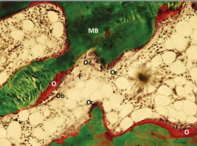
High-turnover disease encompasses the spectrum of hyperparathyroid bone disease (from mild hyper-parathyroid-related bone disease to osteitis fibrosa) and mixed uremic bone disease. The first one is attributable to secondary hyperparathyroidism and is characterized by an increased bone formation rate, a high number of osteoclasts and osteoblasts, an increased number of re-sorption lacunae and increased amount of osteoid al-beit with normal mineralization (Figure 1). In severe forms there can also occur marrow fibrosis (osteitis fi-brosa)17. Mixed uremic bone disease combines in-creased bone remodelling and abnormal mineraliza-tion with low to normal volume (Figure 2)17,31.
Low-turnover disease includes osteomalacia and adynamic bone disease. Osteomalacia was formerly associated with aluminum toxicity and common in dialy -sis patients, but its incidence has significantly decreased
and nowadays is rarely seen32. Other potential etiolo-gies of osteomalacia can also occur in CKD patients, including 25-hydroxyvitamin D deficiency, metabolic acidosis and hypophosphatemia17. It is characterized by increased osteoid volume and thickness, increased osteoid maturation time, longer mineralization lag time and low to medium bone volume (Figure 3).
In adynamic bone disease the main findings include the absence or reduced number of osteoclasts and os-teoblasts, markedly reduce bone formation rate and acti vation frequency, along with normal to decreased amount of osteoid, normal mineralization and low or normal volume (Figure 4)17. This may be the result of a relative hypoparathyroidism caused by an overtreat-ment of hyperparathyroidism with vitamin D and cal-cium supplementation and/or calcimimetics17. Its prevalence is steadily increasing, especially in patients on peritoneal dialysis23,32,33. Other risk factors comprise advanced age, corticosteroid use and diabetes mellitus 32.
In spite of being a valuable tool, bone biopsy is un-derused due to a variety of factors: invasiveness of the procedure, the lack of technical training, the limited number of specialized centres with expertise to inter-pret and process bone samples and costs17. Moreover, the clinical relevance of the information provided by bone biopsy in this context is underestimated by most of practising physicians.
mAnAgement ImplIcAtIons from bone bIopsy result
The KDIGO working group recommends to treat osteoporosis in patients with stages 13a CKD as the gene -ral population, as long as there are no accompanying biochemical abnormalities indicative of the presence of CKD-MBD, since there is evidence from clinical trials that included patients with similar renal
impair-fIgure 1. Iliac crest biopsy histology with goldner staining. showing hyperparathyroid-related bone disease. (F: fibrosis; MB: mineralized bone; O: osteoid; Ob: osteoblasts; Oc: osteoclasts)
fIgure 2. Mixed uremic bone disease with an increased extent and thickness of osteoid seams, activated osteoblasts and fibrosis (F: fibrosis; MB: mineralized bone; O: osteoid; Ob: osteoblasts)
tAble I. hIstomorphometrIc fIndIngs In dIfferent metAbolIc bone dIseAses
Bone disease Turnover Mineralization Volume
Renal osteodystrophy
Osteomalacia Low Abnormal Low to normal
Adynamic bone disease Low Normal/Abnormal Low to normal
Hyperparathyroid bone disease High Normal Low, normal or high
Mixed uremic osteodystrophy Normal to high Abnormal Low to normal
ment, that efficacy and safety of antiosteoporotic thera -pies are similar in both groups22,34.
However, establishing the best therapeutic strategy in patients with severe CKD (stages 3b-5) or with CKD--MBD, who have sustained a low-trauma fracture, is more difficult34-36.
In first place, it is essential to make the correct diagno sis and distinguish between the various forms of ROD and osteoporosis, especially if the physician is
considering using anti-osteoporotic pharmacologic agents designed to improve bone strength and reduce the risk of fracture.
Nonetheless, there is an impressive lack of evidence for the efficacy and safety of these therapeutic agents in patients with more advanced CKD stages, which are the ones who present ROD more frequently.
Although available data shows a reduction in the rate of hip fractures and stabilization and even a slight de-fIgure 3. Osteomalacia showing increased extent and
thickness of osteoid seams, without active bone cells (MB: mineralized bone; O: osteoid)
fIgure 4. Adynamic bone disease showing marked reduction of bone volume and trabecular connectivity and absence of osteoid. (T: trabeculae)
Bone biopsy
Low bone turnover
Anabolic agents
Teriparatide Abaloparatide
Normal to high bone turnover
Antiresorptive agents
Biphosphonates Denosumab
Raloxifene
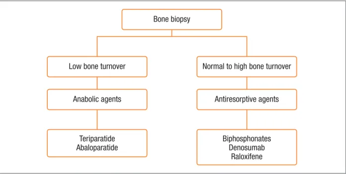
fIgure 5. A conceptual model for selection of anti-fracture treatment based on bone biopsy histomorphometry. Drugs are men-tioned as examples for each mode of action.
crease in the rate of hip and vertebral fractures between 2004 and, respectively, 2010 and 200937,38, in hemodialysis patients, the risk of fracture is still very high in this group of patients: 4–13 times higher in dialysis patients compared to the general population6. This suggests that these patients have unmet needs in relation to bone disease.
Overall treatment decisions should be guided by the presence of fractures and results from BMD as mea-sured by DXA, and ideally the choice of agent should be based on transiliac bone biopsy (Figure 5)34,35.
It should be highlighted that before initiating any antifracture therapy, CKD-MBD should be managed as recommended22, and nonpharmacologic measures should be implemented in all patients, including adequate nutrition and sun exposure, fall prevention, regu -lar weight-bearing exercise, avoidance of excessive al-cohol intake and smoking cessation39.
low bone turnover
As previously stated, low bone turnover states, name-ly adynamic bone disease, are steadiname-ly increasing33. Al-though there are no objective data to confirm it, ad-ministering an antiresorptive therapy in these context would, in theory, be harmful and deteriorate bone quali ty21,35.
Conceptually, anabolic agents, such as teriparatide, a recombinant form of PTH could have a beneficial effect and improve bone remodelling in patients with low turnover bone disease32,40. However, the use of teri-paratide is limited to up to 24 months and should be followed by the prescription of an antiresorptive, but that the latter will be contraindicated in the patient with low turnover bone disease, which renders teriparatide use difficult and perhaps explains the absence of en-thusiasm for this drug in this context. Also the evidence supporting teriparatide use in patients with CKD 3-5D is anedoctal and comes from case reports and small obser vational studies41-43. Palcu et al. 41reported a case of a patient under dialysis with multiple fractures and low turnover bone disease on biopsy that was treated with teriparatide 20 g/day for 24 months, with reso-lution of pain, increased BMD in femoral neck and to-tal hip and improvements in histomorphometric pa-rameters.
Cejka et al. administered teriparatide 20 g/day for 6 months to 7 patients with on haemodialysis with biopsy-proven adynamic bone disease, and showed a
significant increase in lumbar BMD in 6 of the 7 pa-tients42. In another study, lumbar spine BMD also in-creased after teriparatide was administered once week-ly to patients on diaweek-lysis with hypoparathyroidism and a T score <-2.5 at the spine, hip, or forearm or a T score between -2.5 and -1.0 with a prevalent fragility frac-ture43.
It is important to highlight that all but one of the cases included in the cited studies 41-43were associated with true hypoparathyroidism, as well as the exception related to functional hypoparathyroidism.
Nevertheless, teriparatide effect on fractures is un-known. Regarding safety, patients may be more prone to hypercalcemia and cardiovascular security has not been evaluated36,39.
Abaloparatide, an analogue of PTH-related peptide, is another promising osteoanabolic agent, with less risk of hypercalcemia. However, is has not been studied in patients with CKD-MBD39.
normAl to hIgh turnover
On the other hand, the presence of normal to high bone turnover on bone biopsy would support treatment with an antiresorptive agent 32,40.
Bisphosphonates, which are the most commonly used antiresorptive drugs, have a high affinity for bone hydroxyapatite, and persist in the skeleton for many years. As their clearance depends on renal excretion, excessive accumulation of the drug in the skeleton in CKD is a particular concern44. There is limited data on bisphosphonates effects on BMD, fractures and safety in CKD 3-5D36. A randomized trial in stage 3–4 CKD comparing 18 months of alendronate 70 mg/week with placebo reported an increase in lumbar spine T-score45. However, this was a small study with 51 patients in which the primary endpoint was the effect of alen-dronate on vascular calcification, with BMD evaluated as secondary endpoint. Also, patients with creatinine clearance <25mL/min were excluded, resulting that mean GFR in the patients included was 35.1mL/ /min/1.73m2. Thus, the population would be mostly in CKD stage 3. It is also important to remember that the observed effect on spine BMD as measured by an-teroposterior DXA could potentially be affected by the presence of extensive abdominal aortic calcifications common in these patients45. Considering a dose re-duction by 50% and shorter duration of treatment (2--3 years) has been recommended when administering
these drugs34,35.
Denosumab, the monoclonal antibody against re-ceptor activator of NF- B ligand (RANK-L), also inhibits osteoclast activity and proliferation, but has the advan -tage of not having GFR restrictions as it is not cleared by the kidney44. In small open-label uncontrolled pilot study, 12 patients on haemodialysis associated with severe secondary hyperparathyroidism (iPTH > 1000pg/mL) and low BMD (T-score < −1.0) received a single 60 mg subcutaneous dose of denosumab and after 6 months both lumbar spine and femoral neck BMD significantly increased46. Jamal et al. 47showed that 73 and 2817 women in the FREEDOM Trial were in CKD stage 4 and 3, respectively. There was no in-teraction between treatment effect and kidney func-tion, and adverse events did not differ by CKD stage. Denosumab increased BMD at the spine and hip and re-sulted in lower incidence of vertebral fractures in all patients except for CKD 4. Moreover, the incidence of nonvertebral fractures was lower among those ran-domized to denosumab compared with placebo but was not statistically significant for stages 3 and 4 CKD. It should be noted that the number of patients with CKD 4 was limited and there were no patients with stage 5.
There are some safety concerns with denosumab. The risk of hypocalcaemia, which can be more severe and prolonged, seems to be higher36. Therefore, prior to its administration, these patients must be supple-mented with vitamin D, have adequate calcium intake and during treatment serum calcium levels must be carefully monitored39. It appears that infections and diarrhoea are also more common in these patients36.
Raloxifene, a selective oestrogen receptor modulator with antiresorptive action, has been studied in post-menopausal women with varying degrees of CKD severity, including stage 5D48-50. One study48, which was a subgroup analysis of a randomized, controlled trial, reported a significantly higher annualized percentage change in femoral neck BMD in the raloxifene group than in the placebo group. However, the other 2 stu dies did not report any statistically significant differen -ces in femoral neck BMD absolute scores, T-scores, or
Z scores 49,50. One study compared raloxifene with placebo and reported reduced risk of vertebral fractu -res in postmenopausal women48. However this effect was only statistically significant for patients with GFR > 45 mL/min/1.73m2.
None of these studies reported statistically signifi-cant differences between raloxifene and placebo in
lumbar spine BMD, or incident nonvertebral fractures. Regarding safety, there is the special concern of in-creased risk for venous thrombosis when using this drug 51.
conclusIon
CKD patients are particularly prone to fragility frac-tures, not only in the context of osteoporosis, whose diagno sis represents a true challenge in advanced stages, but also due to the presence of ROD. Tetracy-cline double-labelled transiliac bone biopsy with quan-titative histomorphometric analysis is currently the only tool to differentiate between the various forms of ROD and to establish the diagnosis of osteoporosis, often by exclusion.
Although it has been recognized that management of patients with stages 1-3a CKD with fragility fractures can be similar to that of the general population, in more advanced CKD a bone biopsy should be considered prior to initiating treatment, in order to accurately re -cognize the type of bone disease present.
Additionally, there is little evidence to guide anti-fracture treatment in these patients, as this drugs have not been developed or conveniently studied in CKD--MBD, so there is an urgent need for further research in this area.
correspondence to
Francisca da Rocha Aguiar
Rheumatology Department, Centro Hospitalar São João, Porto, Portugal
E-mail: [email protected]
references
1. Pimentel A, Urena-Torres P, Zillikens MC, Bover J, Cohen-Solal M. Fractures in patients with CKD-diagnosis, treatment, and prevention: a review by members of the European Calcified Tis-sue Society and the European Renal Association of Nephrology Dialysis and Transplantation. Kidney Int 2017;92(6):1343--1355.
2. Alem AM, Sherrard DJ, Gillen DL, Weiss NS, Beresford SA, Heckbert SR, et al. Increased risk of hip fracture among patients with end-stage renal disease. Kidney Int 2000;58(1):396-399. 3. Kim SM, Long J, Montez-Rath M, Leonard M, Chertow GM. Hip Fracture in Patients With Non-Dialysis-Requiring Chronic Kid-ney Disease. J Bone Miner Res 2016;31(10):1803-1809. 4. Wakasugi M, Kazama JJ, Narita I. High rates of death and
hos-pitalization follow bone fracture among hemodialysis patients. Kidney Int 2014;86(3):649.
5. Diamond T, Elder GJ. Is there a practical role for bone biopsy in chronic kidney disease? Nephrology (Carlton) 2017;22 Sup-pl 2:22-26.
Exp Nephrol 2017;21(Suppl 1):46-52.
7. Kanis JA, Johnell O, De Laet C, Johansson H, Oden A, Delmas P, et al. A meta-analysis of previous fracture and subsequent fracture risk. Bone 2004;35(2):375-382.
8. NIH Consensus Development Panel on Osteoporosis Preven-tion, Diagnosis, and Therapy, March 7-29, 2000: highlights of the conference. South Med J 2001;94(6):569-573.
9. Babayev R, Nickolas TL. Bone Disorders in Chronic Kidney Dis-ease: An Update in Diagnosis and Management. Semin Dial 2015;28(6):645-653.
10. Kanis JA. Assessment of fracture risk and its application to screening for postmenopausal osteoporosis: synopsis of a WHO report. WHO Study Group. Osteoporos Int 1994;4(6):368-381. 11. K/DOQI clinical practice guidelines for chronic kidney disease: evaluation, classification, and stratification. Am J Kidney Dis 2002;39(2 Suppl 1):S1-266.
12. Ishani A, Paudel M, Taylor BC, Barrett-Connor E, Jamal S, Canales M, et al. Renal function and rate of hip bone loss in old-er men: the Osteoporotic Fractures in Men Study. Osteoporos Int 2008;19(11):1549-1556.
13. Jassal SK, von Muhlen D, Barrett-Connor E. Measures of renal function, BMD, bone loss, and osteoporotic fracture in older adults: the Rancho Bernardo study. J Bone Miner Res 2007;22 (2):203-210.
14. Fried LF, Shlipak MG, Stehman-Breen C, Mittalhenkle A, Seliger S, Sarnak M, et al. Kidney function predicts the rate of bone loss in older individuals: the Cardiovascular Health Study. J Gerontol A Biol Sci Med Sci 2006;61(7):743-748.
15. Nickolas TL, Stein EM, Dworakowski E, Nishiyama KK, Ko-mandah-Kosseh M, Zhang CA, et al. Rapid cortical bone loss in patients with chronic kidney disease. J Bone Miner Res 2013;28(8):1811-1820.
16. Zheng CM, Zheng JQ, Wu CC, Lu CL, Shyu JF, Yung-Ho H, et al. Bone loss in chronic kidney disease: Quantity or quality? Bone 2016;87:57-70.
17. Evenepoel P, Behets GJS, Laurent MR, D'Haese PC. Update on the role of bone biopsy in the management of patients with CKD-MBD. J Nephrol 2017;30(5):645-652.
18. Graciolli FG, Neves KR, Barreto F, Barreto DV, Dos Reis LM, Canziani ME, et al. The complexity of chronic kidney disease-mineral and bone disorder across stages of chronic kidney dis-ease. Kidney Int 2017;91(6):1436-1446.
19. Moe S, Drueke T, Cunningham J, Goodman W, Martin K, Ol-gaard K, et al. Definition, evaluation, and classification of renal osteodystrophy: a position statement from Kidney Disease: Im-proving Global Outcomes (KDIGO). Kidney Int 2006;69(11): 1945-1953.
20. KDIGO clinical practice guideline for the diagnosis, evaluation, prevention, and treatment of Chronic Kidney Disease-Mineral and Bone Disorder (CKD-MBD). Kidney Int Suppl 2009(113): S1-130.
21. Miller PD. Bone disease in CKD: a focus on osteoporosis diag-nosis and management. Am J Kidney Dis 2014;64(2):290-304. 22. Ketteler M, Block GA, Evenepoel P, Fukagawa M, Herzog CA, McCann L, et al. Executive summary of the 2017 KDIGO Chronic Kidney Disease-Mineral and Bone Disorder (CKD-MBD) Guideline Update: what's changed and why it matters. Kidney Int 2017;92(1):26-36.
23. Carvalho C, Alves CM, Frazao JM. The role of bone biopsy for the diagnosis of renal osteodystrophy: a short overview and fu-ture perspectives. J Nephrol 2016;29(5):617-626.
24. Sprague SM, Bellorin-Font E, Jorgetti V, Carvalho AB, Malluche HH, Ferreira A, et al. Diagnostic Accuracy of Bone Turnover Markers and Bone Histology in Patients With CKD Treated by Dialysis. Am J Kidney Dis 2016;67(4):559-566.
25. Evenepoel P, Bover J, Urena Torres P. Parathyroid hormone metabolism and signaling in health and chronic kidney disease. Kidney Int 2016;90(6):1184-1190.
26. Gal-Moscovici A, Sprague SM. Role of bone biopsy in stages 3 to 4 chronic kidney disease. Clin J Am Soc Nephrol 2008;3 Sup-pl 3:S170-174.
27. Vidal B, Pinto A, Galvao MJ, Santos AR, Rodrigues A, Cascao R, et al. Bone histomorphometry revisited. Acta Reumatol Port 2012;37(4):294-300.
28. Trueba D, Sawaya BP, Mawad H, Malluche HH. Bone biopsy: in-dications, techniques, and complications. Semin Dial 2003;16(4):341-345.
29. Torres PU, Bover J, Mazzaferro S, de Vernejoul MC, Cohen-So-lal M. When, how, and why a bone biopsy should be performed in patients with chronic kidney disease. Semin Nephrol 2014;34(6):612-625.
30. Ott SM. Renal Osteodystrophy-Time for Common Nomencla-ture. Curr Osteoporos Rep 2017;15(3):187-93.
31. Kulak CA, Dempster DW. Bone histomorphometry: a concise re-view for endocrinologists and clinicians. Arq Bras Endocrinol Metabol 2010;54(2):87-98.
32. Brandenburg VM, Floege J. Adynamic bone disease-bone and beyond. NDT Plus 2008;1(3):135-147.
33. Malluche HH, Mawad HW, Monier-Faugere MC. Renal os-teodystrophy in the first decade of the new millennium: analy-sis of 630 bone biopsies in black and white patients. J Bone Min-er Res 2011;26(6):1368-1376.
34. Miller PD. Fragility fractures in chronic kidney disease: an opi -nion-based approach. Cleve Clin J Med 2009;76(12):715-723. 35. West SL, Patel P, Jamal SA. How to predict and treat increased fracture risk in chronic kidney disease. J Intern Med 2015;278 (1):19-28.
36. Wilson LM, Rebholz CM, Jirru E, Liu MC, Zhang A, Gayleard J, et al. Benefits and Harms of Osteoporosis Medications in Pa-tients With Chronic Kidney Disease: A Systematic Review and Meta-analysis. Ann Intern Med 2017;166(9):649-658. 37. Arneson TJ, Li S, Liu J, Kilpatrick RD, Newsome BB, St Peter
WL. Trends in hip fracture rates in US hemodialysis patients, 1993-2010. Am J Kidney Dis 2013;62(4):747-754.
38. Wagner J, Jhaveri KD, Rosen L, Sunday S, Mathew AT, Fishbane S. Increased bone fractures among elderly United States hemodialysis patients. Nephrol Dial Transplant 2014;29(1): 146-151.
39. Khairallah P, Nickolas TL. Management of Osteoporosis in CKD. Clin J Am Soc Nephrol 2018.
40. Cejka D, Haas M. Should teriparatide ever be used for adynamic bone disease? Semin Dial 2011;24(4):431-433.
41. Palcu P, Dion N, Ste-Marie LG, Goltzman D, Radziunas I, Miller PD, et al. Teriparatide and bone turnover and formation in a hemodialysis patient with low-turnover bone disease: a case re-port. Am J Kidney Dis 2015;65(6):933-936.
42. Cejka D, Kodras K, Bader T, Haas M. Treatment of Hemodialy-sis-Associated Adynamic Bone Disease with Teriparatide (PTH1--34): A Pilot Study. Kidney Blood Press Res 2010;33(3):221--226.
43. Sumida K, Ubara Y, Hoshino J, Mise K, Hayami N, Suwabe T, et al. Once-weekly teriparatide in hemodialysis patients with
hypoparathyroidism and low bone mass: a prospective study. Osteoporos Int 2016;27(4):1441-1450.
44. Khairallah P, Nickolas TL. Management of Osteoporosis in CKD. Clin J Am Soc Nephrol 2018;13(6):962-969.
45. Toussaint ND, Lau KK, Strauss BJ, Polkinghorne KR, Kerr PG. Effect of alendronate on vascular calcification in CKD stages 3 and 4: a pilot randomized controlled trial. Am J Kidney Dis 2010;56(1):57-68.
46. Chen CL, Chen NC, Hsu CY, Chou KJ, Lee PT, Fang HC, et al. An open-label, prospective pilot clinical study of denosumab for severe hyperparathyroidism in patients with low bone mass undergoing dialysis. J Clin Endocrinol Metab 2014;99(7):2426--2432.
47. Jamal SA, Ljunggren O, Stehman-Breen C, Cummings SR, Mc-Clung MR, Goemaere S, et al. Effects of denosumab on fracture and bone mineral density by level of kidney function. J Bone Miner Res 2011;26(8):1829-1835.
48. Ishani A, Blackwell T, Jamal SA, Cummings SR, Ensrud KE. The effect of raloxifene treatment in postmenopausal women with CKD. J Am Soc Nephrol 2008;19(7):1430-1438.
49. Haghverdi F, Farbodara T, Mortaji S, Soltani P, Saidi N. Effect of raloxifene on parathyroid hormone in osteopenic and osteo-porotic postmenopausal women with chronic kidney disease stage 5. Iran J Kidney Dis 2014;8(6):461-466.
50. Hernandez E, Valera R, Alonzo E, Bajares-Lilue M, Carlini R, Capriles F, et al. Effects of raloxifene on bone metabolism and serum lipids in postmenopausal women on chronic hemodial-ysis. Kidney Int 2003;63(6):2269-2274.
51. Adomaityte J, Farooq M, Qayyum R. Effect of raloxifene thera-py on venous thromboembolism in postmenopausal women. A meta-analysis. Thromb Haemost 2008;99(2):338-342.