Bond energy/bond order relationships for N–O linkages
and a quantitative measure of ionicity:
the role of nitro groups in hydrogen-bonding
Robert A. W. Johnstone,*a Rui M. S. Loureiro,a,c M. Lurdes S. Cristiano,*b and Gaël Labata
a
Department of Chemistry, University of Liverpool, Liverpool L69 3BX, UK.
b
CCMAR e DQB, Universidade do Algarve, Campus de Gambelas, 8005-139 Faro, Portugal
c
Hovione FarmaCiencia SA, Sete Casas, 2674-506 Loures, Portugal E-mail: [email protected], [email protected]
Abstract
The nitro group is active in metabolic systems and can be found as an integral part of a number of useful curative drugs and many toxic substances. The basis for much of this activity is not fully understood. It is not necessarily caused directly by through-bond electronic effects but may also be due to direct H-bonding to nitro or to indirect interference by the nitro group with existing H-bonding. An unusual effect of a nitro substituent on kinetic results from urethane addition/elimination reactions (Scheme 1) has been ascribed to some form of self-association, which was neither specified nor quantified. To investigate self-association phenomena caused by a nitro group, a bond energy/bond order formula for N–O bonds has been developed and then used to interpret relative amounts of covalent and ionic contributions to total N–O bond energy. Calculated bond energies were then used to obtain enthalpies of formation for H-bonds to nitro groups in crystals and in solution. Similar results from solution data reveal that direct H-bonding to nitro is much weaker than in crystals, unless intramolecular H-bonding can occur. The results revealed that the 'self-association' effects observed for nitro substituents in urethanes (Scheme 1) were not caused by nitro participating directly in intermolecular bonding to NH of another urethane but by an indirect intramolecular action of the nitro group on pre-existing normal NH– O amide/amide type H-bonding.
Keywords: Bond Energy/Bond Order relationships, ionicity, N-O linkages, nitro groups and
hydrogen-bonding, addition/elimination of arylurethanes
Introduction
Unusual behaviour of the nitro group can be seen easily in results from kinetic experiments on the elimination/addition reaction of substituted arylurethanes (Scheme 1). For a wide variety of
substituents R, the enthalpies and entropies of the reaction shown in Scheme 1 have very similar magnitudes (60.6 ± 3.1 kJ.mol-1 and 125.6 ± 7.9 J.mol-1K-1) except for the nitro substituent, for which the corresponding values are very much greater (89.0 kJ.mol-1 and 228.6 J.mol-1K-1).1
Scheme 1
Self-association of nitrourethanes was suggested as the reason for the difference. Normal NH–O mutual H-bonding effects between NH-C=O amidic sections of urethanes are expected to be moderately strong but there seems no obvious reason for the nitro group to induce exceptionally strong association except perhaps through additional amide H-bonding to the nitro group itself. Accordingly, H-bonding to nitro has been examined in both the solid phase and in solution. In the crystal state, nitro/amide H-bonding can be strong enough to disrupt normal amide/amide bonding. In solution, there appears to be no evidence for strong intermolecular H-bonding between an aromatic nitro group and any H-donor, except when intramolecular H-bonding is favourable. Results from experimental data have now shown that, in solution, a nitro group does not take part significantly in direct bonding to amide but it does uniquely and indirectly promote dimeric amide/amide bonding in urethanes, rather than linear amide/amide H-bonding. There are no direct through-bond electronic effects of the nitro group on amide/amide self-association.
In exploring the existence of direct H-bonding to a nitro group, it was decided to examine the strength of any such bonding quantitatively in both crystal and solution phases. For this purpose, N–O bond energy/bond order relationships were developed and were then applied to bond length data from X-ray measurements. The results gave good estimates for the magnitudes of enthalpies typical of H-bonding. H-Bond energies in solution were obtained directly by 1H-NMR experiments.
Results and Discussion
Following Pauling's adoption of a semi-empirical approach to understanding molecular bonding and reactivity2a there have been efforts to extend those ideas and to make them more quantitatively accurate outside the complications of formal ab initio or semi-empirical molecular orbital and valence bond theories. Coulson discussed the dilemmas involved in treating molecules as collections of almost independent bonds in relation to quantum methods, which consider molecular orbitals as encompassing all atoms to varying extents at any one instant.3a For isolated bonds in heteronuclear diatomic molecules, Coulson demonstrated that a total wave
function (Ψ) should comprise a covalent part (Ψcovalent) and an ionic part (Ψionic) to allow for ionic contributions to total bond energy. The two terms are related by the expression, Ψ = Ψcovalent + λΨionic, which allows for the temporal probability that two electrons in a bond may both be near one of the atoms making up the bond. The probability of finding both electrons near one atom increases with increasing difference in electronegativities of the two atoms making up the bond and introduces ionic character into a covalent bond. The parameter λ is a measure of this ionicity. At that time there was no method available for obtaining reliable values for λ.3b Later, in ground-breaking work, Johnston and Parr demonstrated that, for many molecular types, the energies of individual bonds could be expressed simply in terms of localised energy/distance (Morse) functions, without the encumbrance of having to deal with the whole molecule.4 More energy terms could be added to accommodate Coulombic (electrostatic) attractive or repulsive forces. Based on these later developments, the whimsically named concept of Complete Neglect of Practically Everything was described, whereby individual bond lengths in molecules could even be used to understand mechanisms of reaction in ground state molecules.5a,b Although this bond energy/bond order (BEBO) approach is almost purely empirical and probably has no respectable standing in quantum mechanics, it does have the enormous advantage for practising chemists of providing accurate quantification of bond breaking/bond forming energies of reaction. It echoes the intuitive thinking used when describing reactivity in terms of resonance structures. As shown here, assessment of H-bond energy is one possible application.
The structure of a nitro group is pictorially best represented as a resonance hybrid 1 and not as the purely covalent form 2, which would imply a pentavalent nitrogen atom in modern terminology rather than simply the fact that all five valence electrons on nitrogen are utilised in forming the NO2 group. There are many indications of the importance of ionicity in a bond. For example, the isocyanates that take part in reaction (1) have significant contributions to the overall structure 3 from electrically charged resonance forms 4, 5. These ionic forms introduce additional attractive electrostatic forces into bonding, which lead to shortening (compression) of the covalent bond length so that fractional bond orders arise.
Non-integral bond orders do not necessarily imply changes in hybridisation states because, although the bond lengths typical of single, double or triple bonds can shorten considerably due to electrostatic forces, the corresponding bond angles typical of sp, sp2 and sp3 hybridisation change only little, if at all. For two electric charges (z+ = z- = 0.5 e) acting over a typical bond length of 1.6 Å, the Born-Landé equation shows that there would be 196 kJ.mol-1 of potential energy due to Coulombic forces alone.6 For comparison, a typical covalent single C–C bond with little or no ionicity has bond energy of 370 kJ.mol-1. In examining the formation of largely
electrostatic H-bonds to the nitro group, it was first necessary to account quantitatively for changes in ionicity of N–O bonds. This was achieved by developing bond energy/bond order formulae for N–O bonds.
The burgeoning fund of readily available accurate bond length data from X-ray and neutron diffraction spectroscopy on crystals, supported by microwave and ultraviolet spectroscopy on vapours provides an excellent foundation for exploring bond energy/bond order relationships.5b Significant changes in bond length and therefore in bond energy can be used to interpret reactivity, such as that reported for selective hydrogenolysis of diaryl ethers and diarylamines.7
The present work extends this bond length methodology by developing suitable bond length/bond order and bond energy/bond order formulae for N–O linkages. These formulae are then used to explore N–O bonding in several types of compounds in addition to those containing specifically a nitro group. These formulae were used to establish the magnitude of the ionicity parameter λ and to provide a quantitative measure of the relative importance of covalent and ionic contributions to total bond energy. Such derived ionic and covalent bond energies were used to predict electron densities, which were checked against similar information provided by κ refinement of X-ray structures. Another particularly valuable result lies in the ability of the method to calculate H-bond energies from bond length data obtained from crystals. Changes in bond energy in an H-acceptor can also be detected by infrared spectroscopy and can be used to assess enthalpies of H-bonding in solution.
Derivation of bond length/bond order and bond energy/bond order formulae
Bond length/bond order formula for N–O bonds. Development of this formula required a
selection of suitable bond lengths for N–O single, double and triple bonds. It has long been recognised that all bonds must contain part covalent and part ionic energy2a,3c but the choice of which bond lengths to choose for single, double and triple bond order markers can be guided by utilising the sums of single, double and triple covalent radii of atoms, in this case those of N and O.8 In addition, an assessment can be made as to the likely degree of ionicity of any bond selected to represent a single, double or triple bond. For example, a simple C–C double bond such as that in ethene is unlikely to have much ionic character and this is confirmed by its bond length of 1.338 Å, which is almost identical to the sum of covalent double bond radii of two carbon atoms (1.334 Å).
The details of which N–O bond lengths should be used to construct a suitable bond length/bond order formula are annotated.9 From these data, the formula Rn = 1.460 – 0.35*ln(n)
was derived, in which 1.460 is the bond length for a typical single N–O bond and Rn (Å) is any
other N–O bond of order n. Use of this formula allows fractional bond orders to be calculated quickly from bond length data. As a simple example, pyridine N-oxide has an N–O bond length Rn = 1.304 Å11d and insertion of this value into the formula gives a fractional bond order, n =
1.56. Hence, attractive ionic (Coulombic) forces in this single N–O bond lead to bond compression by 0.156 Å from a typical single N–O bond length of 1.460 Å (n = 1). Bond angles about the nitrogen atom do not change from those expected of the sp2 state. Therefore, in terms
of total bond energy, the N–O link appears to be about half way between a single and a double bond because of the ionicity in the bond. Other examples of N–O bonding are discussed below to show how individual covalent and ionic bond energies can be separated from total bond energies by using fractional bond orders.
Bond energy/bond order formula for N–O bonds. Once single, double and triple bonds had
been chosen to construct the bond length/bond order formula, it was necessary to assign relevant bond energies. Full details are appended.15 The resulting set of bond orders (n) and bond energies (Dn) give a good linear relationship, Dn = 256*n (kJ.mol-1). Thus, for pyridine N-oxide with n =
1.56 for the N–O bond, total bond energy is estimated to be 256*1.56 = 399.4 kJ.mol-1.
Total bond energy as a sum of covalent and ionic terms. The covalent energy contribution to
a resonance structure can be evaluated simply by considering the number of formal covalent bonds without concern for ionic contributions and then using the bond energy/bond order formula. For example, in pyridine N-oxide, the N–O linkage is notionally a single covalent bond (n = 1), which would be expected to have bond energy (Ecovalent) of just 256*1 = 256 kJ.mol-1. However, the total bond energy, Etotal (equivalent to Dn, as described above) from both covalent
and ionic contributions is obtained from the fractional bond order, n =1.56, viz., Etotal = D1.56 =
1.56*256 = 399.4 kJ.mol-1. Assuming no other bond energy contributions are present then, by difference, the ionic bond energy, Eionic = Etotal – Ecovalent = 143.4 kJ.mol-1. Thus, for any given N–O bond length, the total, covalent and ionic bond energies can be calculated. It may be noted that, in the wave function, Ψ = Ψcovalent + λΨionic, if the resulting energies Ecovalent and Eionic are significantly different, as they invariably are, then the functions Ψcovalent, Ψionic must be orthogonal or nearly so. The overlap integral, ∫Ψcovalent*Ψionic dτ, is everywhere equal or close to zero and the function Ψionic adds ionic bonding energy but does not interact resonantly with Ψcovalent.3d Once the resulting values of Eionic, Ecovalent and Etotal have been obtained, the percentage ionicity of the bond and the parameter λ can be evaluated accurately.2b, 3c, 3d For example, for the N–O bond in pyridine-N–Oxide, the percentage ionicity, I = 100*Eionic/Etotal = 100*143.4/399.4 = 35.9%. Since I = 100*λ2/(1+λ2) then λ = 0.75.3g The considerable percentage of ionic character (ionicity) reflects a large ionic contribution to the N–O bond in pyridine N-oxide and a total wave function Ψ = Ψcovalent + 0.75Ψionic.
Calculation of bond orders and ionicities in some simple molecules containing N–O bonds
Formally, nitrous acid (HNO2) has one single (HO–N; a) and one double bond (N=O; b), for which the bond lengths are respectively a = 1.432 and b = 1.170 Å.18a From the bond length/bond order formula, the respective bond orders are na = 1.08, nb = 2.29. The latter fractional bond order (2.29 in place of an expected 2.0) shows that the b bond has much more ionicity than does a (fractional bond order 1.08 instead of 1.0). The disparity in bond lengths and fractional bond orders illustrates one facet of N–O bonding in NO2 groups that appears to be quite general, viz., the two N–O bonds in NO2 are not necessarily equal in length, even when no hydrogen is present as it is in HNO2. Similarly, nitric acid has a single bond (HO≠N; 1.41 Å; n = 1.19) and two double bonds (N=O; 1.20 Å; n = 2.1).18b The single N–OH bond in nitric acid is
more ionically compressed than the comparable one in nitrous acid but the N=O bonds in nitric acid are not compressed to the same extent. Again, there is differential compression in the N–O bonds and significant ionicity. Similar differences in N–O bond lengths also extend to nitrite or nitrate anions. Both oxygen atoms of nitrite anion can co-ordinate to a single metal cation with overall near 2Cv symmetry. For example, in nitrito bis-2,2'-bipyridyl copper(II) nitrate, the N–O bond lengths of the chelating nitrite ligand are 1.234 and 1.207 Å (n = 1.91, 2.1 respectively) viz., the N–O bonds are of different lengths and each appears to be almost a double bond.19
In contrast to nitrites and nitrates with their sp2 symmetry, trisodium orthonitrate (Na3NO4) has been shown to crystallise with regular tetrahedral symmetry in the NO43- group.20 The four N–O bonds have identical lengths, despite there being only three formal negative charges. The result indicates considerable ionicity (polarity) in each bond and the tetrahedral arrangement proves that pd-π hybridization cannot be involved.20 From the bond length of 1.390 Å, the bond order is 1.22, viz., there is considerable electrostatic compression in each single N–O bond. The bond energy (Etotal; n = 1.22) for each bond is 1.22*256 = 312.7 kJ.mol-1 and the covalent bond energy (Ecovalent; n = 1) is 1*256 = 256 kJ.mol-1. The ionic component of the bond energy is Eionic = Etotal – Ecovalent = 56.7 kJ.mol-1, giving a percentage ionicity I = 100*Eionic/Etotal = 18.1%. This degree of ionicity indicates that there must be 0.181 e of electric charge on each oxygen atom. There are also three electrons for the NO43- group, which must be shared amongst the four identical N–O bonds (0.75 e per bond). Total charge density per oxygen atom is 0.931 e, which is very close to the 1.0 e expected for each oxygen on the basis of the regular tetrahedral symmetry. The closeness of the observed and calculated electron densities gives confidence that the magnitude deduced for Eionic provides a reasonable estimate of ionic bond energy.
The above brief examination of some N–O bond lengths in various 'nitro' compounds demonstrates that, where more than one N–O bond appears in NO2, NO3, or NO4 groups, the individual bond lengths may be equal or may be quite disparate, due to differing proportions of covalent and ionic bond energy. This conclusion is significant when considering the two notionally identical N–O bond lengths of aromatic nitro compounds.
N–O bonds in NO2 groups in crystalline organic compounds
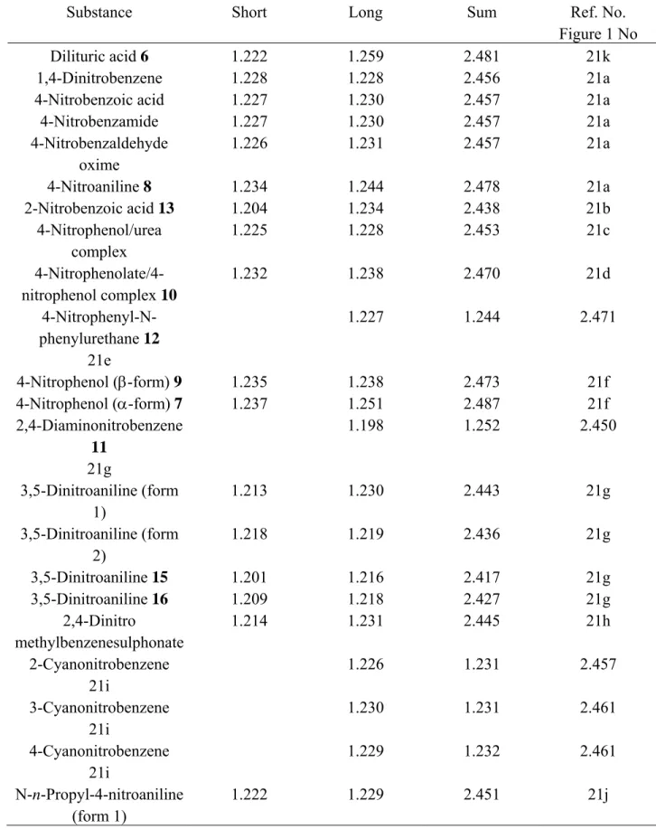
Modern X-ray and neutron diffraction methods have provided bond lengths of considerable accuracy, often with only small error limits in the fourth decimal place. To investigate variations in length for N–O bonds, a random selection of 28 NO2 groups was made from X-ray structures of aromatic nitro-compounds (Table 1).21a-k For each compound, N–O bonds were classified as short or long depending on their relative lengths in any one nitro group. The sum of N–O bond lengths for each NO2 group was obtained and then each half sum was chosen as a measure of the average N–O distance for any NO2 group. The mean of all half sums was 1.227 ± 0.0015 Å and this value was selected as the standard N–O bond length for aromatic nitro groups.
Table 1. Sums of N–O bond lengths (Å) of selected aromatic nitro-compounds, obtained from
X-ray structures
Substance Short Long Sum Ref. No.
Figure 1 No
Dilituric acid 6 1.222 1.259 2.481 21k
1,4-Dinitrobenzene 1.228 1.228 2.456 21a
4-Nitrobenzoic acid 1.227 1.230 2.457 21a
4-Nitrobenzamide 1.227 1.230 2.457 21a 4-Nitrobenzaldehyde oxime 1.226 1.231 2.457 21a 4-Nitroaniline 8 1.234 1.244 2.478 21a 2-Nitrobenzoic acid 13 1.204 1.234 2.438 21b 4-Nitrophenol/urea complex 1.225 1.228 2.453 21c 4-Nitrophenolate/4-nitrophenol complex 10 1.232 1.238 2.470 21d 4-Nitrophenyl-N-phenylurethane 12 1.227 1.244 2.471 21e 4-Nitrophenol (β-form) 9 1.235 1.238 2.473 21f 4-Nitrophenol (α-form) 7 1.237 1.251 2.487 21f 2,4-Diaminonitrobenzene 11 1.198 1.252 2.450 21g 3,5-Dinitroaniline (form 1) 1.213 1.230 2.443 21g 3,5-Dinitroaniline (form 2) 1.218 1.219 2.436 21g 3,5-Dinitroaniline 15 1.201 1.216 2.417 21g 3,5-Dinitroaniline 16 1.209 1.218 2.427 21g 2,4-Dinitro methylbenzenesulphonate 1.214 1.231 2.445 21h 2-Cyanonitrobenzene 1.226 1.231 2.457 21i 3-Cyanonitrobenzene 1.230 1.231 2.461 21i 4-Cyanonitrobenzene 1.229 1.232 2.461 21i N-n-Propyl-4-nitroaniline (form 1) 1.222 1.229 2.451 21j
Table 1. Continued
Substance Short Long Sum Ref. No.
Figure 1 No N-n-Propyl-4-nitroaniline (form 2) 1.222 1.235 2.457 21j N-n-Butyl-4-nitroaniline (form 1) 14 1.201 1.225 2.426 21j N-n-Butyl-4-nitroaniline (form 1) 1.215 1.222 2.437 21j N-n-Pentyl-4-nitroaniline 1.222 1.229 2.451 21j 4-Nitroacetanilide (form 1) 1.222 1.233 2.455 21e 4-Nitroacetanilide (form 2) 1.226 1.230 2.456 21e
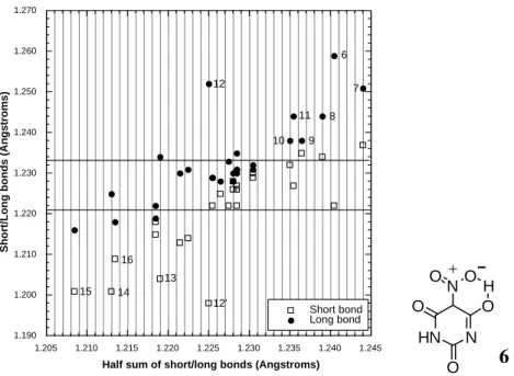
A chart of the individual short/long bond lengths and each corresponding half sum was constructed (Figure 1). Examination of the latter allows several conclusions to be drawn. (i) In atomic terms, there is a large spread in N–O bond lengths ranging from about 1.19 to 1.26 Å. (ii) Nine of the 28 examples have pairs of N–O bonds that are nearly equal to each other and to the average of 1.227 Å. (iii) Another group of 5 has slightly more disparate N–O bond lengths, which still lie reasonably close to the average. (iv) A third set of 7 nitro groups can be seen (Figure 1; compounds 6-1221a,d,e,f,g), in which each NO2 group possesses one N–O bond that is near the average length but for which the other is exceptionally long. As an example of this deviation, compound 6 (dilituric acid) 21k has one N–O distance (1.222 Å) close to the average but another, which is very much longer (1.259 Å). This set of 7 compounds is considered below in relation to H-bonding. (v) Finally, in the remaining set of 4 (Figure 1; compounds 13-1621a,g,j) each NO2 group has one N–O bond, which is near to the standard but also has one N–O bond that is exceptionally short. For example, N-n-butyl-4-nitroaniline 9 has one bond of 1.225 Å (near the average) but another, which is exceptionally short (1.201 Å). In fact, Figure 1 suggests that any NO2 group will have at least one N–O bond that lies within the 'standard' range of 1.227 ± 0.006 Å (four standard errors from the mean). However, a significant proportion of NO2 groups possesses one N–O bond that is either much longer or shorter than this standard length. These particularly long and short bonds are discussed separately below in relation to either attractive H-bonding or repulsive electrostatic forces.
1.190 1.200 1.210 1.220 1.230 1.240 1.250 1.260 1.270 1.205 1.210 1.215 1.220 1.225 1.230 1.235 1.240 1.245 Short bond Long bond Short/
Long bonds (Angstroms)
Half sum of short/long bonds (Angstroms)
6 7 8 9 10 11 12 12' 13 14 15 16 HN N NO O O H O O 6
Figure 1. For each NO2 group the two N–O bonds are shown as short (open squares) and long
(black dots). Each pair is related vertically. Ten pairs fall within four standard errors of the mean (1.226 Å) and lie between the horizontal lines, drawn at 1.220, 1.230 Å. These bonds are regarded as normal for "standard" NO2 groups and are not discussed further. Compounds 6-12 (long bonds) and 13-16 (short bonds) are discussed in the main text. All compounds can be identified from the list in footnote reference 21.
Standard N–O bond energies in NO2 groups
For purposes of illustration, the bonds in 1,4-dinitrobenzene21a serve as a suitable basis. Each of its two nitro groups has N–O bonds of equal length (1.2281(6), 1.2283(6) Å) and, from the bond length/bond order formula, they have the same bond order, n = 1.94. Simple resonance theory would suggest that each bond should have an order, n = 1.5, as implied by structure 1. The observed compression of the bond length to give n = 1.94 must be due an extra large ionic component. From the bond energy/bond order formula, for the two N–O bonds in an NO2 group, the total energy, Etotal = 2*1.94*256 = 993.4 kJ.mol-1 and the covalent energy, Ecovalent = 3*256 = 768 kJ.mol-1. By difference, Eionic = 225.4 kJ.mol-1. Thus, the percentage ionicity for a standard NO2 group is 100*225.4/993.4 = 22.6% and the effective charge on the nitro group is 0.226 e. This is almost identical to that found from an X-ray study with κ refinement, which gave 0.224 e as the effective charge.21a If it is assumed that the electric charge on each oxygen atom of the NO2 group is q and that the resultant balancing charge on the nitrogen is 2q then the mean charge 3q/2 = ±0.226 e and q = ± 0.149 e. Taking into account the relative electronegativities of nitrogen and oxygen, the electric charge on oxygen must be -0.149 and on nitrogen +0.298 e. These atomic charge densities were not given for 1,4-dinitrobenzene. However, in the same publication it was reported that, in 4-nitrobenzoic acid, having standard N–O bond lengths of 1.2268(7) and 1.2296(7) Å, the charge density on oxygen was -0.16 e and, on nitrogen, +0.31
e.21a These experimental and calculated magnitudes for the charges on oxygen and nitrogen in a standard NO2 group are almost identical, giving confidence in the accuracy and meaning of the terms Etotal, Ecovalent and Eionic, obtained from the N–O bond energy/bond order formula.
This result also suggests that any observed major variation from the standard N–O bond length of 1.227 Å may be used to estimate the strengths of external attractive or repulsive forces acting on the oxygen atoms of an NO2 group. A common attractive force found in crystals and operating at very short distances of only 2.2-2.8 Å is that due to H-bonding. It is shown below that such attractive H-bond forces give rise to N–O bond lengthening, which can be used to estimate the strength of the H-bond. In contrast, repulsive forces causing bond contraction seem to be mostly due to electronegative centres lying inside the normal van der Waals approach distance of about 3.4 Å.22
Hydrogen-bonding and long N–O bond lengths in NO2 groups
The oxygen atoms of an NO2 group are very weakly basic, the pKa for ArNO2H+ being approximately –11, which indicates that the group will be a poor H-acceptor. From sparse literature, it seems that H-bonding between a nitro group and known H-donors is weak in solution.23a,24,25 However, in crystals there are many instances in which the distance between a nitro oxygen acceptor atom and a hydrogen atom donor (HA) is very short, the hydrogen atom lying between donor and acceptor in such a way that the acceptor-hydrogen-donor angle is usually near to 180o. Typically, these close NO–H–A interactions in crystals are about 2.2-2.8 Å apart and are often noted generally as "hydrogen-bonds". In the present work, the only H-bonds considered to be significant are those, which have enthalpies greater than about 6-8 kJ.mol-1, particularly those involving alcohols, phenols, acids or amides.
There is little doubt that much of the energy for H-bonding arises through ionicity effects23h,3e but these do not need to be considered separately. Changes in N–O bond lengths are easily related to changes in the total bond energy (Etotal). Thus, Etotal (without H-bonding) > Etotal (with bonding) and the difference between them should be a measure of the enthalpy of the H-bond. From bond energy/bond order relationships, any reduction in Etotal should appear as an increase in length of the N–O bond.
Compounds 6-12 of Figure 1 exhibit longer than usual N–O bonds. The X-ray structural data for these compounds21 confirm that, in each case, one oxygen of a nitro group lies very close to a hydrogen atom of a good hydrogen donor. Table 1 lists these compounds, the relevant N–O bond lengths and Etotal for each N–O bond. In comparison with a standard N–O bond, the long N–O bonds in compounds 6-12 have smaller bond orders and smaller values for Etotal. By subtraction of Etotal(standard N–O bond) from Etotal(long N–O bond), H-bond enthalpies were calculated (Table 2).26 It is satisfying that these H-bond energies fall exactly in the range 8-46 kJ.mol-1 normally observed for typical H-bond enthalpies.23a,24
Entry 1 (Table 2) relates to dilituric acid 6, which is particularly interesting for its intramolecular hydrogen bond between a nitro group and an adjacent hydroxyl. From studies on H-bonding in ortho substituted aromatic compounds such an intramolecular bond to NO2 would
be expected to be strong.23b A bond energy calculation for dilituric acid 6 shows that the H-bond strength to NO2 is about 44 kJ.mol-1 (Table 1).
Table 2. Estimated hydrogen-bond energies N–O from bond energy/bond orders
Compound number 1 Bond length (Rn, Å) Bond order 2 Total bond energy (kJ.mol-1) ∆Dn 3 (kJ.mol-1) ∆Dn (kcal.mol-1) standard nitro 1.227 1.951 499.2 6 1.259 1.776 454.7 -44.5 -10.7 7 1.251 1.817 465.2 -34.1 -8.2 8 1.244 1.854 474.6 -24.6 -5.9 9 1.238 1.886 482.8 -16.4 -3.9 10 1.238 1.886 482.8 -16.4 -3.9 11 1.244 1.854 474.6 -24.6 -5.9 12 1.252 1.812 463.4 -35.8 -8.6 21N–O1 1.249 1.827 467.7 -31.5 -7.5 21N–O2 1.241 1.869 478.5 -20.7 -4.9
1Compounds are numbered as in Figure 1. Their names and reference numbers are as follows. Dilituric acid 6 [ref.Vj]; α-form of nitrophenol 7 [Vf]; nitroaniline 8 [Va]; β-form of nitrophenol 9 [Vf]; nitrophenolate complex 10 [Vd]; 2,diaminonitrobenzene 12 [Vg]; 4-nitrophenyl-N-phenylurethane 11 (19) [Ve]. 2Obtained from the bond length/bond order formula given in the main text. 3The difference (∆Dn) between the standard N–O bond energy (499.2 kJ.mol-1) and that of the compound having a long N–O bond; this is the hydrogen-bond energy.
Evidence for competitive nitro/amide and amide/amide H-bonding in crystals
Except for intramolecular bonding, the nitro group is not known to be a strong H-bonding acceptor in solution but, in the crystal state, it appears to be acting as a stronger base. This dichotomy of the NO2 group was apparent from an investigation of a small series of crystal structures of nitro compounds, in which intermolecular H-bonding between groups other than nitro was expected to be strong. Compounds were chosen to have amide and/or phenolic groups present because strong H-bonding between them is well known, as with amide/amide, phenol/phenol and phenol/amide interactions.23a,24 With its weak H-bonding performance in solution, the nitro group would not be expected to compete with these strongly H-bonding systems, which have H-bond strengths ranging between about 15 and 40 kJ.mol-1.22a,24 The compounds selected for X-ray structure examination were three urethanes 12, 17, 18 RC6H4OCONHPh; R = 4-NO2, 4-H, 4-OCH3 respectively), two acetanilides 19, 20 (RC6H4NHCOCH3; R = H, NO2) and a 1:1 molecular complex 21 of acetamide and 4-nitrophenol.27
The two acetanilides 19, 20 formed structures, in which there were polymeric chains of single amide/amide bonds (N-H…O=C), with N-H-O bond angles of 164o. In nitroacetanilide 20, similar polymeric amide/amide bonding was observed and the NO2 group did not exhibit any H-bonding character. Two urethanes 17 and 18 also had extended chain-like filaments composed of single amide/amide (N–H…O=C) bonds, with N–H–O bond angles of 162o. In marked contrast, the nitrourethane 12 was completely different. There was no chain-like amide/amide bonding but there was nitro/amide bonding (N–H…O=N-), with an N–H–O bond angle of 165o. The N–H–O distance was only 2.292 Å and the N–O bond of the NO2 group was long (1.252 Å), typical of strong H-bonding in the crystal state (entry 7, Table 1). Thus, in the crystalline state, urethanes
17, 18 and anilides 19, 20 behave like many amides with continuous amide/amide type bonding.
In contrast, nitrourethane 12 presents no amide/amide H-bonding at all but does exhibit strong nitro/amide association.
A most remarkable example of H-bonding to an NO2 group occurs in the 1:1 molar complex
21, which crystallises well from a variety of solvents and has a high, sharp (congruent) melting
point.27 Major features of its X-ray structure are illustrated in representational form in diagram
22.28 The continuous repeating structure has an acetamide dimer formed by (N–H) hydrogen
atom bonding to the carbonyl oxygen of a second acetamide molecule (single amide/amide bond). Also, it has the hydrogen from the phenolic OH groups bonding to the carbonyl of the two acetamide (phenol/amide bond) and one nitro groups chelating to the acetamide second hydrogen (a nitro/bis-amide bond). Thus, for each structural unit, there are six hydrogen bonds, each of bond energy about 24-28 kJ.mol-1. The total H-bonding energy for one unit of complex 21 is equal to about half that of a strong covalent bond. The remarkable stability of complex 21 is due to its multiple H-bonds, including an unusual chelated H-bond to a nitro group, similar to that found in some nitrito copper complexes.19 The two N–O bonds of the nitro group have different bond lengths (for one NO2 1.249, 1.231 Å and for the second 1.241, 1.236 Å) and different H-bonding distances (2.212Å to the long N–O bond and 2.284 Å to the shorter N–O bond).
A bond energy/bond order calculation shows that, for the two N–O bonds in complex 21, the total intermolecular H-bond energies are 31.5 and 20.7 kJ.mol-1. These values are similar to those
obtained for the other compounds (Table 2). These X-ray structural results provide graphic confirmation of the variable nature of the NO2 group as an H atom acceptor in the crystal state. In suitable circumstances the nitro group can compete with amide or phenol acceptors.
It may be noted that, in dipotassium nitroacetate, the dianion (O2CCHNO2)2- 23 has a nitro
group which is richer in electrons because of its conjugation to an adjacent carbanion centre. With extra electron density on the nitro oxygen atoms, the nitro group behaves as a much stronger basic acceptor and forms chelates to potassium ions that are indistinguishable from the chelates formed by the carboxylate group. In the crystal of salt 23, a regular array of potassium ions is present, with each K+ ion being co-ordinated to 8 oxygen atoms arising from both nitro and carboxylate groups.29 While this chelation to potassium is not H-bonding, it does illustrate how the bonds in the NO2 group can respond to changes in its ionicity, in this case making it similar to a carboxylate group as a cation-acceptor.
The above few structures reveal that, in the solid state, a nitro group can act as a strong H-acceptor, forming H-bonds of comparable strength to those found in amide/amide or amide/phenol association.
H-Bonding of nitro compounds onto solid supports
Chromatography on silica and similar inorganic adsorbents has been used to investigate the role of H-bonding on the relative rates of migration of various types of compound on various supports.23d,30 It has been proposed that some chromatographic surfaces possessing OH groups, such as SiOH on silica gel, should effect separations based on H-bonding. The presence of some substituent groups such as nitro in molecules can have a major impact on adsorption and movement through a chromatographic column. For similar molecular structures, relative movement through a silica gel column can be influenced by substituents through their different H-bonding characteristics. In the present work, Rf values for two sets of urethanes were compared by thin layer chromatography (TLC) on silica gel plates. Three urethanes 12, 17, 18 comprised one set and two analogues 24, 25 having long hydrocarbon tails provided the second set. The compounds are identical in the central urethane 'amide' system but they also possess additional H-bonding capabilities in their substituents (H, OCH3, NO2) and in the nature of the group attached to the urethane nitrogen atom (Ph or C12-alkyl chain). In toluene, the order of increasing movement along the TLC plate was H > OCH3 ≈ NO2 and, in dichloromethane the order was the same. The results indicate that urethane 17, having no substituent group moves fastest along the TLC plate. Urethanes 18, 12 having methoxy and nitro groups, which are known to form weak H-bonds to OH groups, moved slower along the plate but at similar rates, showing that their energies of H-bonding to SiOH are similar. For urethanes 24, 25 the bulky coiled hydrocarbon section makes contact between the small amide group and the silica surface difficult and reduces amide H-bonding to the silica surface. The more distant nitro and methoxy groups then have greater influence at the surface. Thus, for compounds 24, 25, the Rf values are more closely associated with the NO2 and OCH3 substituents, for which Rf = 0.15, 0.09 respectively in toluene and Rf = 0.82, 0.67 in dichloromethane. Methoxyurethane 18 moves
significantly more slowly than does urethane 12 bearing a nitro group, indicating that, for these compounds, H-bonding from nitro to SiOH is indeed somewhat weaker than similar bonding from the methoxy group.
Relative H-bonding energies of NO2 and OCH3 groups can be estimated from earlier work. Enthalpies of H-bonds have been correlated with infrared vibration frequency shifts (∆νs) in CH3OD.25 Nitrobenzene causes only about half the frequency shift of the OD bond in CH3OD (∆ νs = 52 cm-1) than does methoxybenzene (anisole; ∆ νs = 94 cm-1). The shifts indicate enthalpies of approximately 8.8 and 16.3 kJ.mol-1 respectively for H-bonding from nitrobenzene and anisole to CH3OD.23 The order of elution found during TLC of urethanes 12, 17, 18, 24, 25 is consistent with these values and implies that the strength of weak intermolecular H-bonding from SiOH to NO2 along the silica surface is only about half that of an OCH3 group. In the transition from the crystal state to that of a biphasic solid/liquid system, there is a large drop in the enthalpy of H-bonding to a nitro group. This change is apparent also in monophasic solution (discussed below).
N–O bond energy and H-bonding to the nitro group in solution
There has been very little quantitative research into H-bonding from donors AH to the nitro group in solution but what there is indicates that aliphatic or aromatic NO2 groups act only as weakly basic hydrogen acceptors.24,25 Typical enthalpies of AH/NO2 bonding range from about 1 to 8 or 9 kJ.mol-1. Only with intramolecular bonding, in which the NO2 acceptor and an AH hydrogen donor group can form part of a six-membered ring as with 2-nitroalcohols, is there stronger H-bonding of about 8-16 kJ.mol-1.31
H-bonding to NO2 examined by infrared spectroscopy. Most infrared spectroscopic research into H-bonding has concerned itself with shifts (∆νs) in the H-donor (A-H) stretching vibrations (νs) near 3600-3200 cm-1 or their overtones that appear when H-bonding is present but accurate quantitative interpretation of these shifts can be difficult to make.23c Nevertheless, certain criteria are useful for estimating the enthalpies of H-bonding. These are (a) the shift (∆νs/cm-1) of the monomer A–H vibration to the beginning of the dimeric or oligomeric H-bonded A–H vibration, (b) the ratio between this shift and the original frequency (∆νs/νs) and (c) the ratio of the shift to the peak half-width (∆νs/ν1/2). With (a), observed shifts (∆νs) correlate well with enthalpies of H-bonds over a range of about 4-35 kJ.mol-1. For (b), the ratio is almost constant for individual types of donor/acceptor H-bonding and, for (c), the ratio (∆νs/ν1/2) is about 1.6 for hydroxyl bonding to various bases.23e
In the present work, these criteria were used to investigate changes in the N–H stretching frequency of urethanes 12, 17, 18, under conditions for which there was no possibility for intramolecular H-bonding. Infrared spectra were measured between 4000 and 3000 cm-1 in tetrahydrofuran (THF). In such a solvent, H-bonding from the urethane N–H to the oxygen of THF almost entirely eliminates any amide/amide type self-association between urethane molecules. Amide/THF H-bonding is expected to dominate any other weaker association effects. Therefore, a strong direct effect of the-NO2 group through H-bonding to the urethane N–H, should appear as a significant change between urethanes 17, 18 having no nitro group and nitrourethane 12.
Experimentally, it was found that, throughout three orders of magnitude range of solution concentrations, at any one concentration, the spectra for the three urethanes were essentially identical in the region between 3600 and 3200 cm-1. At the lowest concentration (9.4 x 10⁻⁵ M), the H-bonded N–H frequency near 3250 cm-1 was only just observable, while at the highest concentration (9.4 x 10⁻² M), the non-bonded N–H frequency near 3600 cm-1 was only just apparent. For the respective urethanes 12, 17, 18, the shifts (∆νs) were 274, 260 and 277 cm-1; half-widths (ν1/2) were 146, 149, 146 cm-1, the ratios (∆νs/νs) were 0.078, 0.079 and 0.078 and the ratios (∆νs/ν1/2) were 1.9, 1.8, 1.9. Within the limits of experimental error, these values are essentially identical and show that, for each urethane 12, 17, 18, the enthalpy of intermolecular H-bonding between the urethane N–H and THF is almost constant and is approximately equal to 30 kJ.mol-1. This value would be expected for such a strong N–H–O type bond. There was no evidence for any significant contribution from specific intermolecular N–H/O2N bonding.
In an unusual approach to investigating H-bonding to a nitro group, work with nitro-alcohols has been reported.31 Instead of measuring infrared band shifts in donor A–H vibrations near 3600-3000 cm-1, the effects of H-bonding on infrared bands of the nitro acceptor near 1550 and 1350 were examined in detail.31 Generally, only small shifts in these bands were found for a range of x-nitro-alcohols (x = 1-4), having the hydroxyl and nitro substituents separated by different numbers of carbon atoms. With 2-nitroalcohols, in which intramolecular H-bonding is easy, as illustrated in structure 26, it was found that the symmetric and asymmetric O–N–O stretching vibrations of the nitro group were both shifted significantly to lower frequencies. The average shift observed for the symmetric vibration was from 1567 to 1549 cm-1 and for the asymmetric mode it was 1340 to 1315 cm-1. With the bond energy/bond order relationship developed here, it is now possible to use these results to quantify the strength of this intramolecular H-bonding between nitro and hydroxyl groups in solution. Assuming the N–O bonds in a nitro group act as simple harmonic oscillators then E
∝
k∝
ω2, where E is the bond energy, k is the bond force constant and ω is the bond vibration frequency in cm-1. If E1, E2 are the respective bond energies before and after H-bonding and ω1, ω2 are the corresponding vibration frequencies then E1/E2 = (ω1/ω2)2. As shown above for standard N–O bonds in the nitro group, E1 = 993.4 kJ.mol-1 and therefore E2 = E1*(1549/1567)2 = 970.7 kJ.mol-1. Intramolecular H-bonding (NO–H–O) causes a reduction in the nitro group bond energy of 22.7 kJ.mol-1 (5.4 kcal.mol-1). This is of similar magnitude to the changes found for H-bondingenthalpies in the crystal state (Table 1). It is notable that this relatively high value for H-bonding to the nitro group in solution is found only when intramolecular bonding can occur easily. From the bond length/bond order and bond energy/bond order formulae, a bond energy change of 22.4 kJ.mol-1 for a 2-nitroalcohol 26 in solution corresponds to an increase in N–O bond length from 1.227 to 1.235 Å. N RHC C H2 O H O O 26
It is clear that, in solution, intermolecular H-bonding to the nitro group is weak (1-8 kJ.mol-1) but, if the nitro group and an H-donor lie in close proximity so that, including the H atom, a
six-membered ring 26 can be formed, then H-bonding increases to about 23 kJ.mol-1.
H-bonding to NO2 examined by ultraviolet spectroscopy. Again, THF was used as the solvent. At the dilutions used for UV/visible spectroscopy, the large excess of THF ensures that only amide/THF association is important. Therefore, any unusual effects observed in UV/visible spectra with change in concentration should not be complicated by intermolecular H-bonding changes between urethane molecules. Solutions of each of the urethanes 12, 17, 18 in THF were
prepared at concentrations ranging from 10-7 to 10-1 M and their UV/visible spectra were
recorded in the range 240 (THF cut-off) to 400 nm. For urethanes 17, 18 at a concentration of
1.94x10⁻³ and 1.94x10⁻⁴ M respectively, a new charge transfer band appeared at 290, 298 nm
respectively. At 0.194 M, the εmax for this band was 9.5 for the unsubstituted urethane 17 and
14.0 for the methoxy substituted urethane 18. These bands increased steadily in intensity up to the maximum concentration studied. In contrast, the nitrourethane 12 showed a new charge
transfer band near 300 nm at very low concentration (1.95x10⁻⁵ M) and this increased in intensity
very rapidly with increase in concentration. At 1.94x10⁻⁴ M, the band had an εmax of 16,000. At
the highest concentration examined, the width of the charge transfer band completely dominated the spectrum from about 280 to 320 nm. At the same time, the initial 'nitro band' centred at about 270 nm increased more slowly with concentration and also moved hypsochromically until it merged with the 240 nm cut-off of the THF solvent.
These results indicate that, for urethanes 17, 18 the charge transfer band is weak and only
becomes moderately noticeable at about 1.94x10⁻² M. It is the sort of band expected for simple
intermolecular contact charge transfer between aryl rings. It might also be an intramolecular
charge transfer band between the aryl rings attached to the urethane group. In either case, the
very small εmax suggests that the amount of interaction (π-orbital overlap) is very small.32 In
marked contrast, the charge transfer band for the nitrourethane 12 was not only a thousand times
more intense but it was also observable at the lowest concentrations of 10-6 to 10-7 M. The very
high extinction coefficient is consistent with good π-orbital overlap. Since the band is observable even at very low concentrations and increases extremely rapidly with increasing concentration
there must be also very close easy contact between the aromatic rings, in keeping with intra- rather than intermolecular charge transfer. The concomitant shift and weakening in the 'nitro' band is consistent with polarisation changes in the direction of the nitro group and the 'borrowing' of intensity by the charge transfer band from the ground state structure of the urethane.32
Intermolecular charge transfer is unlikely to be observed as a major difference between urethanes 12, 17, 18 because normal molecular contacts should be little different for these similar structures. Contact dipole/dipole attractions are similar in all three urethanes and are not expected to be much different from dispersion forces in solution. However, the observed εmax values suggest that there is good overlap between the aryl rings in nitrourethane 12 but not such good overlap in urethanes 17, 18. Therefore, it is necessary to consider the possibility that the charge transfer band arises intramolecularly between the aryl rings. This source of the band would require the rings to lie close to each other with their planes parallel.
The amide system in a urethane has a small barrier to rotation about the carbonyl/N bond and two (trans or cis) conformations 27, 28 can be expected to be relatively stable states.33 In conformation 27, the aryl rings are arranged trans to each other and intramolecular charge transfer effects are impossible. In conformation 28, the aryl rings are cis to each other and the planes of the aryl rings can lie parallel and close to each other, facilitating intramolecular charge transfer.
A charge transfer band near 300 nm corresponds to energy of 4.14 eV.34 Resonance theory can be used to calculate the expected position of the band, knowing the ionisation energy, the electron affinity and the Coulombic attraction energy.35,36 Alternatively, using the position of the band and approximately known values for the ionisation energy and electron affinity, the calculation affords the Coulombic energy. For nitrourethane 12, this energy is 5.9 eV.34 Using known dipole moments, other calculations suggest a Coulombic attraction of 4.5-5.0 eV, giving an average of about 5 eV (480 kJ.mol-1).37 Allowing for dielectric constant in THF solution, Coulombic energy becomes 63 kJ.mol-1. For solution phases, this easily exceeds strong H-bonding energy and rivals moderately high barriers to rotation about an amide C–N bond. Thus, the position and strength of the charge transfer band and consideration of intramolecular dipolar forces indicate that conformation 28 is stabilised in urethanes 12, 17, 18 so that the aromatic rings lie close and parallel to each other. The intensities of the observed charge transfer bands indicates that any such stabilisation must be much more important for the nitrourethane 12 than for the other two urethanes 17, 18.
In an ordinary amide system, the trans conformation 27 gives rise to chain or polymer like amide/amide association 29, in which each H-bond has energy of about 8-14 kJ.mol-1 and entropy about 20 J.mol-1K-1. In contrast, the cis configuration 28 leads to dimers 30, having much greater enthalpies of about 40 kJ.mol-1 and to a loss in entropy of about 75-110 J.mol-1K-1. If the charge transfer band does stabilise dimer conformation 28, then amide/amide H-bonding is favoured and the expected H-bond enthalpy will be about 40 kJ.mol-1. If conformation 28 is not stabilised, H-bond enthalpy derived from amide/amide chain linking will be much smaller at
about 8-14 kJ.mol-1. Hence, the differences in amide/amide H-bond enthalpy and entropy between conformations 27 and 28 are about 30 kJ.mol-1 and 90 J.mol-1K-1.
R O N O H R O N H O 27 28
An alternative view would suggest that the much stronger dimeric amide/amide H-bonding in conformation 28 forces the aryl rings of the urethane to lie close together and the charge transfer band arises as a result of this imposed proximity. The exceptional intensity of the charge transfer band in nitrourethane 12 would then be due to the large dipolar effect of the nitro group. The two viewpoints may be distinguished by direct measurement of the magnitude of H-bonding in urethanes 12, 17, 18.
H-bonding to NO2 examined by 1nmr spectroscopy. The strength of amide/amide
H-bonding for urethanes 12, 17, 18 was measured by obtaining the association constant K from chemical shift data for the N-H proton in dry CHCl3.23f,38 The only strong H-bonding expected was either through two bonds (formation of dimers 30) or through single bonds (chain-like or linear bonding 29).
The measured equilibrium constants K at 298 K were respectively 27.0, 1.21, 1.22 mol-1. Thus, association constants for the unsubstituted urethane 17 and the 4-methoxy substituted urethane 18 were almost identical and represent very weak self-association. The value of K for the 4-nitro substituted urethane 12 is exceptionally large and is very close to that found for typical amide/amide self-association to form dimers.23a,24
The K value for the nitrourethane 12 suggests an enthalpy of 29-34 kJ.mol-1 and an entropy of 79-106 J.mol-1K-1 for its bond energy. The K values for urethanes 17, 18 indicate H-bonding energy of only 2-4 kJ.mol-1 and an entropy of 8-16 J.mol-1K-1.39 Therefore, the amide/amide H-bonding in the nitrourethane 12 is approximately 27-30 kJ.mol-1 greater than that for the other two urethanes and the entropy is about 70-90 J.mol-1K-1 greater.
It was also noted that, even at low concentrations, the chemical shifts of protons in the phenyl ring attached to the nitrogen atom of the nitrourethane 12 were moved up-field slightly from the values observed for the same aryl ring in urethanes 17, 18.
The results confirm that amide/amide bonding in the nitrourethane 12 is typical of amide/amide dimer formation 30, whereas the H-bond energies for urethane 17, 18 are quite small and are typical of linear amide/amide bonding 29. There are no direct links from the substituents to the H-bonding amide system and therefore there is no direct through-bond electronic effect to cause a change in H-bond energies. The marked differences in behaviour between urethanes 17, 18 and the nitrourethane 12 do show that the charge transfer band found in UV spectra of the urethanes does not appear because of amide/amide dimeric bonding but must be a cause of the bonding. The nitro group influences the type of amide/amide H-bonding
behaviour through strong dipolar attraction between the aryl rings of the nitrourethane 12. As demonstrated by the weak charge transfer bands, for urethanes 17, 18 this attractive force is largely absent. R O N O H R O N O H O H N R O N H O R O N H O 30 29
There is an extra factor to be considered in comparing amide/amide H-bonding in urethanes and simple amides. The bond angles in the aryl-O-CO-NH-aryl chain of any urethane allow the two aryl rings to approach each other in a parallel fashion in cis conformation 30. In contrast, the bond angles and bond lengths in the shorter aryl-CO-NH-aryl simple amide system prevent the terminal aryl rings attached to C=O and NH getting close to each other, let alone lying parallel in a cis conformation. Therefore, no parallel behaviour between nitroaryl urethanes and nitroaryl amides is expected.
H-bonding to NO2 examined by kinetic rate measurements. Enthalpies and entropies for
equilibrium reaction (1) have been reported for a range of urethanes, RC6H4OCONHPh, in which R represents a variety of substituents in the 3- and 4-positions and includes compounds 12, 17,
18.1 Equilibrium constants (K = k1/k-1) were also given. If the enthalpies and entropies are
averaged it is found that, except for 4-nitrourethane 12, these energies are very similar with ∆HR = 60.6 ± 3.1 kJ.mol-1 and ∆SR = 125.6 ± 7.9 J.mol-1K-1. 4-Nitrourethane 12 is strange, with values ∆HR = 89.0 kJ.mol-1 and ∆SR = 228.6 J.mol-1K-1, which lie well outside the deviation from the mean and are half as large again than the averages. From consideration of heats of formation, it is clear that all of the above substituted-aryl urethanes, including nitrourethane 12, would be expected to have almost the same enthalpy of reaction.41
The large increase in entropy for nitrourethane 12 is particularly revealing. Equilibrium reaction (1) can be expected to show a large entropy change because one molecule becomes two or vice versa. The entropy change for nitrourethane 12 is almost double the average entropy change, of all the other urethanes, indicating that, in the nitro case, one entity becomes four or vice versa during the reaction. If the nitrourethane forms dimers then one dimer becomes four molecules as reaction proceeds and the larger entropy change is explained. Dimer formation through H-bonding in solution has been measured here by 1H-nmr spectroscopy and is discussed above.
The exceptional effect of a nitro substituent on equilibrium reaction (1) can be investigated also by consideration of pK values. For this series of reactions of very similar compounds, it is likely that the transition state will have similar structure for all urethanes. In the elimination step of reaction (1), phenols are produced. If the transition state is one in which the phenol (acidity
constant Ka) is almost formed then it would be expected that pKa and pK should be related linearly.42 It can be shown that the kinetic data for reaction (1) do give a linear correlation over three orders of magnitude of pKa.43 For nitrourethane 12, the pK/pKa point lies exactly on this correlation line and proves that, in the transition state, the nitrourethane behaves like the other urethanes and is unexceptional. These data reinforce the view that the enthalpy of reaction for nitrourethane 12 differs significantly from that for any other similar urethane (reaction 1) because of its strong association in solution.
It was shown above that amide/amide bonding is dimeric for nitrourethane 12 and gives H-bond enthalpies, which are about twice those for urethanes 17, 18. In the latter, amide/amide association appears to be weakly chain-like. It is notable that the range of differences in enthalpy and entropy for H-bonding in nitrourethane 12 (∆H = 29-34 kJ.mol-1 and ∆S = 79-106 J.mol-1K -1) covers exactly the excess of enthalpies and entropies of reaction observed in kinetic experiments (∆H = 29 kJ.mol-1 and ∆S = 103 J.mol-1K-1). It must be concluded that the anomalous behaviour of nitro-substituted urethane 12 is due to its association to form dimers through amide/amide H-bonding. In turn, the required favourable conformation required for dimerisation is promoted by the structure of the urethane and by strong intramolecular dipolar binding between the large dipole of the nitroaryl group and a corresponding aligned dipole in the phenyl ring attached to the N atom of the amide section. Therefore, the nitro group promotes the cis conformation, which in turn favours dimeric amide/amide H-bonding. The nitro group plays no significant role by directly forming its own H-bonds to amide.
Further evidence for strong dipolar attraction between aligned dipoles can be found in the crystal state from X-ray structure determinations.44 For three structures (nitroaniline, 4-nitrophenol and 3,5-dinitroaniline) the stacking separation of the aryl planes is on average 3.48 Å and they are arranged so that their dipoles (about 5.4 D) are opposed in adjacent rings. This arrangement produces a Coulombic dipolar attraction of about 17 kJ.mol-1, a similar magnitude to that of a moderately strong H-bond.45
Conclusions
Bond length/bond order and bond energy/bond order formulae have been developed. The bond energies calculated from fractional bond orders allow bond energy to be separated quantitatively into covalent and ionic terms. The latter can be interpreted to give ionicities of bonds and electron densities, which compare well with values found in X-ray crystallographic work. Also, various tests of the formulae have shown that H-bond energies to the aromatic nitro group as base acceptor can be calculated for both crystal and solution states. For the crystal, the H-bond energy to nitro can rival that of amide/amide self-association. In some cases, normal amide/amide bonding is completely disrupted, being replaced by nitro/amide bonding. However, in solution, H-bonding to nitro is very much weaker than that in the crystal and can often be ignored in the presence of stronger H-bonding forces.47
Equilibrium constants for the elimination/addition reaction of urethanes in solution show clearly that the presence of a nitro group leads to an unusually large reaction enthalpy and entropy but does not lead to any exceptional effect on the transition state. Results of experiments on H-bonding of urethanes in solution show that the presence of a nitro substituent facilitates amide/amide dimerisation. The average reaction enthalpy and entropy for the urethane addition/elimination reaction (1) must be increased significantly to allow for the indirect effect of a nitro group on self-association.
It is clear that, given the right stereochemical situation, a nitro compound can affect the mode of H-bonding in other systems, either increasing or decreasing its strength. From this viewpoint, the pronounced metabolic action of some nitro compounds may be due to direct or indirect interference by the nitro group with normal modes of H-bonding in enzymes or DNA strands.
Experimental Section
General. TLC was performed on silica gel (Merck Kieselgel F254) at 25 oC. 1H-NMR spectra
were measured in CDCl3 (unless stated otherwise) on an AVANCE 400 MHz instrument. Chemical shifts (δ) are shown relative to TMS and coupling constants (J) are expressed in Hertz. IR spectra were recorded on a Perkin Elmer RXI spectrometer, using a cell spacer of 0.05 mm. Melting points were measured on a Gallenkamp apparatus and are uncorrected.
Synthesis. Urethanes. Compounds 12, 17, 181 were prepared by a documented procedure48 and
checked by mp, MS and 1H-NMR. Complex of 4-nitrophenol and acetamide. Prepared from a mixture of 4-nitrophenol and acetamide (1:1 molar) by a reported method and recrystallised, mp 371.9-372.9 K (toluene).27 The X-ray structure is reported elsewhere.28
Relative movement of urethanes on a silica gel surface. For urethanes 18, 17, 12 eluted with
toluene (polarity index 2,4), the Rf values were 0.37, 0.20 and 0.19 respectively and, in DCM, (polarity index 3.1) they were 0.82, 0.74. 0.74. For urethanes 24, 25 the respective Rf values were 0.15. 0.09 (toluene) and 0.82, 0.67 (DCM).
Infrared spectra of urethanes 12, 17, 18 in THF. Solutions of the following concentrations
were prepared (mg/mL, molarity): 0.02[9.4x10⁻⁵], 0.2[9.4x10⁻⁴], 1.0[4.7x10⁻³], 2.0[9.4x10⁻³], 5.0[23x10-3], 10.0[0.047], 20.0[0.094]. All spectra were measured at 25 °C. The spectrum of THF itself interfered over much of the spectral range except in the N-H bonding region at 3600-3200 cm-1. The NH stretching frequency for non-bonded urethanes appeared as two closely spaced overlapping bands near 3550, for which a mean value was used for the vibration frequency νs (cm-1). The width of this peak at half height (ν1/2) was measured (cm-1). The vibration frequencies for H-bonded NH began as a sharp, large peak near 3250, followed by several smaller bands. The shift (∆νs) was obtained from the difference between the non-bonded peak near 3550 and the first large H-bonded peak near 3250 cm-1. Results (cm-1) are given here for solutions of 4.7 x 10⁻³ M, in which the non-bonded and H-bonded NH peaks were of similar
heights and widths: urethane 12 (R = NO2), non-bonded NH (3575, 3500), mean (νs) = 3538, H-bonded NH (3261), shift (∆νs) = 277, half-width (ν1/2) = 146, ∆νs/νs = 0.078, ∆νs/ν1/2 = 1.9; urethane 17 (R = H), non-bonded NH (3580, 3511), mean (νs) = 3545, H-bonded NH (3271), shift (∆νs) = 274; half-width (ν1/2) = 146, ∆νs/νs = 0.078, ∆νs/ν1/2 = 1.9; urethane 18 (R = CH3O), non-bonded NH (3569, 3497), mean (νs) = 3533, H-bonded NH (3273), shift (∆νs) = 260, half-width (ν1/2) = 149, ∆νs/νs = 0.079, ∆νs/ν1/2 = 1.8.
Ultraviolet spectra of urethanes 12, 17, 18 in THF. For each urethane, solutions of
concentrations ranging from 1.94x10⁻¹ to 1.94x10⁻⁷ in THF were prepared. At each concentration, absorption was measured from about 240 nm (cut-off by THF solvent) to 500 nm.
H-Bond energies for urethanes 12, 17, 18 by 1H-NMR. Solutions of each urethane were
prepared in chloroform and examined in the region δ 5-9 at 25 °C. Concentrations ranged from about 10-4 to 10-1 M. The NH proton was easily identified as a much broader peak than those due to protons in the aromatic rings. At each concentration, the δ position of the NH peak was noted. At low concentrations, the slope (dδ/dc) of the δ value plotted against the concentration (c) was linear. This linear portion was extrapolated to zero concentration to give δo, the NH position at infinite dilution. At higher concentrations, the δ value did not change with change in concentration; this maximum value δm was noted.23f The constant for self-association (K) was obtained from the formula, K(mol-1) = (dδ/dc)/(δo-δm). Values for the association constant K for urethanes 12, 17, 18 were respectively 27.0, 1.21, 1.22 mol-1, from which free energies at 298 K are -8.19, -0.50 and -0.52 kJ.mol-1.
X-ray structures. Crystal structures of several compounds were determined. Results have been
fully reported and discussed elsewhere: 1:1 molar complex 21;28 4-nitroacetanilide 20;21e urethanes 12,21e 17,21e 18.21e
Acknowledgments
The authors thank Luis M. T. Frija for his assistance.
References and Notes
1. Lateef, A. B.; Reeder, J. A.; Rand, L. J. Org. Chem. 1971, 36, 2295.
2. Pauling, L. The Nature of the Chemical Bond, 3rd Edn.; Cornell University: Press, 1960 (a) Ch. 3, 4, pp 6-8. (b) pp 79-85.
3. Coulson, C. A., Valence, Clarendon Press: Oxford, 1952 (a) Ch. V-VII, IX. (b) pp 142-135. (c) pp 122-132. (d) pp 73-75. (e) pp 301-306. (f) pp 131-132. (g) p 123.
4. (a) Johnston, H. S. Adv. Chem. Phys. 1960, 3, 131. (b) Johnston, H. S.; Parr, C. J. Am. Chem. Soc. 1963, 85, 2544.
5. (a) Bürgi, H.; Dunitz, J. D. J. Am. Chem. Soc. 1987, 109, 2924. (b) For some leading references to X-ray structure determination in relation to reactivity see, Bent, H. Chem. Rev.
1968, 68, 587. (c) Burgi, H. B. Angew. Chem., Int. Ed. Engl. 1975, 14, 460. (d) Bye, E.;
Schweitzer, W. B.; Dunitz, J. D. J. Am. Chem. Soc. 1982, 104, 5893. (e) Dunitz, J. D. J. Am. Chem. Soc. 1988, 110, 5133.
6. Johnson, D. A. Some Thermodynamic Aspects of Inorganic Chemistry, 2nd Edn.; Cambridge University Press: Cambridge; 1982, pp 32-34.
7. (a) for C–N bonds see: Brigas, A. F.; Clegg, W.; Dillon, C. J.; Fonseca, C. F. C.; Johnstone, R. A. W. J. Chem. Soc., Perkin Trans. 2, 2001, 1315. (b) for C–O bonds see: Alves, J. A. C.; Barkley, J. V.; Brigas, A. F.; Johnstone R. A. W. J. Chem. Soc., Perkin Trans. 2 1997, 669. (c) Brigas, A. F.; Goncalves, P. M.; Johnstone, R. A. W. J. Chem. Soc., Perkin Trans. 1
1997, 2603.
8. Schomaker, V.; Stevenson, D. P. J. Am. Chem. Soc. 1941, 63, 37.
9. Covalent radii of N and O have been proposed.8 From these radii, bond lengths for N–O covalent single, double and triple bonds can be estimated to be 1.46, 1.20 and 1.06 Å respectively. These values are used as guides for selecting suitable measured N–O bond lengths from the literature. Microwave spectra of hydroxylamine10a,11a give 1.46 and 1.45 Å respectively as the N–O bond length while a mean distance of 1.463 Å for all such N–O bonds has been listed.12 The mean single N–O bond length of 1.460 Å was chosen for bond order, n = 1. For N–O double bonds, nitrosomethane provides 1.22 Å,11b nitrosodimethylamine gives 1.235 Å11b and hyponitrous acid monomer indicates 1.211 Å.10b An average of 1.220 Å was used as a typical N–O covalent bond length for bond order, n = 2. For NO•, containing a triple bond, the single "radical" electron occupies a π antibonding orbital and therefore this molecule should have a bond order of 2.5 rather than 3. It has been calculated to be 2.413 and the mean bond length is 1.151 Å10c. Similarly, NO+, which has a triple bond, also has a notional positive charge on oxygen. The bond order is estimated to be 3.1 and the bond length is 1.062 Å.14 These last two bond orders (n = 2.4, 3.1) were used instead of a triple bond order of 3 because there is no simple triply bonded NO molecule and therefore there is no bond length for it. A plot of bond length (Rn; Å) versus the natural
logarithm of the bond order (ln[n]) gave the linear correlation of, Rn = 1.460 – 0.35*ln(n)
with a correlation coefficient of 0.9999.
10. Interatomic Distances; Sutton, L. E. Ed.; London Chemical Society, Special Publication No. 11,1958 and Interatomic Distances Supplement; Sutton, L. E. Ed.; Special Publication No. 18,1965, (a) M25. (b) M24s. (c) M46, M25s.
11. Handbook of Chemistry and Physics, 76th Edn.; Lide, D. R. Ed.; CRC Press: Boca Raton, 1995, 9-1. (a) 9-20. (b) 9-36. (c) 9-55. (d) 9-11. (e) 9-67. (f) 9-70.
12. Allen, F. H.; Kenwood, O.; Watson, D. G.; Brammer, L.; Orpen, A. G.; Taylor, R. J. Chem. Soc., Perkin Trans. 2 1987, S1-S19 and see ref 11, 9-1.
13. Jug, K. J. Am. Chem. Soc. 1978, 100, 6581. 14. Miescher, E. Helv. Phys. Acta 1956, 29, 135.
15. For N–O single bonds there are few relevant directly measured bond strengths, D. A value can be estimated accurately from the mean bond energies of H2N–NH2 and HO–OH.2b Thus, D(H2N–NH2) = 275 kJ.mol-1 and D(HO–OH) = 213 kJ.mol-1 giving a mean of 244 kJ.mol-1 for the N–O single bond strength in H2N–OH11e viz., D1 = 244 kJ.mol-1. Similarly, data from
HN=NH and O=O were used to estimate the double N=O bond energy. The heat of formation, ∆Hf(HN=NH) is 205 kJ. mol-1.16 The heat of formation for NH is ∆Hf(HN) = 352 kJ. mol-1.11f From the equation, HNNH ⇒ 2 x NH, the bond energy D(HNNH) = 2* ∆Hf(HN) – ∆Hf(HNNH) = 2*352 – 205 = 499 kJ.mol-1. D(O=O) = 498 kJ.mol-1 and the mean of D(O=O) and D(HN=NH) gives D2 = 498 kJ.mol-1.11c This value is used for D(N=O). For the NO 'triple' bond n = 2.59 and the bond energy D2.5 is 631 kJ.mol-1.11c A 'triple' bond
(order 3.1)9 exists in NO+. From photoionisation studies, the autoionisation and appearance energies for O+ are 11.4 and 19.5 eV respectively17, giving directly the bond energy, D(NO+) = 8.1 eV = 778 kJ.mol-1. Thus, D3.1 = 778 kJ.mol-1. A plot of bond energy against bond order
gives the straight line, Dn = 1.05*D1*n = 256*n kJ.mol-1 (correlation coefficient, R =
0.9999).
16. Fones, S. N.; Hudson, R. L. J. Chem. Phys. 1958, 28, 719.
17. Weisler, G. L.; Samson, J. A. R.; Ogawa, M.; Cook, G. R. J. Opt. Soc. Am. 1959, 49, 338. 18. Greenwood, N. N.; Earnshaw, A. Chemistry of the Elements; Pergamon Press: Oxford, 1986,
(a) p 532. (b) p 536.
19. For this and other examples see: Procter, I. M.; and Stephens, F. S. J. Chem. Soc. A 1969, 1248.
20. Jansen, M. Angew. Chem., Int. Ed. Engl. 1979, 18, 698.
21. Data for the 28 compounds were taken from the following references (N–O bond lengths appear in parentheses [Å]: (a) Tonogaki, M.; Kawata, T.; Ohba, S.; Iwata, Y.; Shibuya, I. Acta Cryst. 1993, B49, 1031 (1,4-dinitrobenzene [1.228,1.228]; 4-nitrobenzoic acid 13 [1.227,1.230]; nitrobenzamide [1.227,1.230]; nitrobenzaldehyde oxime [1.226,1.231]; 4-nitroaniline 8 [1.234,1.244]). (b) Sakore, T. D.; Tavale, S. S.; Pant, L. M. Acta Cryst. 1967, 22, 720 (2-nitrobenzoic acid [1.204,1.234]). (c) Zhao, Y.-J.; Li, X.-H. Acta Cryst., 2001, E61, o3366 (4-nitrophenol/urea complex [1.225,1.228]). (d) Vembo, N.; Nallu, M.; Spencer, E. C.; Howard, J. A. K. Acta Cryst. 2003, E59, o1192 (4-nitrophenolate/4-nitrophenol complex 10 [1.232,1.238]). (e) Loureiro, R. M. S.; Johnstone, R. A. W.; Labat, G. in preparation (4-nitrophenyl-N-phenylurethane 12 [1.227,1.244]; I, II forms of 4-nitroacetanilide [1.222,1.233; 1.226,1.230]). (f) Kulkarni, G. U.; Kumaradhas, P.; Rao, C. N. R. Chem. Mater. 1998, 3498 (4-nitrophenol α-form 7 [1.237,1.251]; 4-nitrophenol β-form 9 [1.235,1.238]). (g) Glidewell, C.; Cannon, D.; Quesada, A.; Low, J. N.; McWilliam, S. A.; Skakle, J. M. S.; Wardell, J. L. Acta Cryst. 2001, C57, 455 (2,4-diaminonitrobenzene 11 [1.198,1.252]; four polymorphs of 3,5-dinitroaniline [form 1, 1.213,1.230; form 2, 1.218,1.219; form 3 15 , 1.201,1.216; form 4 16, 1.209,1.218]). (h) Cannon, D.; Low, J. N.; McWilliam, S. N; Skakle, J. M. S.; Wardell, J. L.; Glidewell, C. Acta Cryst. 2001, C57, 600 (methyl 2,4-dinitrobenzenesulphenate [1.214,1.231]). (i) Boitsov, S.; Songstad, I.; Törnroos,