ISSN 0001-3765 www.scielo.br/aabc
Membrane and envelope virus proteins co-expressed as lysosome associated
membrane protein (LAMP) fused antigens: a potential tool to develop
DNA vaccines against flaviviruses
RAFAEL DHALIA1, MILTON MACIEL Jr.2, FÁBIA S.P. CRUZ1, ISABELLE F.T. VIANA1, MARIANA L. PALMA1, THOMAS AUGUST2 and ERNESTO T.A. MARQUES Jr.1,2,3
1Fundação Oswaldo Cruz, Centro de Pesquisas Aggeu Magalhães, Departamento de Virologia
Laboratório de Virologia e Terapia Experimental (LaViTE), Av. Professor Moraes Rego s/n Cidade Universitária, Caixa Postal 7472, 50670-420 Recife, PE, Brasil
2Johns Hopkins University, School of Medicine, Department of Pharmacology and Molecular Sciences
725 North Wolfe Street, Biophysics Building, Baltimore, Maryland 21205, USA
3Johns Hopkins University, School of Medicine, Department of Medicine, Division of Infectious Diseases
725 North Wolfe Street, Biophysics Building, Baltimore, Maryland 21205, USA
Manuscript received on August 5, 2008; accepted for publication on March 3, 2009; presented byJERSONL. SILVA
ABSTRACT
Vaccination is the most practical and cost-effective strategy to prevent the majority of the flavivirus infection to which there is an available vaccine. However, vaccines based on attenuated virus can potentially promote collateral side effects and even rare fatal reactions. Given this scenario, the development of alternative vaccination strategies such as DNA-based vaccines encoding specific flavivirus sequences are being considered. Endogenous cytoplasmic anti-gens, characteristically plasmid DNA-vaccine encoded, are mainly presented to the immune system through Major Histocompatibility Complex class I – MHC I molecules. The MHC I presentation via is mostly associated with a cellular cytotoxic response and often do not elicit a satisfactory humoral response. One of the main strategies to target DNA-encoded antigens to the MHC II compartment is expressing the antigen within the Lysosome-Associated Mem-brane Protein (LAMP). The flavivirus envelope protein is recognized as the major virus surface protein and the main target for neutralizing antibodies. Different groups have demonstrated that co-expression of flavivirus membrane and envelope proteins in mammalian cells, fused with the carboxyl-terminal of LAMP, is able to induce satisfactory levels of neutralizing antibodies. Here we reviewed the use of the envelope flavivirus protein co-expression strategy as LAMP chimeras with the aim of developing DNA vaccines for dengue, West Nile and yellow fever viruses.
Key words:dengue, West Nile, yellow fever, Lysosome-Associated Membrane Protein – LAMP, DNA vaccines.
FLAVIVIRUSES AVAILABLE VACCINES
The family Flaviviridae is represented by several viruses of medical importance, such as Japanese ence-phalitis, West Nile, tick-borne enceence-phalitis, yellow fever (Maecker et al. 1998) and dengue (Barrett 2002). Ap-proved human vaccines are available for tick-borne en-cephalitis, Japanese encephalitis and YF (Barrett 2001),
Correspondence to: Dr. Rafael Dhalia E-mail: [email protected]
in-fants less than 9 months old, pregnant women and im-munodeficient subjects (Cetron et al. 2002).
The first human flavivirus-attenuated vaccine was the vaccine against YF (17D). In the last 70 years, more than 400 million people worldwide have been vacci-nated with the 17D vaccine with a remarkable record of safety and efficacy (Putnak et al. 2003). The 17D vac-cine generates both long-lasting neutralizing antibodies and a T cell response (Poland et al. 1981, Reinhardt et al. 1998). However, despite several improvements made in the manufacture and quality control process, in-creased severity of symptoms (Monath et al. 2002) and fatal reactions (Vasconcelos et al. 2001, Lefeuvre et al. 2004) has been systematically associated with the YF virus-attenuated vaccination. Given this scenario, the development of alternative vaccination strategies, such as DNA-based vaccines encoding specific flavivirus se-quences, has been considered (Donnelly et al. 1997, Lewis and Babiuk 1999, Robinson 1999, Schultz et al. 2000) and presents some advantages (Table I).
LYSOSOME-ASSOCIATED MEMBRANE PROTEIN (LAMP) TARGET STRATEGY
Endogenous cytoplasmic antigens, typical of DNA encoded antigens, are mainly presented to the immune system through the Major Histocompatibility Complex (MHC) class I molecules, which are mostly asso-ciated with cellular cytotoxic response. However, such responses do not elicit a satisfactory humoral response very often, which is essential for efficient virus neutral-ization. In fact, the activation of CD4+ T cells is
im-portant to support CD8+T cell responses and the
devel-opment of memory, antibody class switching and clonal expansion of antigen-specific B cells (Rocha and Tan-chot 2004). The activation of CD4+ T cells requires
the Antigen-Presenting Cells (APCs) with antigenic pep-tides loaded into the groove of MHC class II molecules. The loading of the antigenic peptide takes place in in-tracellular organelles rich in MCH class II molecules, termed MIIC (Kleijmeer et al. 1997, Drake et al. 1999). Lysosome-associated membrane protein (LAMP) molecules, which are naturally found in the outer mem-brane of lysosomes (Lippincott-Schwartz and Fam-brough 1986), traffic through the MIIC and the in-frame expression of antigens within LAMP, in plasmid DNA
constructs, is able to drive the new translated chimeri-cal antigen into the MIIC. LAMP/antigen chimeras, like LAMP/HIV Gag (Marques et al. 2003, Chikhlikar et al. 2004, Arruda et al. 2004, 2006) and LAMP/dengue virus 2 (Raviprakash et al. 2000, Lu et al. 2003), have been shown to target the antigens to MIIC and were found to elicit enhanced immune responses when compared to DNA vaccines encoding unmodified native antigens.
Considering that the flavivirus envelope (E) pro-tein is recognized as the major virus surface propro-tein and the main target for neutralizing antibodies, this antigen constitutes a potential target for DNA vaccines develop-ment initiatives. The targeting of flavivirus E protein to the MIIC has been shown to enhance neutralizing anti-body (Turley et al. 2000) production in immunized mice (Raviprakash et al. 2001, Donnelly et al. 2003, Anwar et al. 2005) and in non-human primates (Dr. Kanakatte Raviprakash, personal communication).
DEVELOPMENT OF DNA-BASED VACCINES AGAINST FLAVIVIRUSES: MEMBRANE-ENVELOPE PROTEINS
CO-EXPRESSED AS LAMP FUSED ANTIGENS
The genome of the yellow fever virus, the prototype of the flavivirus family, is organized in a single-stranded positive-sense RNA molecule of ∼10.8Kb, flanked by a 5’ cap and a 3’ non-polyadenylated terminal loop struc-ture. It encodes three genes for structural proteins (Cap-sid, Membrane – M, and E) and seven genes for non-structural (NS) proteins (NS1, NS2a, NS2b, NS3, NS4a, NS4b, and NS5). Co-expression of flavivirus M and E genes in mammalian cells has shown to produce Virus-Like Particles (VLPs) containing M and E proteins (Ravi-prakash et al. 2000, Lu et al. 2003, Wu et al. 2006). The VLP structure preserves viral conformational epitopes that are responsible to induce B-cells neutralizing anti-bodies production and, as a consequence, virus infection neutralization (Wu et al. 2006).
aim-TABLE I
Comparison between virus-attenuated and wild type and LAMP fused DNA-based vaccines.
Attenuated-virus vaccines DNA-based vaccines LAMP-targeted DNA vaccines
Humoral response +++ + ++
CD4 response +++ + +++
CD8 response +++ +++ +++
Safety +++ (?)
Risk depends on the subject, Experiments with rodents and non-human primate trials did age and health status not show significant side effects
Human use ++ (?) (?)
No vaccine approved to No vaccine approved to this date; three veterinary this date
vaccines approved
Development + +++ +++
The development of new Can be tailored-developed Allows the chimerical attenuated strains is time through DNA recombinant expression of whole proteins consuming and greatly based methods, incorporating desired or only T cell epitopes
on guesswork signals to better activate the immune system
Evaluation/testing + +++
Due to the presence of an alive In vitroapproaches, rodents and non-human primates organism, final evaluation in can be used
specific hosts is usually required due to the virus tropism
Adjuvancy ++ ++ ++
Usually, the alive virus Potentially, DNA allows for LAMP works as a activates the immune system the manipulation of the “molecular adjuvant”; it can
through its own adjuvant immune response in many be combined with cytokines properties different ways; it can be used and delivery systems;
in combination with the presence of CpG motifs cytokines and/or delivery systems in the DNA backbone works
the presence of CpG motifs as a TOLL activator in the DNA backbone works
as a TOLL activator
Mass production + +++
Labor intensive and usually Can be produced without the use of animal products dependent on cell culture or and easily purified
embryonated eggs; the production system can lead to the presence
of impurities and undesirable components that can lead to allergy
Transportation/ + +++
distribution Usually requires a cold chain Easy to transport and preserve, and it is stable
for distribution at room-temperature
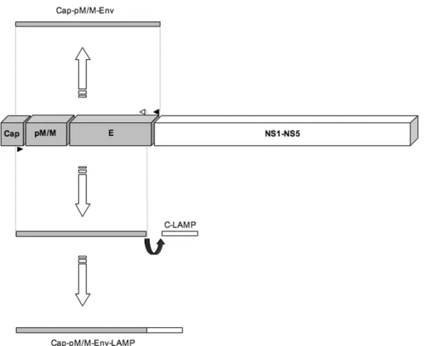
ing to develop DNA vaccines against West Nile, dengue and yellow fever viruses. All wild type DNA constructs comprise a region extending from the capsid reticulum endoplasmic (ER) translocation signal to the E protein transmembrane domain. In parallel, the fused LAMP constructs were obtained through the replacement of E protein transmembrane domain by the C-terminal
do-main of LAMP (Fig. 1). Despite the substitution of the E protein transmembrane domain by LAMP, M-E-LAMP constructs also seem to be able to generate VLPs (Raviprakash et al. 2001).
immuno-Fig. 1 –Membrane-Envelope flavivirus DNA vaccine constructs: co-expression as wild type and as LAMP fused antigens. Polymerase Chain Reaction – PCR amplification was performed to obtain Membrane (M) and Envelope (E) co-expressed flavivirus-based DNA vaccines. To generate the M-E wild type constructs, specific primers were designed to amplify a fragment extending from the capsid reticulum endoplasmic (ER) translocation signal to the E protein transmembrane domain (cap-pM/M-Env or M-E). To generate the M-E/LAMP fused constructs, specific reverse primers were designed to annealed just before the envelope transmembrane domain to allow its substitution by the Carboxi-terminal domain of the Lysosome Associated Membrane Protein – LAMP (Cap-pM/M-Env-LAMP or M-E/LAMP).
genicity, by using dengue virus type 2 as a model. Ex-pression and cellular steady-state localization of the dengue virus antigens were compared to those of the cellular endogenous LAMP by dual fluorescence stain-ing and confocal microscopy. These imagstain-ing studies showed the co-localization of the dengue virus pro-teins in LAMP-containing organelles in DNA-trans-fected NIH3T3 cells. Inin vivoexperiments, M-E and M-E/LAMP encoded DNA plasmids were used in mouse immunization protocols. No mice immunized with M-E DNA seroconverted, while all mice immunized with M-E/LAMP DNA seroconverted on day 30, presenting Plaque-Reduction Neutralization Test – PRTN50 titers
of 49 to 270.
Lu and co-workers (2003) continued the studies on M and E co-expression to evaluate the memory re-sponse. First, the authors evaluated the expression of
Anwar and co-workers (2005) used the M-E/LAMP strategy to develop a West Nile (WN) DNA vaccine. Their Western blot findings showed an increased intra-cellular concentration of the M-E/LAMP chimerical pro-tein in transfected cells when compared to the native protein. This result pointed to a natural cellular secre-tion of E protein that may be retarded by the associ-ation between the LAMP signal and the outer mem-brane of the lysosome. The confocal and immunoelec-tron microscopy analyses showed the typical lysosomal distribution of the WN M-E/LAMP and the co-localiza-tion with endogenous LAMP, MHC class II, and H-2M molecules, the latter of which is closely related to anti-gen presentation. To evaluate the immunoanti-genicity of the WN vaccines, M-E and M-E/LAMP DNAs were injected in mice that were followed for 2 years after the immu-nization. Mice immunized with M-E showed apprecia-ble endpoint titers, but their antibody responses were not sustained after 3 months. In contrast, mice immunized with M-E/LAMP showed high antibody responses, with increasing titers from days 55 to 125, and a significant neutralization titer response at days 90 and 125. To as-sess the presence of memory B cell response, the mice were boosted 19 months after the last immunization, and the blood collected 2 weeks later. While there was no significant neutralization activity in the sera from the M-E DNA immunized group, the sera from the M-M-E/LAMP DNA immunized group showed significant neutraliza-tion titers.
We are currently evaluating a DNA construct ex-pressing the envelope of the YF, fused to LAMP mo-lecules, in murine immunization experiments. Immu-nizations with YF M-E/LAMP encoded DNA showed high titers of neutralizing antibodies that were able to protect the animals from the intracerebral challenge with the YF virus. In addition, when compared to the attenuated 17DD YF virus vaccine, the plasmid DNA expression of the E protein within LAMP induced sim-ilar CD4+and CD8+T cell responses, qualitatively and quantitatively (data not shown).
FUTURE DIRECTIONS
In order to improve the DNA-codified antigen expres-sion in human cells, we are currently developing codon-optimized vaccines by using a genetic algorithm (LETO
1.0 – Entelechonr). Parameters as codon usage,
mes-sage RNA secondary structures, cryptic splice sites, and internal restriction sites, among others, have been modified and/or removed from the native sequences. The blend of LAMP fusion and codon optimization technologies shows promising potential and may lead to new and better DNA vaccines. Our preliminary eval-uations of the novel YF optimized DNA vaccine con-structs address the levels and localization of the protein expression. By immunofluorescence assays, the trans-lation efficiency of the optimized vaccines was consid-ered at least 20 times higher (data not shown). Consid-ering the higher efficiency of the optimized M-E/LAMP DNA constructs (M-EOPT/LAMP), compared to the
non-optimized version, we believe that the non-optimized version will be able to induce much stronger T and B cell re-sponses in animal models. Ultimately, the improvement of the level of antigen expression will allow us to reduce the number and concentration of the DNA vaccine dose used inin vivoimmunization experiments.
ACKNOWLEDGMENTS
The authors are in debt with Miss Meagan Fitzpatrick and Mrs. Claudia Costabile for the helpful editorial re-view of the manuscript.
RESUMO
A vacinação é a estratégia mais prática e o melhor
custo-benefí-cio para prevenir a maioria das infecções dos flavivírus, para os
quais existe vacina disponível. Entretanto, as vacinas baseadas
em vírus atenuados podem potencialmente promover efeitos
colaterais e, mais raramente, reações fatais. Diante deste
ce-nário, o desenvolvimento de estratégias alternativas de
vaci-nação, como vacinas baseadas em DNA codificando
seqüên-cias específicas dos flavivírus, está sendo considerado.
Antí-genos citoplasmáticos endóAntí-genos, caracteristicamente
codifi-cados por vacinas de DNA plasmidial, são majoritariamente
apresentados ao sistema imune através de moléculas do
Com-plexo Maior de Histocompatibilidade de classe I – MHC I.
A via de apresentação MHC I é mais associada à resposta
celular citotóxica e, frequentemente, não elicita uma
respos-ta humoral satisfatória. Uma das principais estratégias para
direcionar antígenos codificados pelas vacinas de DNA para
da Proteína de Associação à Membrana Lisossomal (LAMP).
A proteína do envelope dos flavivírus é reconhecidamente a
principal proteína de superfície viral e o principal alvo para
anticorpos neutralizantes. Diferentes grupos têm
demonstra-do que a co-expressão das proteínas de membrana e demonstra-do
enve-lope dos flavivírus em células de mamíferos, fusionada com a
porção carboxi-terminal de LAMP, é capaz de induzir níveis
satisfatórios de anticorpos neutralizantes. Neste trabalho
re-visamos a estratégia de co-expressão da proteína do envelope
dos flavivírus, como quimeras de LAMP, com o objetivo de
desenvolver vacinas de DNA contra a febre do Oeste do Nilo,
dengue e febre amarela.
Palavras-chave:dengue, febre do Oeste do Nilo, febre amare-la, Proteína de Associação à Membrana Lisossomal – LAMP,
vacinas de DNA.
REFERENCES
ANWARAET AL. 2005. West Nile premembrane-envelope genetic vaccine encoded as a chimera containing the trans-membrane and cytoplasmic domains of a lysosome-associ-ated membrane protein: increased cellular concentration of the transgene product, targeting to the MHC II com-partment, and enhanced neutralizing antibody response. Virology 332: 66–77.
ARRUDA LBET AL. 2004. DNA vaccine encoding human immunodeficiency virus-1 Gag, targeted to the major his-tocompatibility complex II compartment by lysosomal-associated membrane protein, elicits enhanced long-term memory response. Immunology 112: 126–133.
ARRUDALBET AL. 2006. Dendritic cell-lysosomal-associ-ated membrane protein (LAMP) and LAMP-1-HIV-1 gag chimeras have distinct cellular trafficking pathways and prime T and B cell responses to a diverse repertoire of epitopes. J Immunol 177: 2265–2275.
BARRETTAD. 2001. Current status of flavivirus vaccines. Ann N Y Acad Sci 951: 262–271.
BARRETT AD. 2002. Arilvax (PowderJect). Curr Opin Investig Drugs 3: 992–995.
CETRONMSET AL. 2002. Yellow fever vaccine. Recom-mendations of the Advisory Committee on Immunization Practices (ACIP), 2002. MMWR Recomm Rep 51 (RR-17): 1–11.
CHIKHLIKARP ET AL. 2004. Inverted terminal repeat se-quences of adeno-associated virus enhance the antibody and CD8(+) responses to a HIV-1 p55Gag/LAMP DNA vaccine chimera. Virology 323: 220–232.
DONNELLY JJ ET AL. 1997. DNA vaccines. Annu Rev Immunol 15: 617–648.
DONNELLYJJET AL. 2003. Technical and regulatory hur-dles for DNA vaccines. Int J Parasitol 33: 457–467.
DRAKE JR ET AL. 1999. Involvement of MIIC-like late endosomes in B cell receptor-mediated antigen processing in murine B cells. J Immunol 162: 1150–1155.
KLEIJMEERMJET AL. 1997. Major histocompatibility com-plex class II compartments in human and mouse B lym-phoblasts represent conventional endocytic compartments. J Cell Biol 139: 639–649.
LEFEUVREAET AL. 2004. Current Assessment of Yellow Fever and Yellow Fever Vaccine. Curr Infect Dis Rep 6: 96–104.
LEWISPJANDBABIUKLA. 1999. DNA vaccines: a review. Adv Virus Res 54: 129–188.
LIPPINCOTT-SCHWARTZ J AND FAMBROUGH DM. 1986. Lysosomal membrane dynamics: structure and interor-ganellar movement of a major lysosomal membrane gly-coprotein. J Cell Biol 102: 1593–1605.
LIUMA. 2003. DNA vaccines: a review. J Intern Med 253: 402–410.
LUYET AL. 2003. Dengue 2 PreM-E/LAMP chimera tar-geted to the MHC class II compartment elicits long-lasting neutralizing antibodies. Vaccine 21: 2178–2189.
MAECKERHTET AL. 1998. Cytotoxic T cell responses to DNA vaccination: dependence on antigen presentation via class II MHC. J Immunol 161: 6532–6536.
MARQUESETA JR. ET AL. 2003. HIV-1 p55Gag encoded in the lysosome-associated membrane protein-1 as a DNA plasmid vaccine chimera is highly expressed, traffics to the major histocompatibility class II compartment, and elicits enhanced immune responses. J Biol Chem 278: 37926–37936.
MONATHTPET AL. 2002. Single mutation in the flavivirus envelope protein hinge region increases neurovirulence for mice and monkeys but decreases viscerotropism for monkeys: relevance to development and safety testing of live, attenuated vaccines. J Virol 76: 1932–1943.
POLANDJDET AL. 1981. Persistence of neutralizing an-tibody 30-35 years after immunization with 17D yellow fever vaccine. Bull World Health Organ 59: 895–900.
PUTNAKRET AL. 2003. DNA vaccines for flaviviruses. Adv Virus Res 61: 445–468.
RAVIPRAKASHKET AL. 2001. Synergistic neutralizing an-tibody response to a dengue virus type 2 DNA vaccine by incorporation of lysosome-associated membrane pro-tein sequences and use of plasmid expressing GM-CSF. Virology 290: 74–82.
REINHARDTBET AL. 1998. Development of viremia and humoral and cellular parameters of immune activation af-ter vaccination with yellow fever virus strain 17D: a model of human flavivirus infection. J Med Virol 56: 159–167.
ROBINSONHL. 1999. DNA vaccines: basic mechanism and immune responses (Review). Int J Mol Med 4: 549–555.
ROCHABANDTANCHOTC. 2004. Towards a cellular def-inition of CD8+ T-cell memory: the role of CD4+ T-cell help in CD8+ T-cell responses. Curr Opin Immunol 16: 259–263.
SCHULTZJ ET AL. 2000. Immune modulation in cancer using DNA inoculation–antitumour effect of interleukin-12. Dev Biol (Basel) 104: 109–114.
TURLEY SJET AL. 2000. Transport of peptide-MHC class II complexes in developing dendritic cells. Science 288 (5465): 522–527.
VASCONCELOS PFET AL. 2001. Serious adverse events associated with yellow fever 17DD vaccine in Brazil: a report of two cases. Lancet 358 (9276): 91–97.