Acute kidney injury and intra-abdominal
hypertension in burn patients in intensive care
INTRODUCTION
The occurrence of intra-abdominal hypertension (IAH) in surgical patients and those with sepsis or trauma has been widely described in the
literature.(1,2) Abdominal compartment syndrome (ACS) is a complication
resulting from increased intra-abdominal pressure (IAP). High IAP values are not physiologically tolerated and are associated with organ dysfunction, especially of the hemodynamic, respiratory and renal types.(3) Early diagnosis is essential to prevent complications caused by IAH.(4)
Given the importance of the topic, the World Society of Abdominal Compartment Syndrome (WSACS) was founded. This organization prepared a document standardizing definitions and normal IAP values to guide clinical Thalita Bento Talizin1, Meiry Sayuri Tsuda1,
Marcos Toshiyuki Tanita1, Ivanil Aparecida Moro Kauss1, Josiane Festti1, Cláudia Maria Dantas de Maio Carrilho1, Cintia Magalhães Carvalho Grion1, Lucienne Tibery Queiroz Cardoso1
1. Universidade Estadual de Londrina - Londrina (PR), Brazil.
Objective: To evaluate the frequency of intra-abdominal hypertension in major burn patients and its association with the occurrence of acute kidney injury.
Methods: This was a prospective
cohort study of a population of burn patients hospitalized in a specialized intensive care unit. A convenience sample was taken of adult patients hospitalized in the period from 1 August 2015 to 31 October 2016. Clinical and burn data were collected, and serial intra-abdominal pressure measurements taken. The significance level used was 5%.
Results: A total of 46 patients were analyzed. Of these, 38 patients developed intra-abdominal hypertension (82.6%). The median increase in intra-abdominal pressure was 15.0mmHg (interquartile range: 12.0 to 19.0). Thirty-two patients (69.9%) developed acute kidney injury. The median time to development of acute
Conflicts of interest: ConvaTec Inc. donated all AbViser® AutoValve® intra-abdominal pressure monitoring devices used in this study.
Submitted on July 22, 2017 Accepted on October 9, 2017
Corresponding author: Cintia Magalhães Carvalho Grion Divisão de Terapia Intensiva do
Hospital Universitário da Universidade Estadual de Londrina
Rua Robert Koch, 60 - Vila Operária Zip code: 86038-440 - Londrina (PR), Brazil E-mail: cintiagrion@hotmail.com
Responsible editor: Luciano César Pontes de Azevedo
Injúria renal aguda e hipertensão intra-abdominal em paciente
queimado em terapia intensiva
ABSTRACT
Keywords: Intensive care units;
Renal insufficiency; Intra-abdominal hypertension; Burn units; Burns; Multiple organ failure
kidney injury was 3 days (interquartile range: 1 - 7). The individual analysis of risk factors for acute kidney injury indicated an association with intra-abdominal hypertension (p = 0.041), use of glycopeptides (p = 0.001), use of vasopressors (p = 0.001) and use of mechanical ventilation (p = 0.006). Acute kidney injury was demonstrated to have an association with increased 30-day mortality (log-rank, p = 0.009).
Conclusion: Intra-abdominal
hypertension occurred in most patients, predominantly in grades I and II. The identified risk factors for the occurrence of acute kidney injury were intra-abdominal hypertension and use of glycopeptides, vasopressors and mechanical ventilation. Acute kidney injury was associated with increased 30-day mortality.
practice.(5) Normal IAP values range from 0 to 12 mmHg. Sustained increases in IAP above 12mmHg define IAH. ACS is defined as increases in IAP to above 20mmHg associated with organ dysfunction.
Risk factors associated with ACS development can be classified as primary or secondary. Primary factors include causes that are anatomically located in the pelvis and abdomen. Secondary factors are due to other causes, such as sepsis, acidosis, hypothermia, fluid replacement and systemic inflammatory response. In major burn patients, the presence of thermal injury in the abdomen, capillary leak secondary to systemic inflammatory response and aggressive fluid replacement are factors that contribute to increased IAP.(6)
The incidence of IAH in major burn patients is variable in the literature and is associated with the burn area; it is higher in patients with burns covering more than 20% of the body surface area.(7) The use of mechanical ventilation is also associated with an increased incidence of IAH and to a worse prognosis in untreated cases.(8)
In major burn patients, IAH generally occurs in the first 48 hours of the initial resuscitation period. ACS occurs after the acute phase and is associated with episodes
of infectious complications.(9) The kidneys are very
vulnerable organs during the initial treatment of major burns, whether due to the occurrence of IAH, surgical intervention or the presence of nephrotoxic agents. Acute kidney injury (AKI) may result from the reduction in renal blood flow in cases of IAH; in this scenario, urine flow cannot be used as a fluid replacement guide, leading to the loss of an important major burn monitoring parameter.
The objective of this study was to evaluate the frequency of IAH in major burn patients and its association with the occurrence of acute kidney injury.
METHODS
This study was approved by the Research Ethics Committee of the Hospital Universitário Regional do Norte do Paraná - Universidade Estadual de Londrina under CEP 041/2013, CAAE 13327013.8.0000.5231. All study participants agreed with the research and signed an informed consent form.
This was a prospective cohort study. The study population consisted of patients hospitalized in specialized intensive care unit (ICU) beds in the Burn Treatment Center of a university hospital.
A convenience sample was taken of adult burn patients consecutively admitted at the study site. All those admitted between August 2015 and October 2016 were included.
Patients under 18 years of age, those with a burned body surface area of less than 20%, those diagnosed with burn-associated trauma and those who did not consent to participate were excluded. Data pertaining to patients included in the study were collected during their ICU stay, and the date of and outcome at hospital discharge were recorded.
Data collection included clinical, laboratory and demographic data, primary and secondary diagnoses and data on burn type and extent. Data concerning the nephrotoxic drugs used during the ICU stay were also collected. Patient severity was evaluated using the Abbreviated Burn Severity Index (ABSI) score.(10)
The burned body surface was calculated based on the Lund and Browder chart(11) by a plastic surgery specialist at hospital admission. Accumulated fluid balance was defined as the result of the sum of the daily recording of infused fluids and fluids eliminated by the patient within the first 48 hours. IAH was defined, according to WSACS criteria, as a sustained or repeated IAP increase of ≥ 12mmHg. IAH was classified into grades, according to IAP values, and scaled as grade I (12 - 15mmHg), grade II (16 - 20mmHg), grade III (21 - 25mmHg) and grade IV (> 25mmHg). ACS was defined as a sustained IAP value of > 20mmHg associated with new organ failure or dysfunction.(5) AKI was defined as increased baseline creatinine equal to or greater than 0.3mg/dL within 48 hours or greater than or equal to 1.5 times within a 7-day interval.(12)
The initial IAP measurement was taken within 3
hours of admission. If the measurement produced a value within normal limits, the IAP was recorded daily, in the morning, always at the same time, for 7 days or until urinary catheter withdrawal. When the mean was > 12mmHg, it was recorded every 6 hours while it remained high.
The IAP was ascertained from the intravesical pressure. The IAP measurement technique was applied using the AbViser®
measurement system (ConvaTec),(13) which
Autovalve®
device, and 20mL is injected into the bladder, automatically closing the valve to take the IAP reading. The IAP reading taken is shown on a multiparameter monitor at the end of expiration. IAP reading lasts 1 to 3 minutes, and after this period, the valve system opens automatically, and the reading is zeroed. After each reading, it was confirmed that the urine was draining normally.
The results of continuous variables were described using medians and interquartile ranges (ITQ). Categorical data were expressed as frequencies and presented in tables. Categorical variables were analyzed using the chi-squared test. Correlations were ascertained using Pearson’s test to evaluate the degree of dependence between variables. Univariate analysis was performed to identify factors associated with an outcome considered to be AKI. Mortality was described using frequencies. A Kaplan-Meier survival curve analysis was performed, and differences between groups were evaluated using the log-rank test. The significance level used was 5%, and the analyses were performed using the MedCalc program for Windows, version 9.3.2.0 (MedCalc Software, Mariakerke, Belgium).
RESULTS
A total of 68 patients were admitted during the study period. Twenty-two patients were excluded from the study, leaving 46 patients for analysis (Figure 1). Of these, 33 (71.1%) were male; the median age was 40.5 years (ITQ: 28.0 to 53.0). Burns occurred more frequently in domestic accident situations (43.5%), and the median burned body surface area was 30.5% (ITQ: 20.5 to 47.0), as shown in table 1.
Figure 1 - Selection of burn patients admitted to a specialized intensive care unit at a university hospital, 2015-2016. ICF - informed consent form; BBS - burned body surface; IAH - intra-abdominal hypertension.
Table 1 - Characterization of hospitalized burn patients admitted to a specialized intensive care unit
Characteristics N (%)
Age group (years)
18 - 30 13 (28.3)
31 - 50 21 (45.6)
51 - 70 9 (19.6)
≥ 71 3 (6.5)
Gender
Female 13 (28.3)
Male 33 (71.1)
Burn agent
Alcohol 35 (76.1)
Others 11 (23.9)
Burn etiology
Thermal 39 (84.8)
Electrical 3 (6.5)
Scalding 2 (4.3)
Chemical 2 (4.3)
Context of burning
Domestic accident 20 (43.5)
Workplace accident 12 (26.1)
Attempted suicide 6 (13.0)
Attempted homicide 5 (10.9)
Fire 3 (6.5)
Presence of Acute Kidney Injury
Yes 32 (69.9)
No 14 (30.4)
Presence of intra-abdominal hypertension
Yes 38 (82.6)
No 8 (17.4)
Degree of intra-abdominal hypertension
No intra-abdominal hypertension 8 (17.4)
Grade I 17 (37.0)
Grade II 12 (26.1)
Grade III 8 (17.4)
Grade IV 1 (2.2)
Presence of abdominal compartment syndrome
Yes 11 (23.9)
No 35 (76.1)
Use of glycopeptides
Yes 28 (60.9)
No 18 (39.1)
Use of polymyxin
Yes 11 (23.9)
No 35 (76.1)
Use of vasopressors
Yes 33 (71.7)
No 13 (28.3)
Use of mechanical ventilation
Yes 39 (84.8)
No 7 (15.2)
Outcome at hospital discharge
Survival 21 (45.7)
The median accumulated fluid balance 48 hours after hospitalization was 5,233.0 ml (ITQ: 3,562.0 to 8,224.0). In terms of hospital outcome, 21 (45.7%) patients survived. The median length of stay in the ICU was 15 days (ITQ: 6.0 to 26.0), and the median hospital stay was 20 days (ITQ: 11.0 to 32.0).
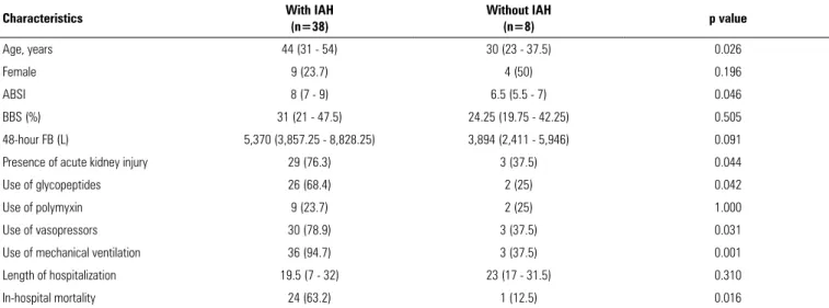
A total of 38 (82.6%) patients developed IAH, with grade I being predominant, with 17 cases (37.0%), followed by grade II, with 12 cases (26.1%). ACS developed in 11 patients (23.9%). Comparison of patient group characteristics revealed that the patients who developed IAH had a higher mean age, more severe burns according to the ABSI, developed AKI more frequently and needed to use glycopeptides, vasopressors and mechanical ventilation. The presence of IAH was also associated with a higher mortality rate (Table 2).
The peak IAP value showed weak positive correlations with the accumulated fluid balance in the first 48 hours (r = 0.29; p = 0.047) and the worst serum creatinine value during the ICU stay (r = 0.47; p = 0.001).
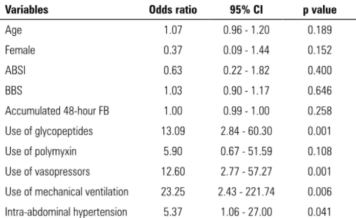
Of the patients studied, 32 (69.9%) developed AKI during the study period. The median peak serum creatinine value of patients during their ICU stay was 1.33mg/dL (ITQ: 1.0 - 2.39). The median time to developing AKI was 3 days (ITQ: 1 - 7). Univariate analysis of AKI risk factors indicated associations with IAH (p = 0.041), use of glycopeptides (p = 0.001), use of vasopressors (p = 0.001) and use of mechanical ventilation (p = 0.006) (Table 3).
The survival analysis (Figure 2) revealed an association between AKI and higher 30-day mortality (log-rank, p = 0.009).
DISCUSSION
The present study demonstrates the high frequency of IAH in major burn patients and its association with the occurrence of AKI. These results highlight the importance of controlling IAP and preventing AKI in burn patients. Furthermore, they suggest that prevention of AKI occurrence in these patients should lead to an improved mortality rate, as there is an association between AKI and worse prognosis.
The clinical characteristics of the patients in this study are similar to those found in data from other countries. In the United States, the majority of burn patients treated between 2006 and 2015 were male and predominantly between 20 and 59 years old. Domestic occurrences were most prevalent, comprising 73% of cases. The predominant etiology was thermal and by scalding, comprising 75% of patients. Mortality was lower in this US study and increased according to age and greater percent body area burned.(14)
In Brazil, males are the most affected, and alcohol is the main agent of burns in adults, predominantly involving domestic accidents.(15) This finding suggests that a high percentage of burns are preventable, with valid prevention measures leading to the avoidance of injury and all of its direct complications and those resulting from
Table 2 - Comparison of clinical characteristics and outcome of burn patients with and without intra-abdominal hypertension admitted to a specialized intensive care unit
Characteristics With IAH
(n=38)
Without IAH
(n=8) p value
Age, years 44 (31 - 54) 30 (23 - 37.5) 0.026
Female 9 (23.7) 4 (50) 0.196
ABSI 8 (7 - 9) 6.5 (5.5 - 7) 0.046
BBS (%) 31 (21 - 47.5) 24.25 (19.75 - 42.25) 0.505
48-hour FB (L) 5,370 (3,857.25 - 8,828.25) 3,894 (2,411 - 5,946) 0.091
Presence of acute kidney injury 29 (76.3) 3 (37.5) 0.044
Use of glycopeptides 26 (68.4) 2 (25) 0.042
Use of polymyxin 9 (23.7) 2 (25) 1.000
Use of vasopressors 30 (78.9) 3 (37.5) 0.031
Use of mechanical ventilation 36 (94.7) 3 (37.5) 0.001
Length of hospitalization 19.5 (7 - 32) 23 (17 - 31.5) 0.310
In-hospital mortality 24 (63.2) 1 (12.5) 0.016
Table 3 - Univariate analysis of acute kidney injury risk factors in burn patients admitted to a specialized intensive care unit
Variables Odds ratio 95% CI p value
Age 1.07 0.96 - 1.20 0.189
Female 0.37 0.09 - 1.44 0.152
ABSI 0.63 0.22 - 1.82 0.400
BBS 1.03 0.90 - 1.17 0.646
Accumulated 48-hour FB 1.00 0.99 - 1.00 0.258
Use of glycopeptides 13.09 2.84 - 60.30 0.001
Use of polymyxin 5.90 0.67 - 51.59 0.108
Use of vasopressors 12.60 2.77 - 57.27 0.001
Use of mechanical ventilation 23.25 2.43 - 221.74 0.006
Intra-abdominal hypertension 5.37 1.06 - 27.00 0.041
95% CI - 95% confidence interval; ABSI - Abbreviated Burn Severity Index; BBS - burned body surface; FB - fluid balance.
Figure 2 - Comparison of 30-day survival between patients with and without acute kidney injury in burn patients admitted to a specialized intensive care unit at a university hospital, 2015-2016. AKI - acute kidney injury. Log-rank, p = 0.009.
treatment. Therefore, it would be appropriate to develop public policies for the prevention of burn accidents and to conduct studies to map the epidemiology of burn accidents in the various regions of the country.
The measurement of IAP has been increasingly performed in the ICU due to the knowledge that has been gained in regard to organ dysfunction resulting from changes in its value.(16) There are variations in techniques used to measure IAP depending on the materials used, but all forms studied involve maintaining the patient in the supine position, without abdominal contraction and with measurement at the end of expiration. The nursing professional who is responsible for setting up the equipment and taking the measurements requires theoretical and practical training to perform this procedure properly.
There is a lack of knowledge among health professionals in regard to IAP measurement methodology(17) and a lack
knowledge about IAH and its clinical implications.(18)
There is still no consensus on a standardized methodology to measure IAP, but there are strong recommendations on the importance of this measurement and its clinical significance for hospitalized patients.(19)
The risk factors found for AKI are related to the pathophysiology of kidney injury. The use of nephrotoxic drugs, such as glycopeptides, is associated with direct kidney injury and the consequent dysfunction of this organ, especially if the patient is in the ICU, where serum levels of the drug are above normal and drug treatment is prolonged.(20,21) Changing organic perfusion in the case of circulatory instability, as evidenced in the literature,(2) is a risk factor for kidney injury. The IAH patient also presents hemodynamic changes with impaired renal perfusion.(9,17) The use of mechanical ventilation with consequent changes in intrathoracic pressure is also associated with the presence of IAH. This risk factor is proportional to the severity of respiratory symptoms and the mechanical ventilation requirement.(22)
An association between AKI and higher 30-day mortality in intensive care patients has been found.(23) IAH is a complication associated with organ dysfunction, especially AKI, which is a major marker of morbidity and worsening prognosis in ICU patients. Several factors are associated with the development of AKI in-hospital, especially in critically ill patients. Constant IAP measurement can provide proactive information, alerting the team about the imminence of IAH and thus preventing increased morbidity in hospitalized patients.
This study has some limitations, such as the small number of patients and the fact that it is a single-center study. The effects of predictor variables for the outcomes studied may have been underestimated and must be interpreted with caution. The strength of this study is the fact that it is one of the few reports on IAP monitoring in burn patients in Latin America and offers unprecedented local data on the occurrence of IAH and AKI in these patients.
CONCLUSION
Objetivo: Avaliar a frequência de hipertensão intra-abdomi-nal no paciente grande queimado e sua associação com a ocor-rência de injúria renal aguda.
Métodos: Estudo de coorte prospectivo, com população de pacientes queimados internados nos leitos de unidade de terapia intensiva especializada. Realizada amostragem de conveniência de pacientes adultos internados no período de 1º de agosto de 2015 a 31 de outubro de 2016. Foram coletados dados clínicos e da queimadura, além de medidas seriadas da pressão intra--abdominal. O nível de significância utilizado foi de 5%.
Resultados: Foram analisados 46 pacientes. Evoluíram
com hipertensão intra-abdominal 38 pacientes (82,6%). A mediana da maior pressão intra-abdominal foi 15,0mmHg (intervalo interquartílico: 12,0 - 19,0). Desenvolveram injúria renal aguda 32 (69,9%) pacientes. A mediana do tempo para
desenvolvimento de injúria renal aguda foi de 3 dias (intervalo interquartílico: 1 - 7). A análise individual de fatores de risco para injúria renal aguda apontou associação com hipertensão intra-abdominal (p = 0,041), uso de glicopeptídeos (p = 0,001), uso de vasopressor (p = 0,001) e uso de ventilação mecânica (p = 0,006). Foi evidenciada associação de injúria renal aguda com maior mortalidade em 30 dias (log-rank, p = 0,009).
Conclusão: Ocorreu hipertensão intra-abdominal em gran-de parte dos pacientes estudados, predominantemente nos graus I e II. Os fatores de risco identificados para ocorrência de injúria renal aguda foram hipertensão intra-abdominal, uso de glico-peptídeos, vasopressor e ventilação mecânica. Injúria renal agu-da esteve associaagu-da à maior mortaliagu-dade em 30 dias.
RESUMO
Descritores: Unidades de terapia intensiva; Insuficiência renal; Hipertensão intra-abdominal; Unidades de queimados; Queimaduras; Insuficiência de múltiplos órgãos
REFERENCES
1. Prado LF, Alves Jr. A, Cardoso ES, Andrade RS, Andrade RS, Fernandes MK. Pressão intra-abdominal em pacientes com trauma abdominal. Rev Col Bras Cir. 2005;32(2):83-9.
2. Holodinsky JK, Roberts DJ, Ball CG, Blaser AR, Starkopf J, Zygun DA, et al. Risk factors for intra-abdominal hypertension and abdominal compartment syndrome among adult intensive care unit patients: a systematic review and meta-analysis. Crit Care. 2013;17(5):R249.
3. Ivatury RR, Diebel L, Porter JM, Simon RJ. Intra-abdominal hypertension and the abdominal compartment syndrome. Surg Clin North Am. 1997;77(4):783-800.
4. Starkopf J, Tamme K, Blaser AR. Should we measure intra-abdominal pressures in every intensive care patient? Ann Intensive Care. 2012;2 Suppl 1:S9.
5. Malbrain ML, Cheatham ML, Kirkpatrick A, Sugrue M, Parr M, De Waele J, et al. Results from the International Conference of Experts on Intra-abdominal Hypertension and Abdominal Compartment Syndrome. I. Definitions. Intensive Care Med. 2006;32(11):1722-32.
6. Ivy ME, Atweh NA, Palmer J, Possenti PP, Pineau M, D’Aiuto M. Intra-abdominal hypertension and Intra-abdominal compartment syndrome in burn patients. J Trauma. 2000;49(3):387-91.
7. Ruiz-Castilla M, Barret JP, Sanz D, Aguilera J, Serracanta J, García V, et al. Analysis of intra-abdominal hypertension in severe burned patients: the Vall d’Hebron experience. Burns. 2014;40(4):719-24.
8. Wise R, Jacobs J, Pilate S, Jacobs A, Peeters Y, Vandervelden S, et al. Incidence and prognosis of intra-abdominal hypertension and abdominal compartment syndrome in severely burned patients: Pilot study and review of the literature. Anaesthesiol Intensive Ther. 2016;48(2):95-109. 9. Malbrain ML, De Keulenaer BL, Oda J, De Laet I, De Waele JJ, Roberts DJ,
et al. Intra-abdominal hypertension and abdominal compartment syndrome in burns, obesity, pregnancy, and general medicine. Anaesthesiol Intensive Ther. 2015;47(3):228-40.
10. Tobiasen J, Hiebert JM, Edlich RF. The abbreviated burn severity index. Ann Emerg Med. 1982;11(5):260-2.
11. Lund CC, Browder NC. The estimation of burns areas. Surg Gynecol Obstet. Chicago. 1944;79(4):352-58.
12. Kidney Disease: Improving Global Outcomes (KDIGO). Acute Kidney Injury Work Group. KDIGO Clinical Practice Guideline for Acute Kidney Injury. Kidney Int Suppl. 2012;2(1):1-138.
13. AbViser® Medical. AbViser® AutoValve® Instructions. Disponível em:
http://abviser.com/wp-content/uploads/2011/04/AbViser-Instructions-Chart-Transducer-Not-Included.pdf. Acesso em 24 nov. 2016.
14. American Burn Association (ABA). National Burn Repository. Report of data from 2006-2015. Chicago: American Burn Association, National Burn Repository; 2016.
15. Cruz BF, Cordovil PB, Batista KN. Perfil epidemiológico de pacientes que sofreram queimaduras no Brasil: revisão de literatura. Rev Bras Queimaduras. 2012;11(4):246-50.
16. Zampieri FG, Almeida JR, Schettino GP, Park M, Machado FS, Azevedo LC. Factors associated with variation in intracranial pressure in a model of intra-abdominal hypertension with acute lung injury. Rev Bras Ter Intensiva. 2011;23(2):164-9.
17. Milanesi R, Caregnato RC. Pressão intra-abdominal: revisão integrativa. Einstein (São Paulo) 2016;14(3):423-30.
18. Japiassú AM, Falcão H, Freitas F, Freitas S, Souza PC, Lannes R, et al. [Measurement of intra-abdominal pressure in the intensive care unit: the opinion of the critical care physicians]. Rev Bras Ter Intensiva; 2007;19(2):186-91. Portuguese.
19. Malbrain ML. You don’t have any excuse, just start measuring abdominal pressure and act upon it! Minerva Anestesiol. 2008;74(1-2):1-2. 20. Perazella MA. Drug use and nephrotoxicity in the intensive care unit,
Kidney Int. 2012;81(12):1172-8.
21. Elyasi S, Khalili H, Dashti-Khavidaki S, Mohammadpour A. Vancomycin-induced nephrotoxicity: mechanism, incidence, risk factors and special populations. A literature review. Eur J Clin Pharmacol. 2012;68(9):1243-55.
22. Soler Morejón Cde D, Tamargo Barbeito TO. Effect of mechanical ventilation on intra-abdominal pressure in critically ill patients without other risk factors for abdominal hypertension: an observational multicenter epidemiological study. Ann Intensive Care. 2012;2 Suppl 1:S22. 23. Gammelager H, Christiansen CF, Johansen MB, Tønnesen E, Jespersen