Research Article
Frequency of TGF-
�
and IFN-
�
Genotype as Risk Factors for
Acute Kidney Injury and Death in Intensive Care Unit Patients
Caren Cristina Grabulosa,
1Marcelo Costa Batista,
1,2,3Miguel Cendoroglo,
1,2,3Beata Marie Redublo Quinto,
1Roberto Narciso,
2Julio Cesar Monte,
1,2Marcelino Durão,
1,2Luiz Vicente Rizzo,
2Oscar Fernando Pavão Santos,
1,2and Maria Aparecida Dalboni
1,41Nephrology Division, Universidade Federal de S˜ao Paulo/UNIFESP, S˜ao Paulo, Brazil 2Hospital Israelita Albert Einstein, S˜ao Paulo, Brazil
3Tuts-New England Medical Center, Boston, MA, USA 4Universidade Nove de Julho/UNINOVE, S˜ao Paulo, Brazil
Correspondence should be addressed to Maria Aparecida Dalboni; dalboni@unifesp.br
Received 2 April 2014; Revised 27 May 2014; Accepted 18 June 2014; Published 23 July 2014
Academic Editor: Raul Lombardi
Copyright © 2014 Caren Cristina Grabulosa et al. his is an open access article distributed under the Creative Commons Attribution License, which permits unrestricted use, distribution, and reproduction in any medium, provided the original work is properly cited.
Genetic variations in TGF-�and IFN-�may interfere with proinlammatory cytokine production and, consequently, may be involved with inlammatory diseases, as acute kidney injury (AKI). We considered that genetic polymorphisms of these cytokines may have a crucial role in the outcome of critically ill patients. To investigate whether the genetic polymorphisms of rs1800470 (codon 10 T/C), rs1800471 (codon 25 C/G) from the TGF-�, and rs2430561 (+874 T/A) from IFN-�may be a risk factor for ICU patients to the development of AKI and/or death. In a prospective nested case-control study, were included 139 ICU patients who developed AKI, 164 ICU patients without AKI, and 244 healthy individuals. We observed a higher frequency to T/A genotype for IFN-�(intermediate producer phenotype) and higher frequency of TT GG and TC GG genotype (high producer) for TGF-� polymorphism in overall population. However, these polymorphisms have not been shown as a predictor of risk for AKI and death. We found an increased prevalence of high and intermediate producer phenotypes from TGF-�and IFN-�, respectively, in patients in ICU setting. However, the studied genetic polymorphism of the TGF-�and IFN-�was not associated as a risk factor for AKI or death in our population.
1. Introduction
Critically ill patients in the intensive care unit (ICU) have a high incidence of acute kidney injury (AKI) [1, 2] and consequently high mortality rates. AKI occurrence is a consequence of the pathophysiology complex of systemic inlammatory response syndrome (SIRS). In SIRS there is the activation of leukocytes, which produce oxidative stress, high adhesion molecules expression, and high serum levels of proinlammatory cytokines as TNF-� and IL-6. hese cytokines have been associated with poor outcome [3, 4] and some inlammatory mediators have also been reported to contribute to the renal vasculature injury and may conse-quent development the AKI [5].
IFN-�is a proinlammatory cytokine. It has been reported that ischemic kidney injury triggers production of several cytokines, such as TGF-�, IFN-�, IL-6, TNF-�, and IL-1�in the inlamed kidney [6–9].
Roedder et al. have been reported in a renal transplant model that lower IFN-�gene expression causes less iniltra-tion of leukocytes and allograt rejeciniltra-tion, demonstrating that this cytokine is associated with inlammation process and kidney injury [10].
TGF-�, another inlammatory cytokine, has been reported to induce chemotaxis for neutrophils, T cells, mon-ocytes, and ibroblasts to the site of the injury. hus, this bio-logic efect of TGF-�1 suggests that this cytokine may also Volume 2014, Article ID 904730, 6 pages
contribute to inlammation and a central role in chronic kidney ibrosis [11–14].
In fact, some authors observed that increased TGF-� activity in vivo results in glomerular extracellular matrix accumulation, proteinuria, tubule-interstitial ibrosis, and the onset of chronic renal failure, suggesting that its activity in promoting the synthesis of extracellular matrix plays a crucial role in ibrotic deposition and the decline in renal function [15,16].
Kopp et al. also related that increased levels of circulating TGF-�1 induced progressive renal disease characterized by mesangial expansion, accumulation of glomerular immune deposits and matrix proteins, and interstitial ibrosis in this transgenic mouse model suggesting that chronically elevated circulating levels of TGF-�1 induce progressive glomeru-losclerosis [15]. herefore, it appears that renal diseases may also result from the inlammation and from inappropriate regulation of TGF-� expression [17]. However, until this moment there is no data about the role of IFN-�and
TGF-�in critically ill patients as risk markers to AKI. Recently, the study of polymorphisms of genes involved in host immune response, including cytokines and other modulators of the inlammatory response, has been a subject of interest, since these genetic markers may be potential determinants of susceptibility to or severity of acute events such as AKI [18, 19]. he reason why some patients with SIRS develop AKI and others do not is still unclear. So, it is possible that polymor-phisms of inlammatory cytokines genes, in particular IFN-� and TGF-�, may be involved in the pathophysiology of AKI in critically ill patients.
hereby, the aim of this study was to investigate the frequency of rs1800470 (codon 10 T/C), rs1800471 (codon 25 C/G) from the TGF-�, and rs2430561 (+874 T/A) from IFN-� polymorphisms genotypes in critically ill ICU patients and to analyze the possible impact of these polymorphisms on AKI and death.
2. Materials and Methods
2.1. Subjects. his study was a prospective, nested case-control which included three hundred and three patients (� = 303) from Hospital Israelita Albert Einstein, Sao Paulo, Brazil, who had been in the ICU for at least 48 hours ago and two hundred forty-four (� = 244) healthy individuals from Hypertension and Cardiovascular Metabolism Center, UNIFESP, Sao Paulo, Brazil, were included (Control group). Overall individuals were over 18 years old. Of the 303 ICU patients, 139 patients developed AKI (AKI group) and 164 patients did not (No AKI group). AKI was conirmed by the Acute Kidney Injury Network (AKIN) and Risk, Injury, Failure, Lesion Renal and End-stage-Renal Disease (RIFLE) [20] criteria deined as an increase of three times of serum creatinine compared with a baseline serum levels of creatinine or the creatinine clearance<60 mL/min/mm3that was calculated by Levey equation (MDRD—Modiication of Diet in Renal Disease) according to K/DOQI [21].
Patients who developed AKI were matched for age and gender to the No AKI and Control group. he criteria of
exclusion at moment of selection of patients were patients who are not considered for resuscitation, kidney transplant patients, patients in dialysis treatment (acute or chronic), and patients who had previously participated in this study.
his study was approved by the Research Ethics Com-mittee of the Universidade Federal de S˜ao Paulo (UNIFESP), reference number: CEP 1520/05, and by the Ethics Committee of the Hospital Israelita Albert Einstein, reference number 06/381.
2.2. Materials. 30 mL of blood in ethylene diamine tetra acetic acid anticoagulant (EDTA) was collected from each individual for renal function analysis (urea and creatinine, Labtest, Lagoa Santa, MG, Brasil and Jafe modiied method [22], resp.), lipid proile (cholesterol, HDL and LDL choles-terol, automated method equipment Cell-Dyn Ruby Abbott Diagnostics), and markers of SIRS (CRP and albumin, Immulite 1000 immunoassay system, IL, USA, and colori-metric method in automated equipment Olympus AU400, Dallas, TX, USA, resp.) and DNA extraction was performed in order to study the polymorphisms of TGF-�and
IFN-� (Genotyping tray, One Lambda, Inc., 21001 Kittridge, St, Canoga Park, CA, USA).
2.2.1. Analysis of Codons 10 and 25 of TGF-�and +874 IFN-� Polymorphisms. Genomic DNA was prepared from periph-eral blood using standard techniques and was extractedbythe DTAB/CTAB method [23]. he polymerase chain reaction (PCR) sequence speciic primer (SSP) technique was used to analyze two SNPs.
he polymorphisms may result from the substitution of a single nucleotide (single nucleotide polymorphisms or SNPs). Most polymorphism of genes of cytokines are of the type microsatellite and SNP and they are located in the promoter region, the introns and untranslated regions (UTR of untranslated region). Polymorphisms located in introns of genes can have signiicant efects on transcription that may be related to changes in the structure of binding sites of promoters or facilitators structure segments (“enhancers”) or blocking (“silencers”) within introns. Consequently, the polymorphism may alter the rate of transduction, inluencing the amount of protein produced by the cell [24].
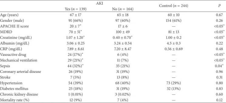
Table 1: Biochemical and epidemiological data according to the development for acute kidney injury (AKI) (� = 303) and healthy individuals (Control) (� = 244).
AKI
Control (� = 244) �
Yes (� = 139) No (� = 164)
Age (years) 67±17 65±18 60±10 0.67
Gender (male) 91 (66%) 97 (60%) 154 (61%) 0.26
APACHE II score 20±7∗ 17±6 — <0.05∗
MDRD 70±51∗ 100±49 81±13 <0.05∗
Creatinine (mg/dL) 1.07±1.20∗ 0.40±0.70∗ 1.00±0.2 <0.05∗
Albumin (mg/dL) 3.06±0.25 3.24±0.54 4.5±0.3 0.22
CRP (mg/dL) 7.89±8.61 7.20±8.47 0.56±0.69 0.48
Vasoactive drug 24 (17%)∗ 6 (4%) — <0.05∗
Mechanical ventilation 29 (21%)∗ 11 (7%) — <0.05∗
Sepsis 44 (32%)∗ 35 (21%) — 0.04∗
Coronary arterial disease 26 (19%) 31 (19%) — 0.96
Stroke 7 (5%) 13 (8%) — 0.31
Hypertension 54 (39%) 68 (40%) 73 (29%) 0.80
Diabetes mellitus 25 (18%) 31 (19%) 32 (13%) 0.83
Chronic kidney disease 1 (0.01%) 3 (0.02%) — 0.60
Mortality rate (%) 12 (9%) 7 (4%) — 0.12
∗
AKI compared to No AKI-Student’s�and chi-square test; ANOVA when appropriate.
combinations of codon 10 and codon 25 genotypes, only one genotype was not present (TC/CC) in these study groups.
IFN-�phenotypes were classiied as high producers (TT genotype), intermediate producers (TA genotype), and low producers (AA genotype).
2.2.2. Statistical Analysis. Data were analyzed using the statistical sotware SPSS (version 20.0, Chicago, IL). he results were expressed as mean and standard deviation while the distribution of genotypes and phenotypes was given as percentages. Clinical characteristics and cytokine gene polymorphisms were compared using the chi-squared test and Fisher’s exact test when needed. To compare the difer-ences between the groups,�test was used for independent samples. To examine whether the genotype frequencies were in Hardy-Weinberg equilibrium, goodness-of-it�2-test was used. Allele frequencies were compared by a2×2contingency table using Fischer’s exact test. All results were considered signiicant at� < 0.05.
3. Results
3.1. Biochemical and Epidemiological Data. he AKI patients had a lower initial renal function (calculated by MDRD equation) compared to No AKI and Control group (70 ±
51 versus 100 ± 49 and 81 ± 13; � < 0.001, resp.) and higher serum levels of creatinine (Table 1). AKI patients were classiied as follows: 59 (41%) as injury, 25 (17%) as failure, 22 (15%) as loss, 33 (23%) as end stage, APACHE score II (20±7versus17±6;� = 0.001), vasoactive drug use (23 (17%) versus 5 (4%),� = 0.0001), mechanical ventilation (29 (21%) versus 9 (7%),� = 0.0001), and sepsis (44 (32%) versus 29 (21%),� = 0.04). Age, gender, coronary artery disease, heart
failure, stroke, hypertension, and diabetes mellitus were not signiicantly diferent between the studied groups (Table 1). In the univariate analysis, only APACHE II score (O.R. 1.07 C.I. 1.04–1.10;� = 0.0001) and mechanical ventilation (O.R. 0.53 C.I. 0.31–0.90;� = 0.02) were risk factors for AKI.
3.2. Biochemical and Epidemiological Data according to Death. With respect to death, an association was observed between a higher APACHE II score (27 ± 7versus18 ± 6;� = 0.0001), higher serum creatinine (1.66 ± 1.49 versus 0.64 ± 0.95;
� = 0.0001), higher CRP (13.7 ± 12.5 versus 7.1 ± 8.0;
� = 0.001), use of vasoactive drug (42% versus 8%,� =
0.0001), mechanical ventilation (53% versus 11%,� = 0.0001), sepsis (68% versus 23%;� = 0.0001), and higher death rates (Table 2).
3.3. Genotype Polymorphisms Frequency of TGF-�and
IFN-� in Patients with and without AKI and Death. Genotype frequencies for TGF-�and IFN-� were similar to previous reports in the Caucasian population and observed Hardy-Weinberg equilibrium.
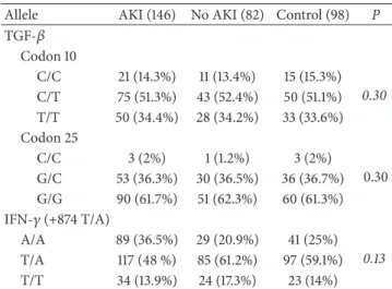
Regarding the frequency of genotypes/alleles of the poly-morphisms of the cytokines TGF-�and IFN-�, we observed a higher prevalence expression of C/C from the TGF-�and T/A from the IFN-� genotypes in both groups (AKI and No AKI patients) (Table 3). However, this prevalence was no signiicant for to develop in AKI or death.
Table 2: Biochemical and epidemiological data according to death
(� = 303).
Death �
Yes (� = 19) No (� = 284)
Age (years) 74±14∗ 65±18 0.04∗
Gender (male) 10 (52%) 178 (62%) 0.38
APACHE II score 27±7∗ 18±6 <0.05∗
Creatinine (mg/dL) 1.66±1.49∗ 0.64±0.95 <0.05∗ Albumin (mg/dL) 3.00±0.02 3.18±0.46 0.43 CRP (mg/dL) 13.7±12.5∗ 7.1±8.0 <0.05∗ Vasoactive drug 8 (42%)∗ 22 (8%) <0.05∗ Mechanical ventilation 10 (53%)∗ 30 (11%) <0.05∗
Sepsis 13 (68%)∗ 66 (23%) <0.05∗
Coronary artery disease 4 (21%) 53 (19%) 0.80
Stroke 1 (5%) 19 (7%) 0.80
Hypertension 4 (21%) 116 (41%) 0.08
Diabetes mellitus 5 (26%) 51 (18%) 0.36
∗
Death compared to No Death-Student’s�and chi-square test.
Table 3: Frequency of allele (%) of TGF-�and IFN-� polymor-phisms in ICU patients according to acute kidney injury (AKI or No AKI) and control group.
Allele AKI (146) No AKI (82) Control (98) P
TGF-� Codon 10
C/C 21 (14.3%) 11 (13.4%) 15 (15.3%)
0.30
C/T 75 (51.3%) 43 (52.4%) 50 (51.1%) T/T 50 (34.4%) 28 (34.2%) 33 (33.6%) Codon 25
C/C 3 (2%) 1 (1.2%) 3 (2%)
0.30 G/C 53 (36.3%) 30 (36.5%) 36 (36.7%) G/G 90 (61.7%) 51 (62.3%) 60 (61.3%) IFN-�(+874 T/A)
A/A 89 (36.5%) 29 (20.9%) 41 (25%)
0.13
T/A 117 (48 %) 85 (61.2%) 97 (59.1%)
T/T 34 (13.9%) 24 (17.3%) 23 (14%)
Chi-square test and� < 0.05.
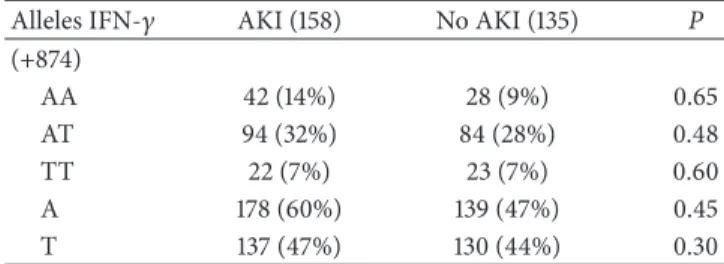
respect to death (Table 4) and sepsis (data not shown). We did not observe any association of haplotypes TGF-�and IFN-� polymorphisms with AKI (Tables5,6, and7).
4. Discussion
his is the irst study aimed to investigate the prevalence of polymorphisms of TGF-� and IFN-� in leukocytes of patients with SIRS and to investigate its possible associations as a risk factor for development of AKI and death. As expected, AKI patients had a high APACHE score II and high incidence of sepsis. However, in respect to these cytokines polymorphisms, we did not show it as risk factors to AKI or death in our population.
We observed a higher prevalence expression of genotype C/C from the TGF-� in both groups (AKI and No AKI
Table 4: Frequency (%) of genotypes of polymorphism from TGF-� and IFN-�in patients according to death.
Genotype Death P
Yes (14) No (279) TGF-�(codon 10 e 25)
TT GG — 73 (26%) n.s.
TC GG 4 (28%) 76 (27%) n.s.
TC GC 8 (57%) 75 (26%) n.s.
CC GG 1 (7%) 19 (6%) n.s.
TT GC 1 (7%) 18 (6%) n.s.
CC GC — 13 (4%) n.s.
CC CC — 0 (0.7%) n.s.
TT CC — 3 (1%) n.s.
IFN-�(+874)
TT 3 (21%) 43 (15%) n.s.
TA 7 (50%) 173 (62%) n.s.
AA 4 (28%) 63 (22%) n.s.
Chi-square test and� < 0.05or Fisher.
n.s. = not signiicant.
Table 5: Frequency (%) of haplotypes codon 10 from the TGF-�in patients in AKI and No AKI.
Alleles TGF-� AKI (158) No AKI (135) P
Codon 10
TT 59 (20%) 38 (12%) 0.30
TC 78 (26%) 84 (28%) 0.25
CC 21 (7%) 13 (4%) 0.48
T 197 (67%) 158 (53%) 0.50
C 121 (41%) 109 (37%) 0.50
Hardy-Weinberg equilibrium.
Table 6: Frequency (%) of haplotypes codon 25 from the TGF-�in patients in AKI and No AKI.
Alleles TGF-� AKI (158) No AKI (135) P
Codon 25
GG 95 (32%) 75 (25%) 0.30
GC 61 (21%) 55 (19%) 0.30
CC 2 (0.6%) 3 (1%) 0.25
G 253 (86%) 206 (70%) 0.20
C 65 (22%) 62 (21%) 0.20
Hardy-Weinberg equilibrium.
Table 7: Frequency (%) of haplotypes +874T/A from IFN-� in patients in AKI and No AKI.
Alleles IFN-� AKI (158) No AKI (135) P
(+874)
AA 42 (14%) 28 (9%) 0.65
AT 94 (32%) 84 (28%) 0.48
TT 22 (7%) 23 (7%) 0.60
A 178 (60%) 139 (47%) 0.45
T 137 (47%) 130 (44%) 0.30
Hardy-Weinberg equilibrium.
model that an activation of TGF-�signaling in the tubular epithelium alone is suicient to cause AKI [15].
In our study, we investigated expression of TGF-� poly-morphisms from DNA extracted from circulating leukocytes from critically ill patients who developed AKI. AKI’s patients did not show more prevalence of TGF-� compared to No AKI. he literature describes that patients with chronic kidney diseases have more ibrosis and it is associated with high TGF-�serum levels. However, the acute mechanism of this cytokine in AKI is unclear. So, to demonstrate whether TGF-� could have a direct efect on kidney function in patients with SIRS, it would be necessary to investigate this in renal cells from these patients.
Regarding IFN-�, we observed a predominant frequency of genotype T/A in the general population. However, these genotypes were not statistically diferent between groups and it was not associated as a risk marker for AKI and death.
In contrast, Karimi et al. reported an association between the TT homozygote for IFN-� (high producer) and acute rejection renal transplantation [29]. hese authors suggest that these conditions are associated with renal injury and may contribute to evolution for AKI [7,9]. However, in our study only 13.7% of AKI patients were high producer phenotype carriers.
Finally, our study had some limitations as follows. (1) he probability to detect polymorphisms is <1% in the overall population; therefore the inclusion of a higher number of patients should be necessary. We included 400 consecutive patients with SIRS in the ICU from a single center, where 303 were eligible for this analysis. hus, it is possible that the sample size may have accounted for the poor correla-tion analysis of these polymorphisms and their associacorrela-tions with outcomes. (2) We investigated the main polymorphism described in the literature for each cytokine, but there are several diferent polymorphisms for these cytokines that could be involved in AKI pathophysiology.
Some authors have described associations between
TGF-�and IFN-�and poor prognosis such as renal ibrosis and inlammation in chronic kidney disease [30, 31]. However, there is still no data about these polymorphisms with AKI. hus, it is possible that the measure of serum levels of these cytokines and the investigation of their polymorphisms together could have contributed to a better understanding of the molecular control and respective protein synthesis, clarifying the possible associations with outcomes.
herefore, in the present study we concluded that the genetic polymorphisms of codons 10 T/C and 25 C/G of the TGF-�and +874 T/A of the IFN-�were not associated as risk factors for AKI or death in our population. To validate if these studied polymorphisms may induce a renal injury in critically ill patients in ICU setting, a multicentric study with a larger sample size should be conducted.
Conflict of Interests
None of the authors has any other conlict of interests related to this paper.
Authors’ Contribution
Miguel Cendoroglo Neto and Marcelo Costa Batista con-tributed with editorial assistance and interpretation of data; Beata Marie Redublo Quinto, Roberto Narciso, Marcelino Dur˜ao, Julio Cesar Monte, Oscar Fernando Pav˜ao dos Santos, and Luiz Vicente Rizzo, provided intellectual content of critical importance to the work described. Maria Aparecida Dalboni contributed as a coordinator of this study and con-tributed to the inal approval of the version to be published.
Acknowledgment
his study was supported by FAPESP (Bras´ılia, Brazil), Grant 07/58363-0.
References
[1] P. Monedero, N. Garc´ıa-Fern´andez, J. R. P´erez-Valdivieso, M. Vives, and J. Lavilla, “Acute kidney injury,”Revista Espa˜nola de Anestesiolog´ıa y Reanimaci´on, vol. 58, no. 6, pp. 365–374, 2011. [2] A. de Mendonc¸a, J. L. Vincent, P. M. Suter et al., “Acute renal
failure in the ICU: risk factors and outcome evaluated by the SOFA score,”Intensive Care Medicine, vol. 26, no. 7, pp. 915–921, 2000.
[3] E. M. Simmons, J. Himmelfarb, M. Tugrul Sezer et al., “Plasma cytokine levels predict mortality in patients with acute renal failure,”Kidney International, vol. 65, no. 4, pp. 1357–1365, 2004. [4] P. V. Giannoudis, P. J. Harwood, P. Loughenbury, M. van Griensven, C. Krettek, and H.-C. Pape, “Correlation between IL-6 levels and the systemic inlammatory response score: can an IL-6 cutof predict a SIRS state?”he Journal of Trauma, vol. 65, no. 3, pp. 646–652, 2008.
[5] J. V. Bonventre and J. M. Weinberg, “Recent advances in the pathophysiology of ischemic acute renal failure,”Journal of the American Society of Nephrology, vol. 14, no. 8, pp. 2199–2210, 2003.
[6] Y. Day, L. Huang, M. J. McDuie et al., “Renal protection from ischemia mediated by A2�adenosine receptors on bone marrow-derived cells,”he Journal of Clinical Investigation, vol. 112, no. 6, pp. 883–891, 2003.
[8] Y. Huang, H. Rabb, and K. L. Womer, “Ischemia-reperfusion and immediate T cell responses,”Cellular Immunology, vol. 248, no. 1, pp. 4–11, 2007.
[9] L. Li, L. Huang, A. L. Vergis et al., “IL-17 produced by neutrophils regulates IFN-�-mediated neutrophil migration in mouse kidney ischemia-reperfusion injury,”Journal of Clinical Investigation, vol. 120, no. 1, pp. 331–342, 2010.
[10] S. Roedder, N. Kimura, H. Okamura, S. Hsieh, Y. Gong, and M. M. Sarwal, “Signiicance and suppression of redundant IL17 responses in acute allograt rejection by bioinformatics based drug repositioning of fenoibrate,” PLoS ONE, vol. 8, no. 2, Article ID e56657, 2013.
[11] J. B. Allen, C. L. Manthey, A. R. Hand, K. Ohura, L. Ellingsworth, and S. M. Wahl, “Rapid onset synovial inlam-mation and hyperplasia induced by transforming growth factor
�,”Journal of Experimental Medicine, vol. 171, no. 1, pp. 231–247, 1990.
[12] W. A. Border and N. A. Noble, “Transforming growth factor� in tissue ibrosis,”he New England Journal of Medicine, vol. 331, no. 19, pp. 1286–1292, 1994.
[13] N. Goes, J. Urmson, V. Ramassar, and P. F. Halloran, “Ischemic acute tubular necrosis induces an extensive local cytokine response: evidence for induction of interferon-�, transforming growth factor-�1, granulocyte-macrophage colony-stimulating factor, interleukin-2, and interleukin-10,”Transplantation, vol. 59, no. 4, pp. 565–572, 1995.
[14] S. R. Keithi-Reddy, F. Addabbo, T. V. Patel, B. V. Mittal, M. S. Goligorsky, and A. K. Singh, “Association of anemia and ery-thropoiesis stimulating agents with inlammatory biomarkers in chronic kidney disease,”Kidney International, vol. 74, no. 6, pp. 782–790, 2008.
[15] J. B. Kopp, V. M. Factor, M. Mozes et al., “Transgenic mice with increased plasma levels of TGF-�1 develop progressive renal disease,”Laboratory Investigation, vol. 74, no. 6, pp. 991–1003, 1996.
[16] Y. Isaka, Y. Fujiwara, N. Ueda, Y. Kaneda, T. Kamada, and E. Imai, “Glomerulosclerosis induced by in vivo transfection of transforming growth factor-�or platelet-derived growth factor gene into the rat kidney,”Journal of Clinical Investigation, vol. 92, no. 6, pp. 2597–2601, 1993.
[17] D. P. Basile, “he transforming growth factor beta system in kid-ney disease and repair: recent progress and future directions,”
Current Opinion in Nephrology and Hypertension, vol. 8, no. 1, pp. 21–30, 1999.
[18] J. Bidwell, L. Keen, G. Gallagher et al., “Cytokine gene poly-morphism in human disease: on-line databases, supplement 1,”
Genes and Immunity, vol. 2, no. 2, pp. 61–70, 2001.
[19] N. Haukim, J. L. Bidwell, A. J. P. Smith et al., “Cytokine gene polymorphism in human disease: on-line databases, supple-ment 2,”Genes and Immunity, vol. 3, no. 6, pp. 313–330, 2002. [20] L. Englberger, R. M. Suri, Z. Li et al., “Clinical accuracy of RIFLE
and Acute Kidney Injury Network (AKIN) criteria for acute kidney injury in patients undergoing cardiac surgery,”Critical Care, vol. 15, no. 1, article R16, 2011.
[21] National Kidney Foundation, “K/DOQI clinical practice guide-lines for chronic kidney disease: evaluation, classiication, and stratiication,”American Journal of Kidney Diseases, vol. 39, no. 2, supplement 1, 2002.
[22] P. Masson, P. Ohlsson, and I. Bjorkhem, “Combined enzymic-Jafe method for determination of creatinine in serum,”Clinical Chemistry, vol. 27, no. 1, pp. 18–21, 1981.
[23] S. Gustincich, G. Manioletti, G. Del Sal, C. Schneider, and P. Carninci, “A fast method for high-quality genomic DNA extraction from whole human blood,”Biotechniques, vol. 11, no. 3, pp. 298–302, 1991.
[24] L. J. Keen, “he extent and analysis of cytokine and cytokine receptor gene polymorphism,”Transplant Immunology, vol. 10, no. 2-3, pp. 143–146, 2002.
[25] C. Perrey, V. Pravica, P. J. Sinnott, and I. V. Hutchinson, “Genotyping for polymorphisms in interferon-�, interleukin-10, transforming growth factor-�1 and tumour necrosis factor-� genes: a technical report,”Transplant Immunology, vol. 6, no. 3, pp. 193–197, 1998.
[26] K. M. Chow, C. C. Szeto, P. Poon, W. Y. Lau, F. M.-M. Lai, and P. K. Li, “Transforming growth factor-�1 gene polymorphism in renal transplant recipients,”Renal Failure, vol. 27, no. 6, pp. 671–675, 2005.
[27] F. He, D. Zhao, F. Deng et al., “Association of TGF-�1 gene polymorphisms in exon1 and blood levels with essential hyper-tension,”Blood Pressure, vol. 19, no. 4, pp. 225–233, 2010. [28] E. Coll, B. Cormand, B. Campos et al., “Association of
TGF-�1 polymorphisms with chronic renal disease,” Journal of Nephrology, vol. 17, no. 6, pp. 794–799, 2004.
[29] M. H. Karimi, S. Daneshmandi, A. A. Pourfathollah et al., “A study of the impact of cytokine gene polymorphism in acute rejection of renal transplant recipients,”Molecular Biology Reports, vol. 39, no. 1, pp. 509–515, 2012.
[30] E. P. Bottinger, “TGF-beta in renal injury and disease,”Seminars in Nephrology, vol. 27, pp. 309–320, 2007.
Submit your manuscripts at
http://www.hindawi.com
Stem Cells
International
Hindawi Publishing Corporationhttp://www.hindawi.com Volume 2014
Hindawi Publishing Corporation
http://www.hindawi.com Volume 2014
INFLAMMATION
Hindawi Publishing Corporation
http://www.hindawi.com Volume 2014
Behavioural
Neurology
Endocrinology
International Journal ofHindawi Publishing Corporation
http://www.hindawi.com Volume 2014
Hindawi Publishing Corporation
http://www.hindawi.com Volume 2014
Disease Markers
Hindawi Publishing Corporation
http://www.hindawi.com Volume 2014
BioMed
Research International
Oncology
Journal ofHindawi Publishing Corporation
http://www.hindawi.com Volume 2014
Hindawi Publishing Corporation
http://www.hindawi.com Volume 2014
Oxidative Medicine and Cellular Longevity
Hindawi Publishing Corporation
http://www.hindawi.com Volume 2014
PPAR Research
The Scientiic
World Journal
Hindawi Publishing Corporation
http://www.hindawi.com Volume 2014
Immunology Research
Hindawi Publishing Corporation
http://www.hindawi.com Volume 2014
Journal of
Obesity
Journal ofHindawi Publishing Corporation
http://www.hindawi.com Volume 2014
Hindawi Publishing Corporation
http://www.hindawi.com Volume 2014 Computational and Mathematical Methods in Medicine
Ophthalmology
Journal ofHindawi Publishing Corporation
http://www.hindawi.com Volume 2014
Diabetes Research
Journal ofHindawi Publishing Corporation
http://www.hindawi.com Volume 2014
Hindawi Publishing Corporation
http://www.hindawi.com Volume 2014
Research and Treatment
AIDS
Hindawi Publishing Corporation
http://www.hindawi.com Volume 2014
Gastroenterology Research and Practice
Hindawi Publishing Corporation
http://www.hindawi.com Volume 2014
Parkinson’s
Disease
Evidence-Based Complementary and Alternative Medicine
Volume 2014 Hindawi Publishing Corporation