Anatomical Variations in Patients with Ménière
Disease: A Tomography Study
Lucas Resende Lucinda
1Daniela Dranka Cristoff
2Luiz Otávio De Mattos Coelho
3Otávio Pereira Lima Zanini
4Rita De Cassia Cassou Guimarães
11Department of Otolaryngology–Head and Neck Surgery, Universidade Federal do Paraná, Curitiba, PR, Brazil
2Instituto Paranaense de Otorrinolaringologia, Curitiba, PR, Brazil 3Department of Radiology, Universidade Federal do Paraná, Curitiba,
PR, Brazil
4Department of Otolaryngology–Head and Neck Surgery, Santa Casa de Curitiba, Curitiba, PR, Brazil
Int Arch Otorhinolaryngol 2018;22:231–238.
Address for correspondence Lucas Resende Lucinda, MD,
Department of Otorhinolaryngology–Head and Neck Surgery, Hospital de Clínicas, Universidade Federal do Paraná. Rua General Carneiro, 181, Curitiba, PR 80060-900, Brazil
(e-mail: [email protected]).
Keywords
►
Ménière disease
►
temporal bone
►
x-ray computed
tomography
Abstract
Introduction
The etiology of Ménière disease (MD), a dif
fi
cult-to-treat condition with
great morbidity, remains controversial in the literature. The possible clinical and
diagnostic impact of anatomical variations of the temporal bone among patients
with MD has been recently studied.
Objective
To identify anatomical variations of the temporal bone associated with the
diagnosis of MD.
Methods
Thirty-seven patients were included, although each ear was considered
separately (
n
¼
74). A case group (
n
A
¼
33) was composed of the affected ears of
patients with de
fi
nite MD and a control group (
n
B
¼
41) was used consisting of the ears
of individuals who did not meet the criteria for MD and of the contralateral ears from
patients with unilateral disease. Tomographic images from the individuals included in
the study were submitted to a blinded and systematic evaluation regarding a broad
variety of anatomical variations of the temporal bone. Obtained data were compared
statistically between the groups and after stratifying the study sample. Signi
fi
cance
level was set at 0.05.
Results
Among the affected ears, it was observed an increased number of
tomo-graphic scans in which the vestibular aqueduct could not be identi
fi
ed (
p
¼
0.01, Fisher
exact test). No statistically signi
fi
cant differences were observed when comparing the
affected and contralateral ears from patients with unilateral MD, between affected ears
from patients with unilateral and bilateral disease or between contralateral ears of
patients with unilateral affection and patients without the disease.
Conclusion
Some anatomical variations might be more frequent in the affected ears
of patients with MD, such as the lower rates of individualization of the vestibular
aqueduct.
received
April 18, 2017
accepted
June 9, 2017
published online
August 28, 2017
DOIhttps://doi.org/ 10.1055/s-0037-1604463.
ISSN 1809-9777.
Copyright © 2018 by Thieme Revinter Publicações Ltda, Rio de Janeiro, Brazil
Introduction
Ménière disease (MD) is a chronic disease affecting the inner ear. It wasfirst described, in 1861, by Prosper Ménière, and it is broadly characterized by intermittent episodes of vertigo, fluctuating hearing loss, tinnitus and aural fullness.1,2
Data concerning the incidence and prevalence of MD are scarce and uncertain.3Prevalence estimates ranges from 10 to 150 per 100,000 individuals.4Most of the studies suggest a slight preponderance of the disease among women (1.3 women per each affected man).5Ménière disease also seems more frequent among adults in their fourth orfifth decades of life and positive family history is extremely common.6,7
The diagnosis of Ménière disease remains eminently clinical.8Its clinical presentation is widely variable. Many patients present with audiological symptoms, some have mostly vestibular complaints and others show a combination of audiovestibular manifestations.9 Bilateral involvement might be seen in 10–50% of the patients, leading to a difficult-to-treat condition with more disabling symptoms and unfavorable prognosis.10 In 2015, the Classification Committee of the Bárány Society, the Japan Society for Equilibrium Research, the European Academy of Otology and Neurotology (EAONO), the Equilibrium Committee of the American Academy of Otolaryngology-Head and Neck Surgery (AAO-HNS) and the Korean Balance Society jointly revised the diagnostic criteria and established a new classi-fication for MD.(►Table 1)11 These criteria remain as the main diagnostic tool for MD in clinical practice.
However, some authors report that evidences of endo-lymphatic hydrops, obtained in particular complementary investigation methods with growing sensibility and specifi -city, might be helpful in patients with MD in its early stage or those with mild and non-specific symptoms.
The possibility of clinical implications of the anatomical variations of the temporal bone among patients with MD has been targeted in some studies. Recent technological advances, unique special resolution and wide availability have made high resolution computed tomography (HRCT) of the temporal bone the main imaging modality in otology.12High resolution computed tomography is capable of detecting anatomical
correlations between the structures of the inner ear, including surrounding vascular components. As a result, it can define, with high sensibility and specificity rates, anatomical varia-tions which, among patients with MD, might serve as clinical markers that could underpin the diagnosis of this condition or play a role in its pathophysiology.
No radiological sign is deemed specific for MD. Previous studies have shown higher prevalence of some anatomical variations in the temporal bones of affected individuals, although they vary significantly in terms of the variables evaluated, the radiological method employed and the stra-tification of the sample. Yet, there are few researches includ-ing patients with bilateral MD.
This present study aims to identify anatomical variations of the temporal bone which might be associated with unilateral and/or bilateral MD.
Methods
This is a case-control study. All included participants (n¼37) attended ambulatories of otoneurology in a public tertiary hospital and a specialized clinic in Curitiba, Brazil, and underwent a computed tomography (CT) of the temporal bone as a complementary evaluation. For the study, each ear was considered separately (nfinal¼74). Hence, a case group (group A,nA¼33) consisted of the affected ears of patients with clinical diagnosis of unilateral or bilateral MD, accord-ing to the new criteria proposed by international societies in 2015, which includes, by definition, suggestive and audio-metrically documented hearing loss. A control group (group B,nB¼41) included the ears of individuals attending the same clinics, although without criteria for either probable or definite MD and presenting a normal audiometry, as well as the contralateral ears of the patients with unilateral MD included in the case group. Patients under 18 years of age or with history of inflammatory otitis media, temporal bone neoplasm or trauma, congenital ear anomalies or previous otologic surgery were excluded from the sample. All included participants signed a free and informed consent form. The project was approved and registered by the Institutional Ethics Committee (CAAE number, 54235316.9.0000.0096).
Table 1 Diagnosis criteria and classification of Ménière disease according to the Classification Committee of the Bárány Society, the Japan Society for Equilibrium Research, the European Academy of Otology and Neurotology (EAONO), the Equilibrium Committee of the American Academy of Otolaryngology-Head and Neck Surgery (AAO-HNS) and the Korean Balance Society11
DIAGNOSIS CRITERIA FOR MÉNIÈRE DISEASE
Definite Ménière disease:
•Two or more spontaneous vertigo episodes, each of them lasting from 20 minutes to 12 hours.
•Audiometrically documented low- to medium- frequency sensorineural hearing loss in one ear, defining the affected ear on at least one occasion before, during or after one of the episodes of vertigo.
•Fluctuating aural symptoms (hearing loss, tinnitus, aural fullness) in the affected ear
•Not better accounted for by another vestibular diagnosis Probable Ménière disease:
•Two or more spontaneous vertigo episodes, each of them lasting from 20 minutes to 12 hours.
•Fluctuating aural symptoms (hearing loss, tinnitus, aural fullness) in the affected ear
The tomographic images were obtained in a matrix of 512512 pixels, in high resolution, between 2014 and 2016. The HRCT scans provided volumetric images in the axial plane, which were transferred to a workstation for post-processing. These data were reformatted in the axial and coronal planes using specific software, yielding images with 1 mm section thickness on each side individually.
Clinical and epidemiological data were collected from the participants and their images were assessed by a single radiologist specialized in temporal bone evaluation, without previous knowledge of their clinical history or physical examination. Anatomical aspects regarding region-specific temporal bone pneumatization, the jugular bulb and the positioning and structure of the cochlear and vestibular aqueducts were systematically analyzed. (►Table 2,►Fig. 1) The results from the case and control groups were con-fronted. A secondary assessment of the case group was performed after its stratification into two subgroups: one (A1,nA1¼13) composed by the affected ears of patients with unilateral MD and another by the ears of those with bilateral disease (A2,nA2¼20). The control group was also divided into two subgroups, which had their data compared as well: one (B1, nB1¼13) formed by asymptomatic contralateral ears of individuals with unilateral MD and another (B2, nB2¼28) consisting of individuals without MD and normal audiometric evaluation. (►Fig. 2). Results from subgroups A1 and B1 were also confronted.
The Pearson chi-square and Fisher exact tests were em-ployed to evaluate categorical variables. Continuous variables were assessed using the Wilcoxon-Mann-Whitney. Statistical analysis was performed using the Statistical Package for Social Science (SPSS). Significance level was set at 0.05.
Results
There was no statistical difference between the study groups in terms of age or sex (p>0.05,data not shown).
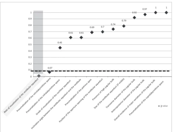
The comparison between the results from the case (A) and control (B) groups is summarized in►Fig. 3. There was a significant correlation between MD in only one of the vari-ables assessed: capacity of individualization of the vestibular aqueduct (VA). Among patients with MD, this structure was observed less frequently (p¼0.01; Fisher exact test).
►Table 3shows the statistical significance level obtained when analyzing subgroups A1 and A2, which were composed by the affected ears of patients with unilateral and bilateral MD, respectively. None of the variables was significantly associated with the occurrence of bilateral disease (p<0.05). The results from the statistical analysis between sub-groups consisting of the affected (A1) and healthy contral-ateral (B1) ears of patients with MD are provided in►Table 4. Similarly, there was no significant difference for any of the variables investigated.
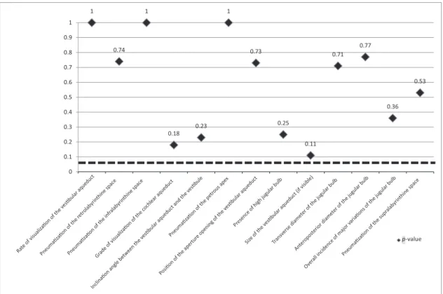
►Fig. 4reports statistical data regarding the results when comparing the control subgroups (B1 and B2). There was a trend toward lower angulation between the vestibular aque-duct and the vestibule in the contralateral ears of patients with unilateral disease, when compared with the ears of individuals without MD (p¼0.11). Nonetheless, none of the variables reached statistical significance (p<0.05).
Discussion
One previous study has suggested that the sigmoid sinus could be placed significantly more anteriorly and medially in
Table 2 Variables individually investigated in each ear of this study’s sample
Anatomical Aspect Categories/Unity of Measurement
for the Variable
Categorical Variables
Height of the jugular bulb considering the basal turn of the cochlea High or normal
Presence of any of the major anatomical variations of the jugular bulb: diverticulum, dehiscence to the middle ear or otic capsule,
contact with cochlear or vestibular aqueducts
Present or absent
Position of the aperture opening of the vestibular aqueduct Above or below the basal turn of the cochlea
Individualization of the cochlear aqueduct More or less than half of its extension
Pneumatization of the retrolabyrinthine space Absent/decreased or increased
Pneumatization of the supralabyrinthine space Absent/decreased or increased
Pneumatization of the infralabyrinthine space Absent/decreased or increased
Pneumatization of the petrous apex Absent/decreased or increased
Continuous Variables
Anteroposterior diameter of the jugular bulb In millimeters (mm)
Transverse diameter of the jugular bulb In millimeters (mm)
Size of the vestibular aqueduct (if visible) In millimeters (mm))
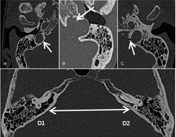
Fig. 1 Computed axial tomography images showing anatomical variations of the temporal bone: (A) High jugular bulb, above the level of the tympanic annulus (arrow); (B) Jugular bulb diverticulum (arrow); (C) Contact between the jugular bulb and the vestibular aqueduct (arrow); (D) Asymmetric pneumatization of the retrolabyrinthine space, deemed increased on the right (D1) and decreased on the left side (D2) (arrow). Source: the author.
74 included ears (37
paents)
Case Group (Group
A, n = 33)
Definite unilateral Ménière disease (Group A1, n = 13)
Definite bilateral Ménière disease (Group A2, n = 20)
Control Group
(Group B, n = 41)
Clinically and audiometrically normal contralateral ears of paents from group A1 (Group
B1, n = 13)
Ears from individuals without definite or probable Ménière disease (group B2, n = 28)
EXCLUDED: paents under 18
years-old or with at least one of the following: chronic inflammatory middle ear disease, temporal bone
neoplasia or trauma, congenital abnormalies or previous ear surgery.
patients with MD. The authors hypothesized that the anom-alous position of the vessel could generate compression of the endolymphatic sac and, therefore, hydrops.13
The jugular bulb consists of a dilation of the internal jugular vein and is located at the posterior part of the jugular foramen. When enlarged, it might be responsible for audiovestibular symptoms.14 Variations in its size and location are common and might occur due to malformations in the surrounding bony structure or in the intracranial venous system itself or secondary to lack of pneumatization of the mastoid portion of the temporal bone. If the jugular bulb is located superiorly to the inferior surface of the tympanic annulus in the middle ear, at the level of the basal cochlear turn or less than 2 mm from the inner auditory channel, it is classified as a high jugular bulb (HJB).15,16Most of the previous studies that aimed to evaluate HJB have shown limitations, such as emphasis on its incidence or small samples.17Radiological and post-mortem studies of the temporal bone report a prevalence of HJB ranging from 3–24% of the population, although this variation might not be clinically apparent.15,17–19 Jugular bulb abnormalities have also been implied in a various range of cochleovestibular symptoms, depending on the impact on the surrounding structures. The jugular bulb might erode into the middle ear or into the cochlear or vestibular aqueducts. Hearing loss, vertigo and tinnitus have also been attributed to high and dehiscent jugular bulbs, as well as to the compressive effects of HJB on the endolymphatic sac.18,20A recent German retrospective controlled study has shown a significantly higher prevalence of jugular bulb abnorm-alities among patients with MD. The authors concluded that the
Rat e of visualiz
aon of the ves bular aqueduct
Pneuma za
on of the r etrolabyrin
thine space
Pneuma za
on of the in fralabyrin
thine space
Grade of visualiz aon of the c
ochlear aqueduct
Inclina on angle be
tween the v
esbular aqueduct and the v esbule
Pneuma zaon of the pe
trous ape x
Posion of the apertur e opening of the v
esbular aqueduct
Presence of high jugular bulb
Size of the v
esbular aqueduct (if visible)
Trans ver
se diame
ter of the jugular bulb
Ante ropos
terior diame
ter of the jugular bulb
Over
all incidence of major v aria
ons of the jugular bulb
Pneuma za
on of the supr alabyrin
thine space
p-value 0
0.1 0.01 0.07 0.45
0.61 0.61
0.69 0.7 0.74
0.79 0.92
0.97 1 1
0.2 0.3 0.4 0.5 0.6 0.7 0.8 0.9 1
Fig. 3 Values (p) for each assessed variable resultant from comparative statistical analysis between the main study groups, case (A) and control (B). Significance level:p-value<0.05.
Table 3 A summary of statistical data (p-value) obtained for each variable after assessing both case subgroups. Comparison between data of affected ears from patients with unilateral disease (subgroup A1) and those of individuals with bilateral disease (subgroup A2)
Variable p-value
Presence of high jugular bulb 0.41 (χ2) Anteroposterior diameter of the
jugular bulb
0.41 (WCX)
Transverse diameter of the jugular bulb 0.17 (WCX) Overall incidence of major variations
of the jugular bulb
0.63 (Fisher)
Rate of visualization of the vestibular aqueduct
0.13 (Fisher)
Size of the vestibular aqueduct (if visible) 1.0 (WCX) Position of the aperture opening of
the vestibular aqueduct
1.0 (Fisher)
Inclination angle between the vestibular aqueduct and the vestibule
0.57 (WCX)
Grade of visualization of the cochlear aqueduct
1.0 (Fisher)
Pneumatization of the infralabyrinthine space 0.73 (Fisher) Pneumatization of the retrolabyrinthine space 0.21 (Fisher) Pneumatization of the supralabyrinthine space 1.0 (Fisher) Pneumatization of the petrous apex 1.0 (Fisher) Abbreviations: Fisher, Fisher exact test; WCX, Wilcoxon-Mann-Whitney test;χ2: chi-square test.
temporal bone in these individuals might be constitutionally different and carry predisposing factors for the development of clinically overt MD.21Redfern et al have also described higher prevalence of abnormalities of the jugular bulb in patients with MD, when compared with the general population.22 In the present study, differences in the prevalence of HJB or major jugular bulb abnormalities among affected ears have not been found.
Ikeda e Sando have shown in a study with post-mortem temporal bones that patients with MD have smaller tube-shaped endolymphatic ducts, in contrast with normal sub-jects, whose ducts were bigger and funnel-shaped.23Welling et al reported that, in MD, the identification of the endo-lymphatic duct on magnetic resonance imaging (MRI) could be significantly less frequent compared with controls. In this study, the authors also compared the measurements of the temporal bone in the region of the endolymphatic duct and found smaller dimensions among affected individuals.24By using a systematic approach to the evaluation of CT scans, Alvarenga et al achieved identification rates of the vestibular aqueduct superior to 90% among both ears of patients with unilateral MD. The rates did not differ statistically from those of control individuals.25In the present study, among affected ears, there was a significantly greater number of CT scans in which the vestibular aqueduct could not be individualized (p¼0.01). However, considering only the exams in which this structure was identified, the absolute measurements did not differ from those of the control arm. No statistically significant difference between the study groups was obtained regarding either the position of the internal 1
0.74 1
0.18 0.23
1
0.73
0.25
0.11 0.71
0.77
0.36 0.53
0 0.1 0.2 0.3 0.4 0.5 0.6 0.7 0.8 0.9 1
p-value
Rate of visualiz
aon of the v esbular aqueduct
Pneuma za
on of the r etrolabyrin
thine space
Pneuma zaon of the in
fralabyrin thine space
Grade of visualiz aon of the c
ochlear aqueduct
Inclina on angle be
tween the v
esbular aqueduct and the v esbule
Pneuma zaon of the pe
trous ape x
Posion of the apertur e opening of the v
esbular aqueduct
Presence of high jugular bulb
Size of the v
esbular aqueduct (if visible)
Trans verse diame
ter of the jugular bulb
Ante ropos
terior diame
ter of the jugular bulb
Over
all incidence of major v aria
ons of the jugular bulb
Pneuma zaon of the supr
alabyrin thine space
Fig. 4 Results obtained after stratifying the control group:p-value associated with each variable, when comparing the results from the contralateral ears of patients with unilateral MD (subgroup B1) and those from individuals without the disease (subgroup B2). Significance level:
p-value<0.05.
Table 4 A summary of statistical data (p-value) obtained for each variable when comparing the results of affected ears from patients with unilateral Ménière disease (subgroup A1) and those of the contralateral ears of these individuals. (subgroup B1)
Variable p-value
Presence of high jugular bulb 1.0 (χ2) Anteroposterior diameter
of the jugular bulb
0.82 (WCX)
Transverse diameter of the jugular bulb 0.45 (WCX) Overall incidence of major
variations of the jugular bulb
0.66 (Fisher)
Rate of visualization of the vestibular aqueduct
1.0 (Fisher)
Size of the vestibular aqueduct (if visible) 0.48 (WCX) Position of the aperture opening
of the vestibular aqueduct
1.0 (Fisher)
Inclination angle between the vestibular aqueduct and the vestibule
0.96 (WCX)
Grade of visualization of the cochlear aqueduct
1.0 (Fisher)
Pneumatization of the infralabyrinthine space 0.69 (Fisher) Pneumatization of the retrolabyrinthine space 1.0 (Fisher) Pneumatization of the supralabyrinthine space 1.0 (Fisher) Pneumatization of the petrous apex 1.0 (Fisher) Abbreviations: Fisher: Fisher exact test; WCX: Wilcoxon-Mann-Whitney test;χ2: chi-square test.
aperture or the inclination of the vestibular aqueduct. Rates of identification of the cochlear aqueduct were also similar. As to the assessment of pneumatization of different portions of the temporal bone, a trend to reduced (p¼0.07, Pearson chi-square test) pneumatization of the retrolabyrinthine space was observed among subjects with MD, although this result could not be deemed statistically significant. No difference was demonstrated in the pneumatization of the petrous apex, infralabyrinthine and supralabyrinthine spaces.
Previous functional studies have shown the presence of abnormal findings in the contralateral ears of patients with unilateral MD.26,27Thesefindings have been corroborated by histopathologic studies, which have identified significantly more damage in the contralateral inner ears of patients with MD compared with those of normal controls.28The incidence of functional contralateral involvement and development of clini-cally apparent bilateral MD increases linearly, so that after 30 years of follow-up, up to 50% of the patients have bilateral disease.29 Increasing evidence of endolymphatic hydrops in asymptomatic contralateral ears might suggest that MD is a systemic disease.30,31 Hence, the recurrent findings in the literature of abnormal anatomical features among both affected and contralateral ears in comparison to normal controls might indicate that MD has a complex pathophysiology, in which the anatomical component might be a marker of disease progres-sion or a contributing factor. Accordingly, the previous existence or the development during follow-up of certain anatomical abnormalities in the asymptomatic contralateral ear could predispose the patient to the progression to bilateral disease, with an unfavorable prognosis. Karatas et al studied the size and width of the endolymphatic duct and the presence of jugular bulb abnormalities in the CT scans of patients with unilateral MD. In this study, the measurements for the endolymphatic duct were reported to be smaller and the prevalence of abnormalities of the jugular bulb was higher among MD patients when compared with healthy controls. However, for none of the variables, there was a significant difference between the results of affected and contralateral non-affected ears.32 Hall et al also described lower periaqueductal pneumatization rates in the CTscans of patients with MD. Similarly, in this study, the results from the contralateral ears did not differ from those of the affected ears.33Using MRI and CT to evaluate the cochlear aqueduct, Park et al found similar bony dimensions among the affected ears of patients with MD when compared with a control group, althoughfluid length within this structure was significantly decreased among case subjects. These findings could not be reproduced when comparing the affected and non-affected ears of patients with MD.34
In this work, no statistically significant difference in the prevalence of any anatomical variations was observed when comparing the affected ears of individuals with unilateral MD with those of patients with bilateral MD. Thus, the anatomical factor, if important in MD’s pathophysiology and/or diagnosis approach, might contribute independently of the existence of involvement of both ears. There was no difference when comparing the results from the affected and the contralateral non-affected ears of patients with unilateral MD and from
these contralateral non-affected ears with the ears of healthy controls. Thisfinding would indicate that the contralateral ears did not differ from the affected ears or from the ears of healthy subjects. Longitudinal and cohort studies might determine more precisely the meaning of these results, as the possibility of progression of unilateral MD to bilateral disease is well known. Thereby, it would be necessary to better stratify contralateral ears longitudinally and check the occurrence of dynamic alterations in the anatomical findings reported ac-cording to the progression of the disease. As a result, it might be possible to establish the role of the anatomical variations as markers of disease progression or risk factors for the develop-ment of bilateral MD, in case of significant identification of these variations, late or precociously in the follow-up of con-tralateral ears that would become affected over time. Experi-mental and histopathological studies might define more clearly the existence of pathophysiological links between anatomical factors and the incidence of MD, which remain elusive.
Conclusion
Some anatomical variations of the temporal bone might be more frequently found in routine CT scans from affected ears of patients with MD, such as the lower rates of individualiza-tion of the vestibular aqueduct. Nonetheless, according to the literature,25the use of the imaging study of the vestibular aqueduct as a supportive diagnostic tool and the role of correlated anatomical variations in the development of MD remain a controversial issue. Longitudinal controlled studies including patients with anatomical variations possibly re-lated to MD might be important to observe if they develop the disease more frequently over time.
Note
This paper has been chosen to be presented in the IFOS-ENT World Congress, which was held in Paris, in June of 2017.
References
1 Sajjadi H, Paparella MM. Meniere’s disease. Lancet 2008;372 (9636):406–414495
2 Meniere P. Maladies de l’oreille interne offrant des symptomes de la congestion cerebral apoplectiforme. Gaz Med de Paris 1961;16:88 3 da Costa SS, de Sousa LCA, Piza MR. Meniere’s disease: overview, epidemiology, and natural history. Otolaryngol Clin North Am 2002;35(03):455–495
4 Schessel DA, Minor LB, Nedzelski J. Meniere's disease and other peripheral vestibular disorders. In: Gaertner RS, Murphy MB, ed. Cummings Otolaryngology Head and Neck Surgery. 4th ed. Philadelphia: Mosby; 2004:3231–3232
5 da Costa SS. Central causes of vertigo. In: Souza SD, Claussen C, ed. Modern concepts of neurology. Mumbai: Prajakta Arts; 1997: 310–331
6 Paparella MM, da Costa SS, Fox R, Yoo TH. Meniere's disease and other labyrinthine diseases. in: Paparella MM, Shumrick DA, Gluckmann J, Meyerhoff WL, ed. Otolaryngology. 3rd edn. WB Saunders, Philadelphia1991:1689–1714
its symptoms (mechanical and chemical). Acta Otolaryngol 1985; 99(3-4):445–451
8 Harcourt J, Barraclough K, Bronstein AM. Meniere’s disease. BMJ 2014;349:g6544
9 Perez-Garrigues H, Lopez-Escamez JA, Perez P, et al. Time course of episodes of definitive vertigo in Meniere’s disease. Arch Oto-laryngol Head Neck Surg 2008;134(11):1149–1154
10 Balkany TJ, Sires B, Arenberg IK. Bilateral aspects of Meniere’s disease: an underestimated clinical entity. Otolaryngol Clin North Am 1980;13(04):603–609
11 Lopez-Escamez JA, Carey J, Chung WH, et al; Classification Commit-tee of the Barany Society; Japan Society for Equilibrium Research; European Academy of Otology and Neurotology (EAONO); Equili-brium Committee of the American Academy of Otolaryngology-Head and Neck Surgery (AAO-HNS); Korean Balance Society. Diag-nostic criteria for Menière’s disease. J Vestib Res 2015;25(01):1–7 12 Jackler RK, Dillon WP, Schindler RA. Computed tomography in
suppurative ear disease: a correlation of surgical and radiographic findings. Laryngoscope 1984;94(06):746–752
13 Paparella MM, Sajjadi H. The significance of the lateral sinus in Meniere's disease. in: J Nadol, ed. Proceedings of the 2nd International Symposium on Meniere's Disease, pathogenesis, pathophysiology, diagnosis and treatment. Kugler & Ghedini Publishers, Amsterdam; 1989:139–146
14 Brook CD, Buch K, Kaufmann M, Sakai O, Devaiah AK. The Prevalence of High-Riding Jugular Bulb in Patients with Suspected Endolym-phatic Hydrops. J Neurol Surg B Skull Base 2015;76(06):471–474 15 Wadin K, Thomander L, Wilbrand H. Effects of a high jugular fossa and
jugular bulb diverticulum on the inner ear. A clinical and radiologic investigation. Acta Radiol Diagn (Stockh) 1986;27(06):629–636 16 Rauch SD, Xu WZ, Nadol JB Jr. High jugular bulb: implications for
posterior fossa neurotologic and cranial base surgery. Ann Otol Rhinol Laryngol 1993;102(02):100–107
17 Woo CK, Wie CE, Park SH, Kong SK, Lee IW, Goh EK. Radiologic analysis of high jugular bulb by computed tomography. Otol Neurotol 2012;33(07):1283–1287
18 Vachata P, Petrovicky P, Sames M. An anatomical and radiological study of the high jugular bulb on high-resolution CT scans and alcohol-fixed skulls of adults. J Clin Neurosci 2010;17(04): 473–478
19 Kuhn MA, Friedmann DR, Winata LS, et al. Large jugular bulb abnormalities involving the middle ear. Otol Neurotol 2012;33 (07):1201–1206
20 Friedmann DR, Eubig J, Winata LS, Pramanik BK, Merchant SN, Lalwani AK. Prevalence of jugular bulb abnormalities and resul-tant inner ear dehiscence: a histopathologic and radiologic study. Otolaryngol Head Neck Surg 2012;147(04):750–756
21 Park JJ, Shen A, Keil S, Kuhl C, Westhofen M. Jugular bulb abnormalities in patients with Meniere’s disease using high-resolution computed tomography. Eur Arch Otorhinolaryngol 2015;272(08):1879–1884
22 Redfern RE, Brown M, Benson AG. High jugular bulb in a cohort of patients with definite Ménière’s disease. J Laryngol Otol 2014;128 (09):759–764
23 Ikeda M, Sando I. Endolymphatic duct and sac in patients with Meniere’s disease. A temporal bone histopathological study. Ann Otol Rhinol Laryngol 1984;93(6 Pt 1):540–546
24 Welling DB, Clarkson MW, Miles BA, et al. Submillimeter magnetic resonance imaging of the temporal bone in Meniere’s disease. Laryngoscope 1996;106(11):1359–1364
25 Alvarenga EHL, Cruz OLM, Yamashita HKL, Lima EJ, Alvarenga AM, Bisinoto SMB. Sistematização do estudo anatômico do aqueduto vestibular por tomografia computadorizada de alta resolução em pacientes com doenças de Ménière unilateral. Radiol Bras 2006; 39(05):345–349
26 Conlon BJ, Gibson WPR. Meniere’s disease: the incidence of hydrops in the contralateral asymptomatic ear. Laryngoscope 1999;109(11):1800–1802
27 Brookes GB, Morrison AW, Richard R. Unilateral Meniere’s dis-ease: is the contralateral ear normal? Am J Otol 1985;6(06): 495–499
28 Kariya S, Cureoglu S, Fukushima H, et al. Histopathologic changes of contralateral human temporal bone in unilateral Ménière’s disease. Otol Neurotol 2007;28(08):1063–1068
29 Friberg U, Stahle J, Svedberg A. The natural course of Meniere’s disease. Acta Otolaryngol Suppl 1984;406:72–77
30 Gürkov R, Pyykö I, Zou J, Kentala E. What is Menière’s disease? A contemporary re-evaluation of endolymphatic hydrops. J Neu-rol 2016;263(Suppl 1):S71–S81
31 Pyykkö I, Nakashima T, Yoshida T, Zou J, Naganawa S. Meniere’s disease: a reappraisal supported by a variable latency of symp-toms and the MRI visualisation of endolymphatic hydrops. BMJ Open 2013;3(02):e001555
32 Karatas A, Kocak A, Cebi IT, Salviz M. Comparison of Endolym-phatic Duct Dimensions and Jugular Bulb Abnormalities Between Meniere Disease and a Normal Population. J Craniofac Surg 2016; 27(05):e424–e426
33 Hall SF, O’Connor AF, Thakkar CH, Wylie IG, Morrison AW. Significance of tomography in Meniere’s disease: periaqueductal pneumatization. Laryngoscope 1983;93(12):1551–1553 34 Park JJ, Shen A, Keil S, Kraemer N, Westhofen M. Radiological