Togni Filho PH et al. / Post-bone marrow transplant: radiation dose reduction
Radiol Bras. 2017 Mar/Abr;50(2):90–96 90
Original Article
Utility of the inspiratory phase in high-resolution computed
tomography evaluations of pediatric patients with bronchiolitis
obliterans after allogeneic bone marrow transplant: reducing
patient radiation exposure
Necessidade da fase inspiratória na avaliação de tomografia computadorizada de alta resolução de pacientes pediátricos com bronquiolite obliterante após transplante alogênico de medula óssea: diminuição da exposição à radiação do paciente
Togni Filho PH, Casagrande JLM, Lederman HM. Utility of the inspiratory phase in high-resolution computed tomography evaluations of pediatric patients with bronchiolitis obliterans after allogeneic bone marrow transplant: reducing patient radiation exposure. Radiol Bras. 2017 Mar/Abr;50(2):90–96.
Abstract
R e s u m o
Objective: To evaluate the utility of the inspiratory phase in high-resolution computed tomography (HRCT) of the chest for the diagnosis of post-bone marrow transplantation bronchiolitis obliterans.
Materials and Methods: This was a retrospective, observational, cross-sectional study. We selected patients of either gender who under-went bone marrow transplantation and chest HRCT between March 1, 2002 and December 12, 2014. Ages ranged from 3 months to 20.7 years. We included all examinations in which the HRCT was performed appropriately. The examinations were read by two radiologists, one with extensive experience in pediatric radiology and another in the third year of residency, who determined the presence or absence of the following imaging features: air trapping, bronchiectasis, alveolar opacities, nodules, and atelectasis.
Results: A total of 222 examinations were evaluated (mean, 5.4 ± 4.5 examinations per patient). The expiratory phase findings were comparable to those obtained in the inspiratory phase, except in one patient, in whom a small uncharacteristic nodule was identified only in the inspiratory phase. Air trapping was identified in a larger number of scans in the expiratory phase than in the inspiratory phase, as was atelectasis, although the difference was statistically significant only for air trapping.
Conclusion: In children being evaluated for post-bone marrow transplantation bronchiolitis obliterans, the inspiratory phase can be ex-cluded from the chest HRCT protocol, thus reducing by half the radiation exposure in this population.
Keywords: Bronchiolitis obliterans; Radiation dosage; Bone marrow transplantation; Tomography, X-ray computed.
Objetivo: Avaliar a necessidade da fase inspiratória na tomografia computadorizada de alta resolução (TCAR) do tórax no diagnóstico de bronquiolite obliterante pós-transplante de medula óssea.
Materiais e Métodos: Estudo retrospectivo, observacional, transversal. Foram selecionados pacientes que realizaram transplante de medula óssea e TCAR do tórax, com idades entre 3 meses e 20,7 anos, de ambos os sexos, durante 12 anos e 9 meses (de 1º de março de 2002 a 12 de dezembro de 2014). Todos os exames foram realizados com qualidade técnica adequada para análise pelos radiologistas. As imagens foram analisadas em consenso por um radiologista com grande experiência em radiologia pediátrica e um radiologista em treinamento, avaliando aspectos específicos das imagens como aprisionamento aéreo, bronquiectasia, opacidade, nódulos inespecíficos e atelectasia, com critérios objetivos.
Resultados: Foram avaliados 222 exames (média de 5,4 ± 4,5 exames por paciente). A fase expiratória demonstrou os mesmos achados que as duas fases em conjunto, exceto por um único nódulo identificado somente na fase inspiratória. A fase expiratória identificou um número estatisticamente superior de aprisionamento aéreo em relação à fase inspiratória e um número maior de atelec-tasia, porém sem diferença significativa.
Conclusão: A fase inspiratória pode ser excluída do protocolo para avaliação de crianças pós-transplante de medula óssea com suspeita de bronquiolite obliterante, reduzindo, assim, pela metade a quantidade de radiação à qual essas crianças são expostas.
Unitermos: Bronquiolite obliterante; Redução de dose de radiação; Transplante de medula óssea; Tomografia computadorizada.
Study conducted in the Department of Diagnostic Imaging, Escola Paulista de Medicina da Universidade Federal de São Paulo (EPM-Unifesp), São Paulo, SP, Brazil.
1. MD, MSc, Attending Physician, Department of Diagnostic Imaging, Escola Pau-lista de Medicina da Universidade Federal de São Paulo (EPM-Unifesp), São Paulo, SP, Brazil.
2. Radiologist, Fellow in Musculoskeletal Imaging, Instituto de Radiologia do Hos-pital das Clínicas da Faculdade de Medicina da Universidade de São Paulo (InRad/ HC-FMUSP), São Paulo, SP, Brazil.
Paulo Henrique Togni Filho1, João Luiz Marin Casagrande2, Henrique Manoel Lederman3
3. Tenured Full Professor, Department of Diagnostic Imaging, Escola Paulista de Medicina da Universidade Federal de São Paulo (EPM-Unifesp), São Paulo, SP, Bra-zil.
Mailing Address: Dr. Paulo Henrique Togni Filho. Departamento de Diagnóstico por Imagem – EPM-Unifesp. Rua Napoleão de Barros, 715, Vila Clementino. São Paulo, SP, Brazil, 04024-002. E-mail: [email protected].
INTRODUCTION
Bronchiolitis obliterans (BO) is a generic term used in order to describe the inflammation of the small airways, defined as those with a diameter of less than 2 mm and with no cartilage in their walls(1). It is an obstructive airway dis-ease, caused by a wide variety of conditions, such as connec-tive-tissue diseases, inhalation of toxins, infections, and drug use(2). BO is associated with high mortality rates, ranging from 21% to 100%(3–9).
BO is the most common noninfectious late pulmonary complication of allogeneic bone marrow transplantation (ABMT) and the one with the worst prognosis, usually oc-curring more than 100 days after transplantation(10–12). In the first study of post-ABMT BO, conducted in 1982(13), lym-phocytic bronchiolitis was found in 10% of the autopsies of patients who died after ABMT.
The clinical course of BO includes irreversible and pro-gressive airway obstruction, and the treatment is aimed at stabilizing the forced expiratory volume in one second (FEV1). According to The International Bone Marrow Trans-plantation Registry, the incidence of BO is 1.7% in the first two years after ABMT, BO having been identified in 6275 patients who underwent ABMT with a compatible donor(9), and the disease is rare among patients who undergo autolo-gous transplantation(14–16).
The symptoms of BO are often insidious at their onset and usually include cough (60–100%), dyspnea (50–70%), wheezing and reduced breath sounds(4,6,17,18). Pulmonary function tests show reduced FEV1 and FEV1/forced vital capacity ratio.
The risk factors associated with post-ABMT BO are shown in Table 1(3–8,17,19–28). The most important associ-ated risk factor is the presence of chronic graft-versus-host disease (GVHD)(4,21).
Chien et al.(28) found an attributable mortality of 9% in 3 years, 12% in 5 years, and 18% in 10 years after ABMT in
patients with airflow obstruction, and it was statistically higher in patients with chronic GVHD (22% in 3 years, 27% in 5 years, and 40% in 10 years).
Given the severity of the disease and the fact that its presence increases the long-term mortality rates between those who undergo ABMT, more studies are needed to bet-ter define the clinical features of BO(28).
The definitive diagnosis of BO is made by biopsy and histopathological examination(11). However, high-resolution computed tomography (HRCT) of the chest plays an impor-tant role in diagnosing bronchiolar diseases, because they present nonspecific symptoms which usually appear only when advanced destruction of the peripheric airways has al-ready become established(1). Although the tomographic pat-terns are nonspecific, they are useful in showing which parts of the lungs are affected(1), and they may also show associ-ated conditions such as coexisting infections, BO organiz-ing pneumonia, and idiopathic pneumonia syndrome(26), thus narrowing down the differential diagnosis.
The current chest HRCT protocol for BO evaluation in pediatric patients is the same at that used for adults, includ-ing an inspiratory and an expiratory phase. In pediatric pa-tients, the concerns about the use of ionizing radiation are even greater, particularly in post-ABMT patients, because they need follow-up CTs from early ages, which increases the risks of radiation-induced cancer(10,29).
Given these concerns in reducing the radiation exposure in these children and the fact that one of the most important imaging features in BO is air trapping secondary to the air-way obstruction, which is best seen in the expiratory phase, the real need for a chest HRCT protocol including the in-spiratory phase when evaluating these patients has yet to be proven.
Our aim was to evaluate the usefulness of the chest HRCT inspiratory phase for the diagnosis of BO in post-ABMT patients, considering additional findings that would not be detected in the expiratory phase and the implications for clinical decision-making.
MATERIALS AND METHODS
This was a retrospective, observational, cross-sectional study conducted in the Diagnostic Imaging Department of the Escola Paulista de Medicina da Universidade Federal de São Paulo and at the Instituto de Oncologia Pediátrica/ Grupo de Apoio a Criança com Câncer (IOP/GRAACC, Pediatric Oncology Institute/Support Group for Children with Cancer) and was approved by the research ethics com-mittee of the institution.
We selected consecutive patients who underwent ABMT and HRCT of both genders, between March 1, 2002 and December 12, 2014. Patient ages ranged from 3 months to 20.7 years. The diagnosis of BO was based on clinical and biochemical data, as well as on the results of functional tests and on patient medical history. All patients were diagnosed at least 90 days after ABMT, the mean time from ABMT to
Table 1—Risk factors associated with post-ABMT BO.
Feature
Consistent
Likely
Possible
Risk factor
Allogeneic hematopoietic stem cell transplantation Chronic and progressive graft-versus-host disease
De novo or quiescent-type chronic graft-versus-host disease Donor with advanced age
Airflow obstruction before ABMT History of viral airway infection Acute graft-versus-host disease Busulfan-based regimens Full-body irradiation
Methotrexate-based graft-versus-host disease prophylaxis Hypogammaglobulinemia
Cytomegalovirus infection Donor with advanced age
Associated diseases (e.g., chronic myeloid leukemia) Gastroesophageal reflux disease
diagnosis being 180 days. Other complications were excluded on the basis of the natural history of the disease and physical examination. None of the patients underwent CT before ABMT, because CT of the chest is not indicated in asymp-tomatic patients with normal chest X-rays. None of our pa-tients had reported pulmonary disease. All examinations were performed at the IOP/GRAACC Diagnostic Imaging Cen-ter, which is a referral center for pediatric cancer. We in-cluded only the studies in which the imaging technique was considered appropriate for reading.
The images were acquired on a dual-slice CT scanner (MX8000 Dual; Philips, Best, The Netherlands) with volu-metric acquisition, a slice thickness of 1 mm, and an inter-slice gap of 8 mm. In most of the cases, the voltage and current were set to 120 kV and 130 mAs, respectively, yield-ing the same dose of radiation (2.4 mSv) in the inspiratory and expiratory phases, regardless of whether the acquisition was dynamic or (in older children) static. The images were reviewed by two radiologists: one was a radiologist with ex-tensive experience in pediatric radiology; and the other was a third-year radiology resident. Initially, the radiologists read the images acquired in both phases (inspiratory and expira-tory), seeking to identify the presence or absence (all quali-tative measurements) of air trapping (Figure 1A), bron-chiectasis (Figure 1B), alveolar opacities (Figure 1C), nod-ules (Figure 1D), and atelectasis (Figure 1E).
We defined air trapping as differing degrees of attenu-ation within the lung parenchyma—decreased attenuattenu-ation
(areas that are darker than the rest of the parenchyma) indi-cating the areas of air trapping. The thickening of the chial walls was assessed subjectively. In the cases of bron-chiectasis, the radiologists applied general criteria such as bronchial diameter ≥ 1.5× that of the adjacent pulmonary artery, bronchial diameter ≥ 2.0 cm, and image of the bron-chus approaching the peripheral lung parenchyma (< 1.0 cm from the adjacent pleural or mediastinal pleura). At another time point, the expiratory phase was analyzed sepa-rately, in order to identify those same imaging features. It was understood that if there were disagreements between the two radiologists, the opinion of the most experienced radiologist would prevail. However, there was no such dis-agreement.
In children who were uncooperative (those under six years of age), the inspiratory and expiratory phases were obtained bilaterally in the lateral decubitus position, the side in contact with the litter corresponding to the expiratory phase and the other side corresponding to the inspiratory phase.
Data were analyzed by descriptive statistics, expressed as absolute and relative frequencies, as well as by inferential statistics, with either the chi-square test or Fisher’s exact test, together with the Z-test for comparisons between two sample proportions.
The statistical analysis was performed with the Statisti-cal Package for the Social Sciences, version 16.0 (SPSS Inc., Chicago, IL, USA), and values of p < 0.05 were considered statistically significant.
Figure 1. Chest HRCT, expiratory phase, showing air trapping (arrow in A), bronchiectasis (arrow in B), alveolar opacities (arrow in C), nonspecific nodules (arrow in
D), and atelectasis (arrow in E).
A B C
E
D
RESULTS
During the study period, 55 patients underwent ABMT and chest HRCT for the evaluation of BO. Of those 55 pa-tients, 15 (27.3%) were excluded because their examinations were technically poor (available only on paper or film) or had not been performed at the IOP/GRAACC. Therefore, the final sample comprised 40 patients were included, rang-ing in age from 3 months to 20.7 years (mean, 9.7 ± 5.4 years). Those 40 patients underwent a total of 222 chest HRCT scans (mean, 5.4 ± 4.5 scans per patient), all of which were reviewed.
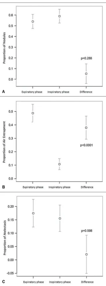
Table 2 shows the main imaging findings after the in-spiratory and expiratory phases had been analyzed (together and separately). The expiratory phase findings were the same as those obtained when the phases were analyzed together, except in one scan, in which a nonspecific nodule (of no clinical significance) was found only in the inspiratory phase (Figure 2A). However, there was no statistically difference between the findings as analyzed by the Z-test (p = 0.288). Nevertheless, the number of examinations in which air trap-ping was identified (Figure 2B) was higher for the expira-tory phase than for the inspiraexpira-tory phase, as was the number of examinations in which atelectasis was present (Figure 2C), although the difference was statistically significant only for the former comparison (p < 0.0001 and p = 0.598, respec-tively).
Tables 3 to 7 show the statistically significant relation-ships between analyzing the phases together and analyzing them separately, in terms of the findings of air trapping, bronchiectasis, opacities, atelectasis, and nonspecific nod-ules, respectively.
Table 2—Frequency of imaging findings when the inspiratory and expiratory phases were analyzed together and separately in 40 patients (n = 222 examina-tions).
Imaging feature
Air trapping Bronchiectasis Opacities
Nonspecific nodules Atelectasis
Phases
Inspiratory + Expiratory Inspiratory Expiratory
N
108 107 35 13 36
%
48.6 48.2 15.8 5.9 16.2
N
108 107 35 12 36
%
48.6 48.2 15.8 5.4 16.2
N
24 107
35 13 32
%
10.8 48.2 15.8 5.9 14.4
Table 3—Comparison between the analysis of both phases together and that of the expiratory phase separately, in terms of the finding of air trapping, in 40 pa-tients (n = 222 examinations).
Phase
Inspiratory + Expiratory
Absent Present Total
Expiratory
Absent
114 0 114
Present
0 108 108
Total
114 108 222
Figure 2. Graphics showing the difference between the inspiratory and expiratory phases in terms of the sample proportions for the findings of nodules (A), air trapping (B), and atelectasis (C).
A
B
The aim of this study was to find statistically significant data proving that the chest HRCT protocol for evaluation of pediatric patients with BO could include a smaller number of phases, thus reducing the level of radiation exposure. Al-though CT of the chest has been the subject of a series of recent publications in the Brazilian radiology literature of Brazil(30–38), there have been, to our knowledge, no studies with a similar aim.
Miglioretti et al.(29) suggested that radiation dose-reduc-ing strategies could drastically reduce the incidence of ra-diation-induced cancer. Our sample included a large num-ber of examinations, which were all performed at the same service, with the same protocols and CT equipment and the same quality, making it more homogeneous and therefore showing more statistically relevant results. However, we excluded some examinations, either because they were per-formed with a different protocol, were not perper-formed at our institution, were technically poor, or were otherwise unsuit-able for radiological analysis, which limited the size of our sample.
It is known that children have an increased lifetime can-cer incidence risk, ten times higher than for adults(39–41), not only because of the longer life expectancy but also because they will probably undergo a larger number of CT scans and other examinations involving ionizing radiation during their life. During the period analyzed in the present study, the patients underwent a median of 13.8 examinations. The ex-posure of the public to radiation from natural sources is 2.4 mSv/year(42), whereas the median effective dose of one chest CT scan in a 5-year-old child is 2.1 mSv(43).
The radiation exposure caused by medical procedures is on the rise and is currently the major artificial source of radiation. In addition, some studies have shown that there have been changes in radiological practices as a result of the creation of new techniques. The use of CT has increased worldwide, from 1–3 procedures/1000 population in the 1977–1980 period to 35 procedures/1000 population in the 1997–2007 period. Although CT accounts for approximately 7% of all radiological procedures world, it accounts for more than 40% of the collective effective dose(42). In the largest population study involving radiation exposure(44), the inci-dence of all types of cancer was found to be higher for the exposed group than for the unexposed group. At our institu-tion, reducing the radiation dose is a major goal, the health care professionals are continuously informed about the ra-diation risks, as well as the need for a more conscientious use of radiological procedures, and protocols are constantly being changed in order to achieve that goal. Recent changes in the adult abdominal CT protocols at our institution— modifications in technical aspects of the examinations and in the number of acquisition phases—have reduced the me-dian level of radiation exposure by half(45), benefiting not only the patients, who are exposed to a lower radiation dose, but also the institution, because the scans have become faster and consequently less expensive(46).
Table 5—Comparison between the analysis of both phases together and that of the expiratory phase separately, in terms of the finding of opacities, in 40 patients (n = 222 examinations).
Phase
Inspiratory + Expiratory
Absent Present Total Expiratory Absent 187 0 187 Present 0 35 35 Total 187 35 222
Table 6—Comparison between the analysis of both phases together and that of the expiratory phase separately, in terms of the finding of atelectasis, in 40 pa-tients (n = 222 examinations).
Phase
Inspiratory + Expiratory
Absent Present Total Expiratory Absent 186 0 186 Present 0 36 36 Total 186 36 222
Table 7—Comparison between the analysis of both phases together and that of the expiratory phase separately, in terms of the finding of nonspecific nodules, in 40 patients (n = 222 examinations).
Phase
Inspiratory + Expiratory
Absent Present Total Expiratory Absent 209 1 210 Present 0 12 12 Total 209 13 222
Table 4—Comparison between the analysis of both phases together and that of the expiratory phase separately, in terms of the finding of bronchiectasis, in 40 patients (n = 222 examinations).
Phase
Inspiratory + Expiratory
Absent Present Total Expiratory Absent 115 0 115 Present 0 107 107 Total 115 107 222 DISCUSSION
Our data show that the inspiratory phase could be ex-cluded from the chest HRCT protocol in children being evaluated for post-ABMT BO. Taking that measure could reduce the radiation exposure in this population by half.
REFERENCES
1. Nobre LF. Doenças das pequenas vias aéreas (bronquiolite). In: Silva CIS, Müller NL, editors. Tórax – Série CBR. Rio de Janeiro, RJ: Elsevier; 2010. p.231–46.
2. Vieira AG. Bronquiolite obliterante em pacientes submetidos a trans-plante alogênico de células tronco hematopoiéticas no Hospital de Clínicas – UFPR no período de 1979 a 2009 [dissertação]. Curiti-ba, PR: Universidade Federal do Paraná; 2012.
3. Holland HK, Wingard JR, Beschorner WE, et al. Bronchiolitis oblit-erans in bone marrow transplantation and its relationship to chronic graft-v-host disease and low serum IgG. Blood. 1988;72:621–7. 4. Chan CK, Hyland RH, Hutcheon MA, et al. Small-airways disease
in recipients of allogeneic bone marrow transplants. An analysis of 11 cases and a review of the literature. Medicine (Baltimore). 1987; 66:327–40.
5. Clark JG, Schwartz DA, Flournoy N, et al. Risk factors for airflow obstruction in recipients of bone marrow transplants. Ann Intern Med. 1987;107:648–56.
6. Clark JG, Crawford SW, Madtes DK, et al. Obstructive lung dis-ease after allogeneic marrow transplantation. Clinical presentation and course. Ann Intern Med. 1989;111:368–76.
7. Schwarer AP, Hughes JM, Trotman-Dickenson B, et al. A chronic pulmonary syndrome associated with graft-versus-host disease af-ter allogeneic marrow transplantation. Transplantation. 1992;54: 1002–8.
8. Dudek AZ, Mahaseth H, DeFor TE, et al. Bronchiolitis obliterans in chronic graft-versus-host disease: analysis of risk factors and treatment outcomes. Biol Blood Marrow Transplant. 2003;9:657– 66.
9. Santo Tomas LH, Loberiza FR Jr, Klein JP, et al. Risk factors for bronchiolitis obliterans in allogeneic hematopoietic stem-cell trans-plantation for leukemia. Chest. 2005;128:153–61.
10. Soubani AO, Uberti JP. Bronchiolitis obliterans following haemato-poietic stem cell transplantation. Eur Respir J. 2007;29:1007–19. 11. Soubani AO, Miller KB, Hassoun PM. Pulmonary complications of
bone marrow transplantation. Chest. 1996;109:1066–77. 12. Whimbey E, Champlin RE, Couch RB, et al. Community
respira-tory virus infections among hospitalized adult bone marrow trans-plant recipients. Clin Infect Dis. 1996;22:778–82.
13. Beschorner WE, Saral R, Hutchins GM, et al. Lymphocytic bron-chitis associated with graft-versus-host disease in recipients of bone-marrow transplants. N Engl J Med. 1978;299:1030–6.
14. Gasparetto EL, Ono SE, Escuissato DL, et al. Tomografia compu-tadorizada de alta resolução nas complicações pulmonares pós-trans-plante de medula óssea: ensaio iconográfico. Radiol Bras. 2005;38: 439–45.
15. Paz HL, Crilley P, Patchefsky A, et al. Bronchiolitis obliterans after autologous bone marrow transplantation. Chest. 1992;101:775– 8.
16. Frankovich J, Donaldson SS, Lee Y, et al. High-dose therapy and autologous hematopoietic cell transplantation in children with pri-mary refractory and relapsed Hodgkin’s disease: atopy predicts id-iopathic diffuse lung injury syndromes. Biol Blood Marrow Trans-plant. 2001;7:49–57.
17. Curtis DJ, Smale A, Thien F, et al. Chronic airflow obstruction in long-term survivors of allogeneic bone marrow transplantation. Bone Marrow Transplant. 1995;16:169–73.
18. Ralph DD, Springmeyer SC, Sullivan KM, et al. Rapidly progres-sive air-flow obstruction in marrow transplant recipients. Possible
association between obliterative bronchiolitis and chronic graft-versus-host disease. Am Rev Respir Dis. 1984;129:641–4. 19. Wyatt SE, Nunn P, Hows JM, et al. Airways obstruction associated
with graft versus host disease after bone marrow transplantation. Thorax. 1984;39:887–94.
20. Urbanski SJ, Kossakowska AE, Curtis J, et al. Idiopathic small air-ways pathology in patients with graft-versus-host disease following allogeneic bone marrow transplantation. Am J Surg Pathol. 1987;11: 965–71.
21. Payne L, Chan CK, Fyles G, et al. Cyclosporine as possible pro-phylaxis for obstructive airways disease after allogeneic bone mar-row transplantation. Chest. 1993;104:114–8.
22. Schultz KR, Green GJ, Wensley D, et al. Obstructive lung disease in children after allogeneic bone marrow transplantation. Blood. 1994;84:3212–20.
23. Philit F, Wiesendanger T, Archimbaud E, et al. Post-transplant obstructive lung disease (“bronchiolitis obliterans”): a clinical com-parative study of bone marrow and lung transplant patients. Eur Respir J. 1995;8:551–8.
24. Yokoi T, Hirabayashi N, Ito M, et al. Broncho-bronchiolitis oblit-erans as a complication of bone marrow transplantation: a clinico-pathological study of eight autopsy cases. Nagoya BMT Group. Virchows Arch. 1997;431:275–82.
25. Sánchez J, Torres A, Serrano J, et al. Long-term follow-up of im-munosuppressive treatment for obstructive airways disease after al-logeneic bone marrow transplantation. Bone Marrow Transplant. 1997;20:403–8.
26. Palmas A, Tefferi A, Myers JL, et al. Late-onset noninfectious pul-monary complications after allogeneic bone marrow transplanta-tion. Br J Haematol. 1998;100:680–7.
27. Ringdén O, Remberger M, Ruutu T, et al. Increased risk of chronic graft-versus-host disease, obstructive bronchiolitis, and alopecia with busulfan versus total body irradiation: long-term results of a ran-domized trial in allogeneic marrow recipients with leukemia. Nor-dic Bone Marrow Transplantation Group. Blood. 1999;93:2196– 201.
28. Chien JW, Martin PJ, Gooley TA, et al. Airflow obstruction after myeloablative allogeneic hematopoietic stem cell transplantation. Am J Respir Crit Care Med. 2003;168:208–14.
29. Miglioretti DL, Johnson E, Williams A, et al. The use of computed tomography in pediatrics and the associated radiation exposure and estimated cancer risk. JAMA Pediatr. 2013;167:700–7.
30. Alves UD, Lopes AJ, Maioli MCP, et al. Changes seen on com-puted tomography of the chest in mildly symptomatic adult patients with sickle cell disease. Radiol Bras. 2016;49:214–9.
31. Torres PPTS, Moreira MAR, Silva DGST, et al. High-resolution computed tomography and histopathological findings in hypersen-sitivity pneumonitis: a pictorial essay. Radiol Bras. 2016;49:112–6. 32. Ribeiro BNF, Ribeiro RN, Zanetti G, et al. Hughes-Stovin syn-drome: an unusual cause of pulmonary artery aneurysms. Radiol Bras. 2016;49:202–3.
33. Mogami R, Goldenberg T, Marca PGC, et al. Pulmonary infection caused by Mycobacterium kansasii: findings on computed tomog-raphy of the chest. Radiol Bras. 2016;49:209–13.
34. Queiroz RM, Gomes MP, Valentin MVN. Pulmonary paracoccidioi-domycosis showing reversed halo sign with nodular/coarse contour. Radiol Bras. 2016;49:59–60.
35. Koenigkam-Santos M, Cruvinel DL, Menezes MB, et al. Quantita-tive computed tomography analysis of the airways in patients with cystic fibrosis using automated software: correlation with spirom-etry in the evaluation of severity. Radiol Bras. 2016;49:351–7. 36. Bastos AL, Corrêa RA, Ferreira GA. Tomography patterns of lung
with early surgical resection of endobronchial metastasis in a fol-low-up of ovarian carcinoma. Radiol Bras. 2015;48:130. 38. Francisco FAF, Rodrigues RS, Barreto MM, et al. Can chest
high-resolution computed tomography findings diagnose pulmonary al-veolar microlithiasis? Radiol Bras. 2015;48:205–10.
39. Committee on the Biological Effects of Ionizing Radiations. Health effects of exposure to low levels of ionizing radiation: BEIR V. Washington, DC: National Academy Press; 1990.
40. International Commission on Radiological Protection. 1990 Rec-ommendations of the International Commission on Radiological Protection. ICRP Publication 60. Ann ICRP. 1991;21(1-3). 41. Brenner D, Elliston C, Hall E, et al. Estimated risks of
radiation-induced fatal cancer from pediatric CT. AJR Am J Roentgenol. 2001;176:289–96.
42. Mettler FA, Bhargavan M, Faulkner K, et al. Radiologic and nuclear
medicine studies in the United States and worldwide: frequency, radiation dose, and comparison with other radiation sources—1950-2007. Radiology. 2009;253:520–31.
43. Huda W. Radiation doses and risks in chest computed tomography examinations. Proc Am Thorac Soc. 2007;4:316–20.
44. Mathews JD, Forsythe AV, Brady Z, et al. Cancer risk in 680,000 people exposed to computed tomography scans in childhood or adolescence: data linkage study of 11 million Australians. BMJ. 2013;346:f2360.
45. Romano RF, Salvadori PS, D’Ippolito G, et al. Readjustment of abdominal computed tomography protocols in a university hospi-tal: impact on radiation dose. Radiol Bras. 2015;48:292–7. 46. Prasad KN, Cole WC, Haase GM. Radiation protection in humans: