DOI: 10.5935/2359-4802.20170094
ORIGINAL ARTICLE
Mailing Address: Andrea Mabilde Petracco
Av. Ipiranga, 7464, sala 524. Postal Code: 91530-000, Jardim Botânico, Porto Alegre, RS – Brazil. E-mail: [email protected]; [email protected]
Assessment of the Relationship of Ankle-Brachial Index With Coronary Artery
Disease Severity
Andrea Mabilde Petracco, Luiz Carlos Bodanese, Gustavo Farias Porciúncula, Gabriel Santos Teixeira, Denise de Oliveira Pellegrini, Luiz Claudio Danzmann, Ricardo Medeiros Pianta, João Batista Petracco
Hospital São Lucas, da Pontifícia Universidade Católica do Rio Grande do Sul, Porto Alegre, RS – Brazil
Manuscript received February 28, 2017; revised manuscript August 12, 2017; accepted August 21, 2017.
Abstract
Background: Peripheral Artery Disease (PAD) is associated with cardiovascular events and can be diagnosed and estimated by use of the Ankle-Brachial Index (ABI). ABI is a worsening factor in the stratification of cardiovascular risk, but its contribution to define the severity of coronary artery disease has not been well established.
Objectives: To compare the ABI value with the coronary atherosclerotic disease severity by use of the Syntax Score (SS) in patients with Acute Coronary Syndrome (ACS).
Methods: This prospective study measured the ABI of all patients with ACS consecutively admitted to the São Lucas Hospital of PUCRS from May to September 2016, and compared the ABI values with the SS and ACS types
of those patients. The analyzes were performed considering the 95%confidence interval (α = 5%).
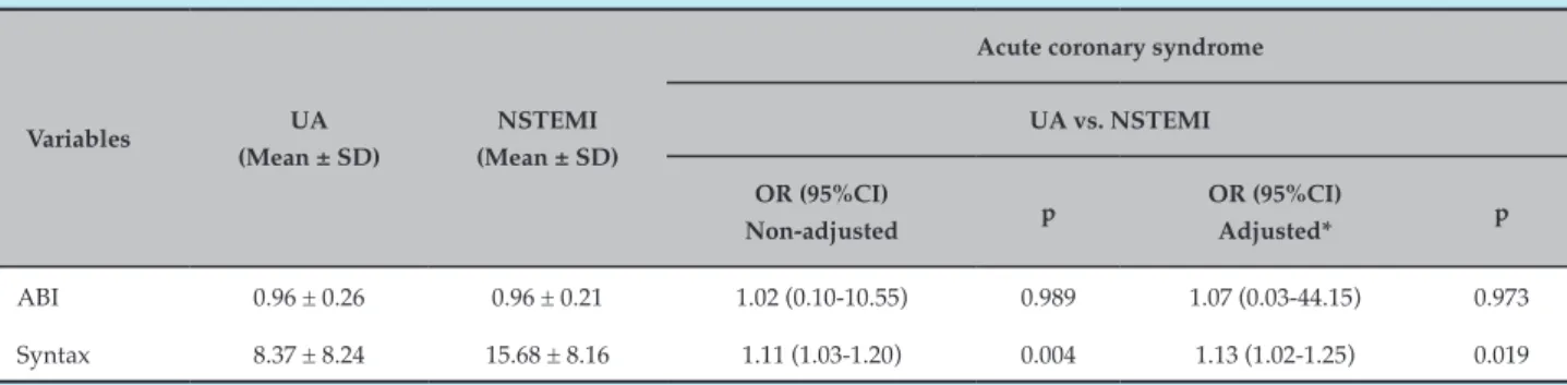
Results: This study assessed 101 patients [mean age, 62.6 ± 12.0 years; 58 men (57.4%)], 74 (82.2%) were hypertensive, 33 (45.8%) had diabetes and 46 (45,5%) had ST-elevation acute myocardial infarction (STEMI). The PAD severity was not related to the anatomical severity of the coronary artery disease (CAD). We found a significant association of intermediate SS with non-ST-elevation acute myocardial infarction (NSTEMI), and of low SS with unstable angina (UA) [OR (95% CI): 1.11 (1.03-1.20) (p = 0.004)], which remained after multivariate analysis adjusted to age, smoking, family history of CAD and previous CAD [(OR 95%): 1.13 (1.02-1.25) (p = 0.019)].
Conclusions: Patients with ABI < 0.9 showed no association with higher disease complexity determined by the SS in patients with ACS. Patients with NSTEMI were more associated with an intermediate risk on the SS. (Int J Cardiovasc Sci. 2018;31(1)47-55)
Keywords: Ankle Brachial Index; Acute Coronary Syndrome, Coronary Artery Disease; Severity of Illness Index; Atherosclerosis, Peripheral Arterial Disease.
Introduction
Cardiovascular diseases are a major cause of death and disability in Brazil and worldwide. Stroke and acute myocardial infarction are the major causes of death secondary to cardiovascular diseases. The identification of risk factors for the development of atherosclerotic disease in the population has received increasing attention,1-2 and the prediction of those factors can contribute to preventive measures and therapeutic strategies.
Different presentations of atherosclerotic disease can coexist in one single individual.3 Peripheral artery disease (PAD) is one of those presentations, usually without clinical symptoms, its diagnosis being established by calculating the ankle-brachial index (ABI).4-6 This non-invasive, easily performed test is considered a worsening factor for cardiovascular risk.2,7-9
coronary artery involvement can be obtained by use of the Syntax Score (SS).10-16
Some studies have assessed the association of CAD with PAD, and the Syntax Score II (SS II) has incorporated the presence of peripheral vascular disease, among other variables, into the SS, enabling better stratification. Some studies have attempted to correlate the severity of PAD, assessed by use of the ABI, with the complexity of CAD.13-14, 17-21 They have found a negative association between ABI and the severity of coronary atherosclerosis, and some of those studies, similarly to ours, have assessed ACS as the presentation form of CAD.18,15,22
Our study used the ABI and the SS to quantify different forms of atherosclerotic cardiovascular disease impairment in patients with ACS, and assessed whether the ABI is related to higher or lower disease severity defined by the SS.
Methods
This is a cross-sectional, descriptive and analytical study. Data were collected prospectively and consecutively in the Coronary Care Unit (CCU) of the Hospital São Lucas of PUCRS (HSL-PUCRS) from all patients admitted due to ACS from May to September 2016. Data were retrieved from the patients’ medical records and the measurements taken from each patient. All patients were invited to participate, and provided either verbal or informed consent.
During the study period, all patients who sought the HSL-PUCRS with chest pain accompanied by changes in their cardiac biomarkers and/or their electrocardiographic findings compatible with the diagnosis of ACS, with no other cause for chest pain were invited to participate in this study. Patients who could not undergo ABI measurement, such as those with lower limb lesion, and those who did not undergo coronary angiography were excluded.
This study research project was submitted to the Ethics Committee in Research of the HSL-PUCRS, being approved (1.316.041).
Data Collection Methodology
ACS: All patients who sought the HSL of the PUCRS due to anginal chest pain and who had enzymatic and/or electrocardiographic changes compatible with the diagnosis of ACS were admitted to the CCU and invited to participate in this study. The ACS classification was
based on the 2014-updated version of the 2007 Brazilian Society of Cardiology Guideline on Unstable Angina (UA) and Acute non-ST-Elevation Myocardial Infarction (NSTEMI), and on the 2015 Brazilian Society of Cardiology V Guideline for the Management of Acute ST-Elevation Myocardial Infarction (STEMI).
ABI: The patient must be placed supine, and systolic blood pressure (SBP) should be measured in the upper arm and at the ankle. The SBP in the upper arm was measured manually with DINAMAP non-invasive technology. The SBP at the ankle was measured by use of the auscultatory technique with the Dopplex SD2 Huntleigh device, an 8-MHz probe at the level of the posterior tibial artery and an aneroid sphygmomanometer with cuff. We chose to take the measure at the left side, because most patients had undergone hemodynamic study via the right lower limb, which had to be at absolute rest. The ABI was calculated by dividing the SBP reading in the lower limb by the SBP reading in the upper limb of each patient. The diagnosis of PAD was established based on the ABI,
considering the cutoff points ≤ 0.9 as presence of disease,
and those > 0.9 to 1.4 as absence of disease. Neither an ABI > 1.40 nor a non-compressible ABI were computed.4,5
Claudication: The diagnosis of claudication was based on the Edinburgh Questionnaire, which was validated for the Brazilian population in the study by the Peripheral Artery Disease Committee of the Brazilian Society of Cardiology “Projeto Corações do Brasil”.23
SS: Coronary angiography was performed according to the Judkins or Sones technique, and analyzed by two interventional cardiologists blinded to the study protocol. In case of disagreement, assessment by a third observer also blinded to the study protocol was requested.
Lesions causing a reduction in coronary diameter ≥ 50% of vessels with diameters ≥ 1.5 mm were assessed
separately with the SS, and they were added to determine each patient’s overall SS. The score was calculated by using the Syntax Score algorithm.16 The cutoff points for statistical analysis attributed to the SS were: low risk (< 22), intermediate risk (22-32), and high risk (> 32).
Statistical analysis
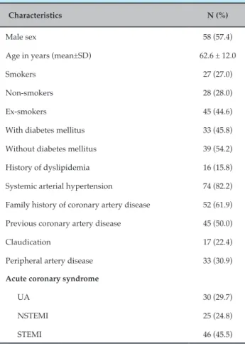
Table 1 – Characteristics of the patients admitted to the coronary care unit of the HSL-PUCRS with acute coronary syndrome from May to September 2016
Characteristics N (%)
Male sex 58 (57.4)
Age in years (mean±SD) 62.6 ± 12.0
Smokers 27 (27.0)
Non-smokers 28 (28.0)
Ex-smokers 45 (44.6)
With diabetes mellitus 33 (45.8)
Without diabetes mellitus 39 (54.2)
History of dyslipidemia 16 (15.8)
Systemic arterial hypertension 74 (82.2)
Family history of coronary artery disease 52 (61.9)
Previous coronary artery disease 45 (50.0)
Claudication 17 (22.4)
Peripheral artery disease 33 (30.9)
Acute coronary syndrome
UA 30 (29.7)
NSTEMI 25 (24.8)
STEMI 46 (45.5)
SD: standard deviation; UA: unstable angina; STEMI: ST-elevation myocardial infarction; NSTEMI: non-ST-elevation myocardial infarction. Note: Number of losses: 1 to smoking, 29 to diabetes mellitus, 11 to systemic arterial hypertension, 17 to family history of coronary artery disease, 11 to previous coronary artery disease, and 25 to the symptom of claudication.
continuous variables, as mean and standard deviation. The association between the categorical variables was performed with Pearson’s chi-square and Fisher exact tests, and the means of the continuous variables were compared by using Student t test for independent samples and ANOVA with Bonferroni adjustment. The variables with a p > 0.2 association were entered into the binary logistic regression model. The analyses were performed considering the 95% confidence interval (α = 5%).
Results
This study assessed 101 patients, with a mean age of 62.6 years (31 - 92 years), 57.4% of whom were of the male sex. Most patients assessed had a low risk according to the SS (83.2%), being classified as normal regarding the ABI (45.5%). Of the 101 patients, 4 had non-compressible ABI, being excluded from the diagnosis of PAD by the ABI method. Thus, the diagnosis of PAD based on the ABI could be considered in 97 patients. Peripheral artery disease was present in 33 patients (30.9%). Regarding the diagnosis of the clinical presentation of ACS, participants most frequently had STEMI (45.5%).
The patients’ clinical characteristics are shown in Table 1. Most patients were ex-smokers (44.6%), had diabetes mellitus (45.8%), systemic arterial hypertension (SAH – 82.2%) and family history of CAD (61.9%). Half of the patients had previous CAD (50.0%) and most had no intermittent claudication (58.4%). There was a large number of losses: 1 to smoking, 29 to diabetes mellitus, 11 to SAH, 17 to family history of CAD, 11 to previous CAD, and 25 to the symptom of claudication. The diagnosis of claudication was established by use of the Edinburgh Questionnaire, and patients with claudication more often had PAD (p = 0.050) (Table 2).
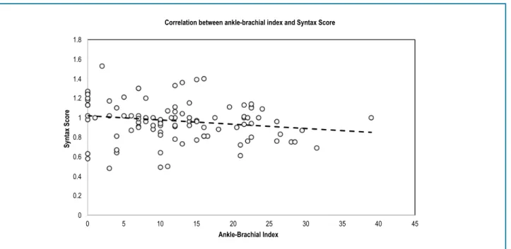
The association between the categorical variables was performed with Pearson’s chi-square and Fisher exact tests, and the means of the continuous variables were compared by using Student t test for independent samples and ANOVA with Bonferroni adjustment. To assess the correlation between the ABI and the SS, Pearson correlation test was used. The variables with a p > 0.2 association were entered into the binary logistic regression model. The analyses were performed considering the 95% confidence interval (α = 5%). The correlation between ABI and SS was not significant (r = -0.184; p = 0.070) (Figure 1).
Patients with NSTEMI were older than those with STEMI (p = 0.021). The SS of patients with NSTEMI was
higher (approximately twice) than that of those with UA (p = 0.004). According to the SS, intermediate risk was more frequent among patients with NSTEMI, and low risk, among patients with UA (p = 0.015). When the SS was reclassified, isolating patients with zero score, those with UA more frequently had zero SS, while those with NSTEMI had an intermediate risk according to the SS (p = 0.004) (Table 3).
Previous CAD was more frequently found among patients with UA, while patients with no previous CAD more often had STEMI (p = 0.001) (Table 3).
Table 2 – Clinical characteristics related to peripheral artery disease of patients admitted to the coronary care unit of the HSL-PUCRS with acute coronary syndrome from May to September 2016
Variables
Peripheral artery disease
p Yes (n = 30) N (%) No (n = 67) N (%)
Male sex 18 (60.0) 36 (53.7) 0.660*
Age in years (mean ± SD) 65.0 ± 12.0 61.6 ± 12.1 0.212¥
Syntax Score (mean ± SD) 14.8 ± 9.2 11.6 ± 8.13 0.093¥
Syntax classification
Low risk 22 (73.3) 58 (86.6)
Intermediate risk 8 (26.7) 8 (11.9) 0.133£
High risk 0 (0.0) 1 (1.5)
Smoking 8 (26.7) 17 (25.8) 0.850*
Diabetes mellitus 11 (47.8) 21 (46.7) 0.928*
Systemic arterial hypertension 22 (81.5) 49 (81.7) 1.000£
Family history of coronary artery disease 17 (68.0) 33 (60.0) 0.493*
Previous coronary artery disease 12 (46.2) 30 (49.2) 0.796*
Claudication 9 (37.5) 8 (16.7) 0.050*
SD: standard deviation. * Chi-square test; ¥: Student t test for independent samples; £: Fisher Exact test. Note: Number of losses: 1 to smoking, 29 to
diabetes mellitus, 11 to systemic arterial hypertension, 17 to family history of coronary artery disease, 11 to previous coronary artery disease, and 25 to the symptom of claudication.
associated with ACS for the clinical form of UA as compared to NSTEMI [OR (95%CI): 1.13 (1.02-1.25); p = 0.019] (Table 4).
When combining UA with STEMI, STEMI with NSTEMI, and UA + NSTEMI with STEMI, after adjusting to age, family history of CAD and previous CAD (variables with p > 0.2 on univariate and bivariate analysis), the ABI and the SS did not maintain the association with the type of ACS clinical presentation.
Discussion
The atherosclerotic disease is multifactorial. The clinical manifestations of patients with ACS are: UA, NSTEMI and STEMI. Peripheral artery disease, an expression of peripheral atherosclerotic disease, is a more severe form, defined as a worsening factor in the cardiovascular risk stratification of patients at intermediate risk.2,9,24
The major objective of this study was to determine the ABI value of patients with ACS, and to relate ABI to
the severity of coronary lesion by use of the SS. Several studies have shown the relationship between ABI and CAD severity, most of them conducted in patients with suspected CAD or unstable CAD.11-13,15,20,21,25,26 Studies using the SS II have shown a negative relationship between the presence of peripheral vascular disease and the SS, but they included no patient with ACS.11,12,14
In our study, we expected to find a lower ABI value when compared to the higher SS in ACS. In our sample, the ABI value was not related to the severity of CAD on the SS. We found a negative correlation between those two indices, but without statistical significance. Some differences between the methodologies might help us understand the result different from that expected.
1.8
1.6
1.4
1.2
1
0.8
0.6
0.4
0.2
0
0 5 10 15 20 25 30 35 40 45
Syntax Score
Correlation between ankle-brachial index and Syntax Score
Ankle-Brachial Index
Figure 1 – Correlation between ankle-brachial index and Syntax Score of patients admitted to the coronary care unit of the HSL-PUCRS with acute coronary syndrome from May to September 2016.
STEMI and those with previous CAD had been excluded.18Our sample had a small number of patients with STEMI, and those with previous CAD were not excluded. When stratifying the presentation forms of ACS, we found no relationship of the SS with the cases of STEMI, which comprised most of our sample. However, the comparison of the SS of patients with NSTEMI and with UA evidenced a relationship of the intermediate SS with cases of NSTEMI and of the zero SS with cases of UA. Such aspects can clarify the fact that there was no significant negative relationship between the SS and the ABI in ACS. In the study by Benyakorn with 213 patients, correlating the ABI with the severity of the coronary artery lesion, patients with ACS who were known to have PAD were excluded, and the ABI cutoff point of 0.7 was used. That author found a strong negative relationship between ABI and SS.17
A multicenter study with 1054 patients, assessing the impact of PAD in patients with ACS and not excluding STEMI, has suggested that the detection of PAD at the bedside might be a useful tool to stratify early risk.27
The well-known low diagnostic power of PAD based on symptomatology was confirmed in our sample with the use of the Edinburgh Questionnaire.23,24 The prevalence of PAD in our sample was three-times that described for the general population. This evidence emphasizes that we assessed patients with diffuse
atherosclerotic disease, and that patients with CAD are prone to develop PAD. A similar finding was observed in the study by Korkmaz, in which the frequency of asymptomatic PAD was higher.18
Although PAD is strongly associated with fatal and non-fatal cardiovascular event,22,28 its severity is not yet used to help stratify the coronary atherosclerotic complexity. Studies comparing the ABI value with the severity of stable CAD have found a negative relationship between them, similarly to the studies on ACS.13,15,17,18, 20-22, 26,27 Comparing our results with those of other studies on ACS, the frequency of patients with STEMI was the most discrepant finding, because we had a greater prevalence of STEMI, which might explain the difference. However, the inverse relationship between those indices seems to be present and significantly repeated in several studies, even with the different methodologies used.
Table 3 – Clinical characteristics according to the clinical forms of acute coronary syndrome of patients admitted to the coronary care unit of the HSL-PUCRS with acute coronary syndrome from May to September 2016
Variables Total sample
N (%)
ACS
p UA (n = 30)
N (%)
NSTEMI (n = 25) N (%)
STEMI (n = 46) N (%)
Male sex 58 (57.4) 14 (46.7) 15 (60.0) 29 (63.0) 0.353*
Age in years (mean ± SD) 62.6 ± 12.0 66.1a ± 10.3 65.0a ± 13.3 59.0b ± 11.6 0.021¥
ABI (mean ± SD) 0.97 ± 0.20 0.96 ± 0.26 0.96 ± 0.21 0.97 ± 0.17 0.996¥
ABI classification
Low to intermediate risk 33 (32.7) 10 (33.3) 10 (40.0) 13 (28.3)
Borderline 19 (18.8) 3 (10.0) 5 (20.0) 11 (23.9) 0.653£
Normal 46 (45.5) 16 (53.3) 9 (36.0) 21 (45.7)
Non-compressible 3 (3.0) 1 (3.3) 1 (4.0) 1 (2.2)
Peripheral artery disease 30 (30.9) 10 (35.7) 11 (24.4) 9 (37.5) 0.434*
Syntax Score (mean ± SD) 12.29 ± 8.59 8.37b ± 8.24 15.68a ± 8.16 13.01ab ± 8.21 0.004¥
Syntax classification
Low risk 84 (83.2) 28 (93.3)** 16 (64.0) 40 (87.0)
Intermediate risk 16 (15.8) 2 (6.7) 9 (36.0)** 5 (10.9) 0.015£
High risk 1 (1.0) 0 (0.0) 0 (0.0) 1 (2.2)
Syntax classification isolating zero
Zero 12 (11.9) 8 (26.7)** 3 (6.5) 1 (4.0)
Low risk 72 (71.3) 20 (66.7) 37 (80.4) 15 (60.0) 0.004£
Intermediate risk 16 (15.8) 2 (6.7) 5 (10.9) 9 (36.0)**
High risk 1 (1.0) 0 (0.0) 1 (2.2) 0 (0.0)
Smoking 27 (27.0) 4 (13.3) 5 (20.0) 18 (40.0) 0.093*
Diabetes mellitus 33 (45.8) 12 (46.2) 10 (55.6) 11 (39.3) 0.557*
Systemic arterial hypertension 74 (82.2) 26 (89.7) 18 (85.7) 30 (75.0) 0.291£
Family history of CAD 52 (61.9) 20 (69.0) 15 (71.4) 17 (50.0) 0.177*
Previous CAD
Yes 45 (50.0) 21 (77.8)** 10 (47.6) 14 (33.3)
0.001*
No 45 (50.0) 6 (22.2) 11 (52.4) 28 (66.7)
Claudication 17 (22.4) 6 (26.1) 6 (33.3) 5 (14.3) 0.254£
ACS: acute coronary syndrome; UA: unstable angina; STEMI: ST-elevation myocardial infarction; NSTEMI: non-ST-elevation myocardial infarction; SD: standard deviation; ABI: ankle-brachial index; CAD: coronary artery disease. * Chi-square test; ¥: ANOVA with post-hoc Bonferroni adjustment; £:
Fisher Exact test **Analysis of adjusted residues. Note: Number of losses: 1 to smoking, 29 to diabetes mellitus, 11 to systemic arterial hypertension, 17 to
Table 4 – Multivariate analysis
Variables UA
(Mean ± SD)
NSTEMI (Mean ± SD)
Acute coronary syndrome
UA vs. NSTEMI
OR (95%CI)
Non-adjusted p
OR (95%CI)
Adjusted* p
ABI 0.96 ± 0.26 0.96 ± 0.21 1.02 (0.10-10.55) 0.989 1.07 (0.03-44.15) 0.973
Syntax 8.37 ± 8.24 15.68 ± 8.16 1.11 (1.03-1.20) 0.004 1.13 (1.02-1.25) 0.019
UA: unstable angina; NSTEMI: non-ST-elevation myocardial infarction; SD: standard deviation; CI: confidence interval; ABI: ankle-brachial index; Syntax: Syntax Score. *Adjusted to age, smoking habit, family history of coronary artery disease and previous coronary artery disease.
when they had PAD, requiring a longer DAPT time.29 That study corroborates the importance of the diagnosis of PAD in patients with CAD submitted to PCI.
The several types of clinical stratification for patients with ACS, such as the GRACE (Global Registry of Acute Coronary Events) risk score, were related to hemodynamic severity, risk of death and major cardiovascular events. So far, we have no definitive clinical score that helps us assess the risk of the CAD complexity found in patients with ACS.
Study Limitations
In our study, we valued only the anatomical presentation of coronary lesions and tried to relate it to the ABI. A comparison of the complexity of the anatomical and functional impairment of those two presentations of atherosclerotic disease might find a more exuberant negative relationship in patients with ACS.
No assessment of myocardial functional impairment was performed in our study, because the patients had ACS according to the diagnostic criteria defined by the guidelines.
Our sample was limited for this initial study, but a larger one in future studies might be able to establish a better relationship between the ABI and the SS in patients with ACS, in addition to contributing to their cardiovascular risk stratification.
Conclusion
Our study showed that patients with an ABI < 0.9 had no association with higher disease complexity determined by the SS in patients with ACS. In addition, patients with NSTEMI were more associated with an intermediate risk on the SS.
Author contributions
Conception and design of the research: Petracco AM, Bodanese LC, Danzmann LC. Acquisition of data: Petracco AM, Porciuncula GF, Teixeira GS, Piantá RM, Pellegrini DO. Analysis and interpretation of the data: Petracco AM, Bodanese LC. Writing of the manuscript: Petracco AM. Critical revision of the manuscript for intellectual content: Petracco AM. Supervision / as the major investigador: Bodanese LC, Danzmann LC, Petracco JB, Piantá RM.
Potential Conflict of Interest
No potential conflict of interest relevant to this article was reported.
Sources of Funding
There were no external funding sources for this study.
Study Association
This article is part of the thesis of master submitted by Andrea Mabilde Petracco, from Faculdade de Medicina da PUCRS.
Ethics approval and consent to participate
1. Polanczyk C. Cardiovascular risk factors in Brazil: the next 50 years! Arq Bras Cardiol. 2005;84(3):199-201. doi: http://dx.doi.org/10.1590/ S0066-782X2005000300001.
2. Xavier HT, Izar MC, Faria Neto JR, Assad MH, Rocha VZ, Sposito AC, et al. [V Brazilian Guidelines on Dyslipidemias and Prevention of Atherosclerosis]. Arq Bras Cardiol. 2013;101(4 Suppl 1):1-20. doi: 10.5935/ abc.2013S010.
3. Murabito JM, Evans JC, Nieto K, Larson MG, Levy D, Wilson PW. Prevalence and clinical correlates of peripheral arterial disease in the Framingham Offspring Study. Am Heart J. 2002;143(6):961-5. PMID: 12075249.
4. Lin JS, Olson CM, Johnson ES, Whitlock EP. The ankle-brachial index for peripheral artery disease screening and cardiovascular disease prediction among asymptomatic adults: a systematic evidence review for the U.S. Preventive Services Task Force. Ann Intern Med. 2013;159(5):333-41. doi: 10.7326/0003-4819-159-5-201309030-00007.
5. Dachun Xu, Jue Li, Liling Zou, Yawei Xu, Dayi Hu, Pagoto SL, et al. Sensitivity and specificity of the ankle--brachial index to diagnose peripheral artery disease: a structured review. Vasc Med. 2010;15(5):361-9. doi: 10.1177/1358863X10378376.
6. Torres AG, Machado EG, Lopes TS, Gentili PC, Vieira AC, Soares LG, et al. Prevalence of ankle-brachial index alterations in patients with asymptomatic peripheral arterial occlusive disease. Rev Bras Cardiol. 2012;25(2):87-93. ID: [i]-629911.
7. O'Hare AM, Katz R, Shlipak MG, Cushman M, Newman AB. Mortality and cardiovascular risk across the ankle-arm index spectrum: results from the Cardiovascular Health Study. Circulation. 2006;113(3):388-93. doi: 10.1161/CIRCULATIONAHA.105.570903.
8. Zheng ZJ, Sharrett AR, Chambless LE, Rosamond WD, Nieto FJ, Sheps DS, et al. Associations of ankle-brachial index with clinical coronary heart disease, stroke and preclinical carotid and popliteal atherosclerosis: the Atherosclerosis Risk in Communities (ARIC) Study. Atherosclerosis. 1997;131(1):115-25. PMID: 9180252.
9. Malachias MV, Souza WK, Plavnik FL, Rodrigues CI, Brandão AA, Neves MF, et al; Sociedade Brasileira de Cardiologia. 7a Diretriz Brasileira de hipertensão arterial. Arq Bras Cardiol. 2016;107(3 supl 3):1-83. doi: http://dx.doi.org/10.5935/abc.20160153.
10. Gomes WJ, Braile DM. SYNTAX Trial: analysis and clinical implications. Rev Bras Cir Cardiovasc. 2008;23(4):3-5. doi: http://dx.doi.org/10.1590/ S0102-76382008000400002.
11. Tajik P, Oude Rengerink K, Mol BW, Bossuyt PM. SYNTAX score II. Lancet. 2013;381(9881):1899. doi: 10.1016/S0140-6736(13)61151-4.
12. Farooq V, van Klaveren D, Steyerberg EW, Meliga E, Vergouwe Y, Chieffo A, et al. Anatomical and clinical characteristics to guide decision making between coronary artery bypass surgery and percutaneous coronary intervention for individual patients: development and validation of SYNTAX score II. Lancet. 2013;381(9867):639-50. doi: 10.1016/S0140-6736(13)60108-7.
13. Sebastianski M, Narasimhan S, Graham MM, Toleva O, Shavadia J, Abualnaja S, et al. Usefulness of the ankle-brachial index to predict high coronary SYNTAX scores, myocardium at risk, and incomplete coronary revascularization. Am J Cardiol. 2014;114(11):1745-9. doi: 10.1016/j. amjcard.2014.09.010.
14. Parissis H. Is the Syntax score II and its principles behind it applicable to the entire spectrum of the real world practice? Int J Cardiol. 2016 Jul 1;214:13-5. doi: 10.1016/j.ijcard.2016.03.159.
15. Kumar CN, Subba Reddy YV, Adi Kesava Naidu O, Srinivas R. Role of ankle brachial index (ABI) diabetes hypertension and waist hip ratio as a predictor of severity of coronary artery disease by SYNTAX score. Indian Heart J. 2014;66(Suppl 2):S30. doi: 10.1016/j.ijh.2014.10.084.
16. Serruys PW, Morice MC, Kappetein AP, Colombo A, Holmes DR, et al; SYNTAX Investigators. Percutaneous coronary intervention versus coronary-artery bypass grafting for severe coronary artery disease. N Engl J Med. 2009;360(10):961-72. doi: 10.1056/NEJMoa0804626. Erratum in: N Engl J Med. 2013;368(6):584.
17. Benyakorn T, Kuanprasert S, Rerkasem K. A correlation study between ankle brachial pressure index and the severity of coronary artery disease. Int J Low Extrem Wounds. 2012;11(2):120-3. doi: 10.1177/1534734612446966.
18. Korkmaz L, Adar A, Erkan H, Ağaç MT, Acar Z, Kurt IH, et al. Ankle-brachial index and coronary artery lesion complexity in patients with acute coronary syndromes. Angiology. 2012;63(7):495-9. doi: 10.1177/0003319711429561.
19. Chen CC, Hung KC, Hsieh IC, Wen MS. Association between peripheral vascular disease indexes and the numbers of vessels obstructed in patients with coronary artery disease. Am J Med Sci. 2012;343(1):52-5. doi: 10.1097/MAJ.0b013e31821fec80.
20. Aykan AÇ, Hatem E, Karabay CY, Gül İ, Gökdeniz T, Kalaycıoğlu E, et al. Complexity of lower extremity peripheral artery disease reflects the complexity of coronary artery disease. Vascular. 2015;23(4):366-73. doi: 10.1177/1708538114550738.
21. Tripathi VD, Sharma RK, Kuila M, Saha A. Correlation in between coronary artery disease severity and peripheral artery disease. Indian Heart J. 2015;67(Suppl 1):S45. doi: 10.1016/j.ijh.2015.10.109.
22. Al-Thani HA, El-Menyar A, Zubaid M, Rashed WA, Ridha M, Almahmeed W, et al. Peripheral arterial disease in patients presenting with acute coronary syndrome in six middle eastern countries. Int J Vasc Med. 2011;2011:815902. doi: 10.1155/2011/815902.
23. Makdisse M, Nascimento Neto R, Chagas AC, Brasil D, Borges JL, Oliveira A, et al. Cross-cultural adaptation and validation of the Brazilian Portuguese version of the Edinburgh Claudication Questionnaire. Arq Bras Cardiol. 2007;88(5):501-6. doi: http://dx.doi.org/10.1590/S0066-782X2007000500001.
24. Makdisse M, Pereira Ada C, Brasil Dde P, Borges JL, Machado-Coelho GL, Krieger JE, et al. Prevalence and risk factors associated with peripheral arterial disease in the Hearts of Brazil Project. Arq Bras Cardiol. 2008;91(6):370-82. doi: http://dx.doi.org/10.1590/S0066-782X2008001800008.
25. Sabedotti M, Sarmento-Leite R, Quadros AS. Índice tornozelo-braquial como preditor de doença coronariana significativa em pacientes submetidos à angiografia coronária. Rev Bras Cardiol Invasiva. 2014;22(4):359-63. doi: 10.1590/0104-1843000000060.
26. Ueki Y, Miura T, Miyashita Y, Shimada K, Kobayashi H, Kobayashi M, et al. Adding ankle-brachial index to SYNTAX score improves prediction of clinical outcome in patients undergoing percutaneous coronary intervention. EuroIntervention. 2015 May 15. Abstracts EuroPCR 2015. [Access in 2017 Jan 10]. Available from: https://www.pcronline.com/ eurotintervention/abstractsEuroPCR2015/ abstracts-europcr-2015/ POS203/adding-index-to-syntax-score.
27. Morillas P, Quiles J, Cordero A, Guindo J, Soria F, Mazón P, et al; Prevalence of Peripheral Arterial Disease in Patients With Acute Coronary Syndrome (PAMISCA) Investigators. Impact of clinical and subclinical peripheral arterial disease in mid-term prognosis of patients with acute coronary syndrome. Am J Cardiol. 2009;104(11):1494-8. doi: 10.1016/j.amjcard.2009.07.014.
28. Pereira C, Miname M, Makdisse M, Kalil Filho R, Santos RD. Association of peripheral arterial and cardiovascular diseases in familial hypercholesterolemia. Arq Bras Cardiol. 2014;103(2):118-23. doi: http:// dx.doi.org/10.5935/abc.20140097.
29. Franzone A, Piccolo R, Gargiulo G, Ariotti S, Marino M, Santucci A, et al. Prolonged vs short duration of dual antiplatelet therapy after percutaneous coronary intervention in patients with or without peripheral arterial disease: a subgroup analysis of the PRODIGY randomized clinical trial. JAMA Cardiol. 2016;1(7):795-803. doi: 10.1001/ jamacardio.2016.2811.