Non-ruminants
Effect of different vitamin D
3metabolites on intestinal calcium
homeostasis-related gene expression in broiler chickens
Felix Shih-Hsiang Hsiao1, Yeong-Hsiang Cheng2, Jin-Cheng Han3, Ming-Huang Chang4, Yu-Hsiang Yu2*
1 Tunghai University, Department of Animal Science and Biotechnology, Taichung, Taiwan. 2 National Ilan University, Department of Biotechnology and Animal Science, I-Lan, Taiwan. 3 Shangqiu Normal University, Department of Animal Science, Shangqiu, Henan, China. 4 National Chia-Yi University, Department of Veterinary Medicine, Chia-Yi, Taiwan.
ABSTRACT - The purpose of this study was to investigate the effects of vitamin D3 metabolites 1α-hydroxycholecalciferol (1α(OH)D3), 25-hydroxycholecalciferol (25(OH)2D3), and 1,25-dihydroxycholecalciferol (1,25(OH)2D3) on growth performance, bone quality, and intestinal calcium homeostasis-related gene expression in broiler chickens. One-day-old broilers were fed a basal diet and basal diet containing different vitamin D3 metabolites. The body weight, feed intake, and feed conversion ratio in control and experimental broilers were measured to assess the growth performance, mineral levels, and bone breaking strength. The duodenum was used to assess calcium homeostasis-related gene expressions by quantitative reverse transcription-PCR. No statistically significant difference was found in growth performance, mineral deposition, or bone breaking strength in broiler chickens after three weeks feeding with vitamin D3. However, supplementation of vitamin D3 metabolites tended to improve feed conversion rate, bone mineral deposition, and breaking strength in broiler chickens. The results demonstrated that vitamin D3 metabolites significantly upregulated calcium homeostasis-related genes, including calbindin, β-glucuronidase, TRPV6, and Na/Pi IIb cotransporter, mRNA levels after 12 h of feeding. The vitamin D3 metabolite 1,25(OH)2D3 was the most effective at regulating calcium homeostasis-associated gene expression after 6 h of feeding. Dietary vitamin D3 metabolites may alleviate the development of TD in broiler chickens and these effects probably occur through regulation of intestinal calcium homeostasis-related gene expression.
Key Words: gene, 1α-hydroxycholecalciferol, 25-hydroxycholecalciferol, 1,25-dihydroxycholecalciferol
© 2018 Sociedade Brasileira de Zootecnia ISSN 1806-9290
www.sbz.org.br
R. Bras. Zootec., 47:e20170015, 2018
https://doi.org/10.1590/rbz4720170015
Received: February 2, 2017 Accepted: October 19, 2017
*Corresponding author: [email protected]
Copyright © 2018 Sociedade Brasileira de Zootecnia. This is an Open Access article distributed under the terms of the Creative Commons Attribution License (http://creativecommons.org/licenses/by/4.0/), which permits unrestricted use, distribution, and reproduction in any medium, provided the original work is properly cited.
Introduction
Tibial dyschondroplasia (TD) is an extremely common skeletal abnormality associated with rapid growth rate in broiler chickens (Leach and Lilburn, 1992). It leads to enormous economic losses worldwide and to animal welfare problems (Pines et al., 2005). Tibial dyschondroplasia is characterized by the formation of a lesion composed of non-vascularized, non-mineralized cartilage that extends from the epiphyseal growth plate into the metaphysic (Leach and Nesheim, 1965). It has been reported that TD is influenced by several factors, including genetics and nutrition (Waldenstedt, 2006). Nutrition plays a major role in the development and maintenance of bone structure, such as calcium, phosphorus, and vitamin D (Fleming, 2008).
Vitamin D3 (cholecalciferol) has been widely used as
a feed additive to improve calcium and phosphorus metabolism and bone development in poultry (Baker et al., 1998). It has also been demonstrated that supplementation of vitamin D metabolites could efficiently prevent TD in broiler chickens (Edwards, 1990; Roberson and Edwards, 1996; Whitehead et al., 2004).
Conversion of vitamin D3 to 25-hydroxycholecalciferol
(25(OH)D3) is catalyzed by 25-hydroxylase in
livers. 25(OH)D3 is further converted to
1,25-dihydroxycholecalciferol (1,25(OH)2D3) by 1α-hydroxylase
in kidney. 1,25(OH)2D3 is the biologically active form of
vitamin D in vivo (Henry et al., 1979; Henry et al., 1985).
It has been shown that 25(OH)D3 significantly improved
body weight and feed efficiency of broiler chickens
compared with those of the birds fed vitamin D3 (Yarger
et al., 1995; Fritts and Waldroup, 2003; Koreleski and Swiatkiewicz, 2005). Recently, we also demonstrated that
broilers fed 1α-hydroxycholecalciferol (1α(OH)D3) had
higher body weight gain, feed intake, and tibia breaking
strength compared with birds fed 25(OH)D3 at 42 days of
found on body weight or feed efficiency in broiler chickens
after supplementation of diet with 1,25(OH)2D3 (Roberson
and Edwards, 1996).
Vitamin D3 is involved in the regulation of levels
of minerals such as calcium and phosphorous. Several mechanisms have been proposed to elucidate the vitamin
D3 metabolism and mechanisms of calcium transport in
the small intestine (Norman et al., 1982; Kumar, 1990). However, little is known about the transcriptional level of intestinal calcium homeostasis-related genes in response
to different vitamin D3 metabolites in broiler chickens.
Therefore, the present study was conducted to investigate the effects of different vitamin D metabolites including
1α(OH)D3, 25(OH)D3, and 1,25(OH)2D3 on intestinal
calcium homeostasis-related gene expression in broiler chickens.
Material and Methods
All experiments were performed in accordance with approved guidelines. The animal protocol was approved by local Institutional Animal Care and Use Committee (IACUC case No. 104-14). The concentration of vitamin D metabolites described earlier (Yarger et al., 1995; Roberson and Edwards, 1996; Chou et al., 2009; Han et al., 2009) was used in the present study. For examination of growth performance, mineral levels, and breaking strength, forty-eight one-day-old broilers (Avian) were randomly allocated to four different treatment groups (n = 12 per group) that were fed: basal diet (control), basal diet plus 5 μg/kg of
1α(OH)D3, basal diet plus 69 μg/kg of 25(OH)D3, and
basal diet plus 5 μg/kg of 1,25(OH)2D3. The basal diets
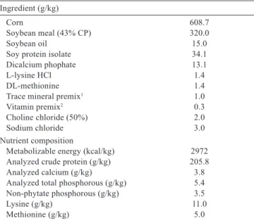
were formulated based on National Research Council recommendations (NRC, 1994) (Table 1). Water and feed
was provided ad libitum over the entire experimental
period. Total individual body weight, feed intake, and feed conversion ratio were recorded every week. At the end of the experiment, broilers were sacrificed by cervical dislocation. The blood was collected and centrifuged to harvest serum for biochemistry. The left tibia was removed for determination of mineral deposition and breaking strength measurement. For examination of calcium homeostasis-related gene expression, sixteen one-day-old broilers (Avian) were randomly allocated to four different treatment groups (n = 4 per group): basal diet (control), basal diet plus 5 μg/kg of
1α(OH)D3, basal diet plus 69 μg/kg of 25(OH)D3, and basal
diet plus 5 μg/kg of 1,25(OH)2D3. After 0, 6, and 12 h of
feeding, the duodenum was excised for RNA extraction. Tissue RNA was extracted from the duodenum of broilers using TRIzol (Invitrogen, Carlsbad, CA,
USA) according to the manufacturer’s instructions and resuspended in diethyl pyrocarbonate-treated water. Reverse transcription was performed with a Transcriptor Reverse Transcriptase kit (Roche Applied Science, Indianapolis, IN, USA). Quantitative reverse transcriptase-polymerase chain reaction (PCR) for each gene (Table 2) was performed using Miniopticon Real-Time PCR Detection System (Bio-Rad, Hercules, CA, USA) and KAPA SYBR FAST qPCR Kit (Kapa Biosystems, Boston, MA, USA). Polymerase chain reaction was performed by 40 cycles at 95 °C for 30 s, 58-60 °C for 60 s, and 72 °C for 30s. Beta-actin mRNA was determined as the internal control gene. The mRNA expression of each gene was normalized to its β-actin mRNA expression in the same sample. Threshold cycle (Ct) values were obtained and relative gene expression was calculated using the formula: (1/2)Ct target genes-Ct β-actin.
The left tibias were collected, cleaned of adhering tissue, weighed, dried to constant weight at 105 °C, defatted, and then ashed in a muffle furnace at 600 °C. Ash was dissolved in concentrated HCl for mineral determination. Calcium and phosphorus content was measured by atomic absorption spectrophotometry (AOAC, 1995a,b). Tibia breaking strength (breaking force divided by bone weight) was measured using TA.XT plus Texture Analyser (Mason Technology, Dublin, Ireland) with the use of a probe (HDP/ 3PB). Tibia was placed at the central position with 10 cm clearance, allowing the comparison of breaking strength values.
Table 1 - Nutrient composition of basal diet Ingredient (g/kg)
Corn 608.7
Soybean meal (43% CP) 320.0
Soybean oil 15.0
Soy protein isolate 34.1
Dicalcium phophate 13.1
L-lysine HCl 1.4
DL-methionine 1.4
Trace mineral premix1 1.0
Vitamin premix2 0.3
Choline chloride (50%) 2.0
Sodium chloride 3.0
Nutrient composition
Metabolizable energy (kcal/kg) 2972
Analyzed crude protein (g/kg) 205.8
Analyzed calcium (g/kg) 3.8
Analyzed total phosphorous (g/kg) 5.4
Non-phytate phosphorous (g/kg) 3.5
Lysine (g/kg) 11.0
Methionine (g/kg) 5.0
CP - crude protein.
1 The trace mineral premix provided the following (per kg of diet): iron, 100 mg; zinc, 100 mg; copper, 8 mg; manganese, 120 mg; iodine, 0.7 mg; selenium, 0.3 mg.
Data were subjected to one-way ANOVA using the General Linear Model procedure of SAS statistical package (Statistical Analysis System, version 9.2) for completely randomized designs. Duncan’s new multiple range test was used to evaluate differences between means (SAS Institute, 2008). P-values ≤ 0.05 were considered statistically significant.
Results
To examine the effect of vitamin D3 metabolites on
growth performance and bone quality, broilers were fed
different vitamin D3 metabolites (1α(OH)D3, 25(OH)D3,
and 1,25(OH)2D3) for three weeks. However, there were
no statistically confirmed differences between the growth
performance and vitamin D3 metabolite supplementation
during the entire feeding period (Table 3). Although the changes in tibia calcium and phosphorus content were not statistically significant, a trend of increased tibia calcium and phosphorus content was observed with the
supplementation of vitamin D3 metabolites (Table 4).
Dietary supplementation of vitamin D3 metabolites had no
significant effect on serum calcium and serum phosphorus content (Table 4). Although they did not reach statistical
significance, vitamin D3 metabolites caused a similar trend
in increasing bone breaking strength in broiler chickens after feeding for three weeks (Table 4). These results
indicate that supplementation of vitamin D3 metabolites
did not significantly improve growth performance and bone quality of broiler chickens.
To examine the effect of different vitamin D3 metabolites
on calcium homeostasis-related gene expression in the
duodenum, broiler chickens were fed 5 μg/kg of 1α(OH)D3,
69 μg/kg of 25(OH)D3, and 5 μg/kg of 1,25(OH)2D3 for
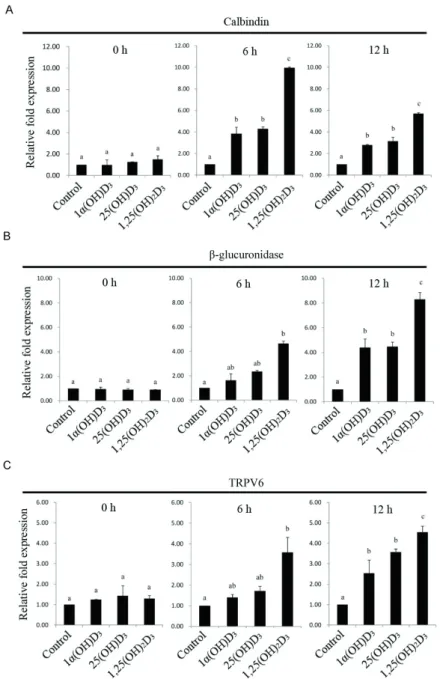
0, 6, and 12 h. The level of calbindin mRNA was rapidly increased at 6 h after feeding broiler chickens additional
amounts of vitamin D3 metabolites (Figure 1A). Among
vitamin D3 metabolites, 1,25(OH)2D3 appeared to be
more efficient than 1α(OH)D3 and 25(OH)D3 at regulating
calbindin mRNA expression at 6 and 12 h (Figure 1A). The level of β-glucuronidase mRNA was induced at 6 h
after feeding 1,25(OH)2D3 compared with other vitamin D3
metabolites (Figure 1B). After 12 h of feeding, 1α(OH)D3
and 25(OH)D3 also promoted β-glucuronidase mRNA
expression and 1,25(OH)2D3 increased the β-glucuronidase
mRNA even further (Figure 1B). Similarly, 1,25(OH)2D3
rapidly induced the level of transient receptor potential cation channel, subfamily V, member 6 (TRPV6) mRNA
at 6 h (Figure 1C). After 12 h of feeding, all vitamin D3
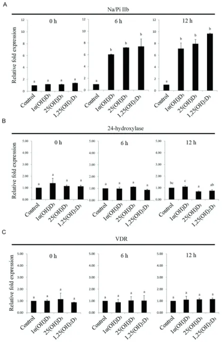
metabolites exhibited increased TRPV6 mRNA expression (Figure 1C). The level of type IIb sodium-dependent phosphate (Na/Pi IIb) cotransporter mRNA was rapidly increased at 6 and 12 h after feeding broiler chickens
additional amounts of vitamin D3 metabolites (Figure 2A).
The level of 24-hydroxylase mRNA was reduced at 12 h
after feeding 25(OH)D3 compared with the control diet
(Figure 2B). However, none of the vitamin D3 metabolites
changed the vitamin D receptor (VDR) mRNA levels in the duodenum of broilers (Figure 2C). Taken together,
these results indicate that vitamin D3 metabolites were
able to rapidly modulate calcium homeostasis-related gene
expression in the duodenum and 1,25(OH)2D3 was the
most effective vitamin D3 metabolite in regulating calcium
homeostasis-associated genes.
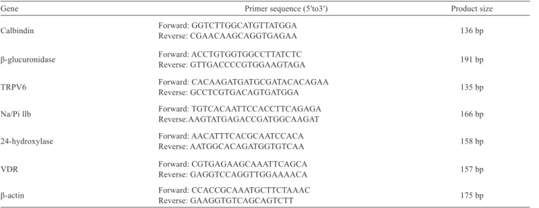
Gene Primer sequence (5′to3′) Product size
Calbindin Forward: GGTCTTGGCATGTTATGGAReverse: CGAACAAGCAGGTGAGAA 136 bp
β-glucuronidase Forward: ACCTGTGGTGGCCTTATCTCReverse: GTTGACCCCGTGGAAGTAGA 191 bp
TRPV6 Forward: CACAAGATGATGCGATACACAGAAReverse: GCCTCGTGACAGTGATGGA 135 bp
Na/Pi llb Forward: TGTCACAATTCCACCTTCAGAGAReverse:AAGTATGAGACCGATGGCAAGAT 166 bp
24-hydroxylase Forward: AACATTTCACGCAATCCACAReverse: AATGGCACAGATGGTGTCAA 158 bp
VDR Forward: CGTGAGAAGCAAATTCAGCAReverse: GAGGTCCAGGTTGGAAAACA 157 bp
β-actin Forward: CCACCGCAAATGCTTCTAAACReverse: GAAGGTGTCAGCAGTCTT 175 bp
Table 2 - Primer sequences for quantitative reverse transcription-PCR
Table 3 - Effect of different vitamin D3 metabolites on growth performance of 21-day-old broilers
Control (n = 12) 1α(OH)D3 (n = 12) 25(OH)D3 (n = 12) 1,25(OH)2D3 (n = 12) P-value
Mean SD Mean SD Mean SD Mean SD
Initial body weight (g) 43 0.3 43 0.3 43 0.4 43 0.2 NS
Final body weight (g) 750 270 800 80 730 5 780 19 NS
0-21days
ADG (g/day) 35 2 38 1 34 0.4 37 2 NS
ADFI (g/day) 45 1 46 0.08 43 1 44 1 NS
FCR (gain/feed) 0.787 0.035 0.825 0.012 0.797 0.006 0.835 0.016 NS
1α(OH)D3 - 1α-hydroxycholecalciferol; 25(OH)D3 - 25-hydroxycholecalciferol; 1,25(OH)2D3 - 1,25-dihydroxycholecalciferol; ADG - average daily gain; ADFI - average daily feed intake; FCR - feed conversion ratio; SD - standard deviation; NS - no significant difference (P>0.05).
1α(OH)D3 - 1α-hydroxycholecalciferol; 25(OH)D3 - 25-hydroxycholecalciferol; 1,25(OH)2D3 - 1,25-dihydroxycholecalciferol; TRPV6 - transient receptor potential cation channel, subfamily V, member 6.
Values were expressed as mean ± standard deviation (n = 4).
a-c - Means followed by different letters are significantly different (P<0.05).
Figure 1 - Effects of different vitamin D3 metabolites on duodenal (A) calbindin, (B) β-glucuronidase, and (C) TRPV6 gene expression in
Table 4 - Effect of different vitamin D3 metabolites on tibia, serum calcium, and phosphate levels and breaking strength in broilers
Control (n = 12) 1α(OH)D3 (n = 12) 25(OH)D3 (n = 12) 1,25(OH)2D3 (n = 12)
P-value
Mean SD Mean SD Mean SD Mean SD
Calcium of bone (%) 13.00 2.36 14.24 1.89 14.62 2.71 13.37 0.83 NS
Phosphate of bone (%) 6.06 1.17 6.99 0.73 7.04 1.42 6.32 0.37 NS
Serum calcium (mg/dL) 8.77 0.17 9.60 0.83 9.13 0.24 8.58 0.40 NS
Serum phosphate (mg/dL) 8.58 1.07 8.67 0.26 7.60 0.08 8.67 0.19 NS
Breaking strength (N) 187.43 18.31 197.67 20.51 208.66 19.92 193.48 8.65 NS
1α(OH)D3 - 1α-hydroxycholecalciferol; 25(OH)D3 - 25-hydroxycholecalciferol; 1,25(OH)2D3 - 1,25-dihydroxycholecalciferol;SD - standard deviation; NS - no significant difference (P>0.05).
1α(OH)D3 - 1α-hydroxycholecalciferol; 25(OH)D3 - 25-hydroxycholecalciferol; 1,25(OH)2D3 - 1,25-dihydroxycholecalciferol; Na/Pi IIb - type IIb sodium-dependent phosphate; VDR - vitamin D receptor.
Values were expressed as mean ± standard deviation (n = 4).
a-c - Means followed by different letters are significantly different (P<0.05).
Figure 2 - Effects of different vitamin D3 metabolites on duodenal (A) Na/Pi IIb, (B) 24-hydroxylase, and (C) VDR gene expression in
Discussion
In this study, we demonstrated that vitamin D3
metabolites could rapidly regulate calcium homeostasis-related gene expression in the duodenum from 6-12 h after feeding, although the growth performance and bone quality of broiler chickens at 21 days of age were not significantly
changed. Among these vitamin D3 metabolites, 1,25(OH)2D3
was the most effective in modulating calcium homeostasis-associated gene expression.
It has been reported that feeding broilers 69 μg/kg
of 25(OH)D3 for three weeks did not affect body weight
gain, feed conversion ratio, or feed intake (Chou et al., 2009). There were no significant effects on body weight or feed efficiency at 21 days of age in broiler chickens after
supplementation of the diet with 1,25(OH)2D3 (Roberson
and Edwards, 1996). Similarly, our results also confirm the previous reports that no significant effects were observed on growth performance of broiler chickens at 21 days
of age after supplementation of the diet with 1α(OH)D3,
25(OH)D3, or 1,25(OH)2D3. Furthermore, 1α(OH)D3
did not alter bone breaking strength, tibia calcium, and phosphorus content in broiler chickens at 21 days of age (Han et al., 2009). Consistently, we also found that vitamin
D3 metabolites did not improve bone breaking strength
and bone mineral deposition after feeding for three weeks.
In contrast, broilers fed 6 μg/kg of 1,25(OH)2D3 for three
weeks exhibited significantly increased bone ash content (Roberson and Edwards, 1996). The contradiction may be due to the differences between composition of basal diet and chicken strains. Together, these findings demonstrate
that vitamin D3 metabolites do not have a significant effect
on growth performance, but mineral deposition and bone breaking strength in broiler chickens may vary after feeding for three weeks.
Calbindin is a vitamin D-induced calcium-binding protein that plays a key role in intestinal intracellular calcium transport. The expression of calbindin in the intestine and
kidney has been shown to be regulated by 1,25(OH)2D3 in
rats and chickens (Brehier and Thomasset, 1990; Hall and Norman, 1990; Ferrari et al., 1992). Supplementation of
1,25(OH)2D3 could induce calbindin mRNA expression in the
duodenum of 1α-hydroxylase knockout mice (Hoenderop et al., 2004). Here, we further demonstrated that vitamin
D3 metabolites are able to induce intestinal calbindin
mRNA expression in vivo. Particularly, the expression
of calbindin peaked at 6 h and declined afterwards, 12 h
after 1,25(OH)2D3 treatment. The intestinal epithelial Ca2+
channel, TRPV6, is primarily activated by β-glucuronidase. It has been found that β-glucuronidase secretion from
isolated intestinal epithelial cells was remarkably increased
by supplementation of 1,25(OH)2D3 (Khanal et al., 2008).
The intestinal TRPV6 gene expression was significantly
increased after incubation for 6 h with 25(OH)D3 or
1,25(OH)2D3 (Balesaria et al., 2009). Supplementation
of 1,25(OH)2D3 significantly upregulated the TRPV6
mRNA expression in the duodenum of 1α-hydroxylase knockout mice (Hoenderop et al., 2004). Our findings
further suggested that vitamin D3 metabolites, including
1,25(OH)2D3, transcriptionally regulate β-glucuronidase
and TRPV6 mRNA expression in vivo. The NaPi-IIb
cotransporter is primarily expressed in the brush-border membranes of the small intestinal epithelium, where it is considered the major sodium-Pi cotransporter (Hilfiker
et al., 1998). Dietary supplementation of 1,25(OH)2D3
significantly regulates the expression of NaPi-IIb cotransporter in the intestine of rats (Katai et al., 1999). In
broilers, 1α(OH)D3 also has a similar effect on regulation
of intestinal NaPi-IIb cotransporter gene expression (Han
et al., 2009). Here, we also demonstrated that vitamin D3
metabolites could rapidly induce the levels of NaPi-IIb cotransporter mRNA in the intestine of broiler chickens. Several genes associated with calcium homeostasis have been shown to be regulated by VDR, such as calbindin and TRPV6 (Bolt et al., 2005; Meyer et al., 2006). Since dietary
1,25(OH)2D3 could be absorbed and bound to intestinal VDR
and then regulate its downstream gene transcriptionally, we speculate that intestinal gene expression may be directly
regulated by VDR in response to vitamin D3 metabolites.
The 24-hydroxylase belongs to cytochrome P450-containing enzyme (CYP24) that is important in the regulation of vitamin D metabolism (Minghetti and Norman, 1988). The expression of 24-hydroxylase in the kidney has
been demonstrated to be regulated by 1,25(OH)2D3 and to
initiate the inactivation of 1,25(OH)2D3 (Armbrecht et al.,
1997). Interestingly, we found that intestinal 24-hydroxylase mRNA expression was reduced by dietary supplementation
of 25(OH)D3 in broiler chickens. However, 1α(OH)D3 and
1,25(OH)2D3 did not cause a significant effect on intestinal
24-hydroxylase mRNA expression. The metabolite
1,25(OH)2D3 plays a central role in regulating mineral
metabolism through direct interaction with intracellular VDR in intestinal and kidney epithelial cells (Haussler et al., 1998). It has been reported that vitamin D analogs auto-regulate the expression of the VDR gene expression in cultured kidney cells (Costa et al., 1985; Santiso-Mere et al., 1993). In contrast, intestinal VDR gene expression
was not affected by 25(OH)D3 or 1,25(OH)2D3 in human
cells (Balesaria et al., 2009). Similarly, we also found
regulation of VDR gene expression in broiler chickens.
How vitamin D3 metabolites differently regulate intestinal
calcium homeostasis-associated gene expression in broiler chickens remains to be investigated in the further studies.
Conclusions
Calbindin, β-glucuronidase, TRPV6, and NaPi-IIb cotransporter gene expression is rapidly responsive to
dietary vitamin D3 metabolites in the duodenum of broilers.
Among vitamin D metabolites, 1,25(OH)2D3 is the most
effective at regulating calcium homeostasis-associated genes in broilers.
Acknowledgments
This work was supported by the Ministry of Science and Technology (NSC 99-2324-B-197-007) of Taiwan. The first and second authors contributed equally to this work.
References
AOAC - Association of Official Analytical Chemists. 1995a. Calcium analysis 968.08. Official methods of analysis. AOAC, Gaithersburg, MD.
AOAC - Association of Official Analytical Chemists. 1995b. Phosphorus analysis 964.06. Official methods of analysis. AOAC, Gaithersburg, MD.
Armbrecht, H. J.; Chen, M. L.; Hodam, T. L. and Boltz, M. A. 1997 Induction of 24-hydroxylase cytochrome P450 mRNA by 1,25-dihydroxyvitamin D and phorbol esters in normal rat kidney (NRK-52E) cells. Journal of Endocrinology 153:199-205. Balesaria, S.; Sangha, S. and Walters, J. R. 2009. Human duodenum
responses to vitamin D metabolites of TRPV6 and other genes involved in calcium absorption. American Journal of Physiology-Gastrointestinal and Liver Physiology 297:G1193-1197.
Baker, D. H.; Biehl, R. R. and Emmert, J. L. 1998. Vitamin D3 requirement of young chicks receiving diets varying in calcium and available phosphorus. British Poultry Science 39:413-417. Bolt, M. J.; Cao, L. P.; Kong, J.; Sitrin, M. D. and Li, Y. C. 2005.
Vitamin D receptor is required for dietary calcium-induced repression of calbindin-D9k expression in mice. Journal of Nutritional Biochemistry 16:286-290.
Brehier, A. and Thomasset, M. 1990. Stimulation of calbindin-D9k (CaBP9K) gene expression by calcium and 1,25-dihydroxycholecalciferol in fetal rat duodenal organ culture. Endocrinology 127:580-587.
Chou, S. H.; Chung, T. K. and Yu, B. 2009. Effects of supplemental 25-hydroxycholecalciferol on growth performance, small intestinal morphology, and immune response of broiler chickens. Poultry Science 88:2333-2341.
Costa, E. M.; Hirst, M. A. and Feldman, D. 1985. Regulation of 1,25-dihydroxyvitamin D3 receptors by vitamin D analogs in cultured mammalian cells. Endocrinology 117:2203-2210.
Edwards, H. M. Jr. 1990. Efficacy of several vitamin D compounds in the prevention of tibial dyschondroplasia in broiler chickens. Journal of Nutrition 120:1054-1061.
Ferrari, S.; Molinari, S.; Battini, R.; Cossu, G. and Lamon-Fava, S. 1992. Induction of calbindin-D28k by 1,25-dihydroxyvitamin D3 in cultured chicken intestinal cells. Experimental Cell Research 200:528-531.
Fleming, R. H. 2008. Nutritional factors affecting poultry bone health. Proceedings of the Nutrition Society 67:177-183.
Fritts, C. A. and Waldroup, P. W. 2003. Effect of source and level of vitamin D on live performance and bone development in growing broilers. Journal of Applied Poultry Research 12:45-52.
Hall, A. K. and Norman, A. W. 1990. Regulation of calbindin-D28K gene expression in the chick intestine: effects of serum calcium status and 1,25-dihydroxyvitamin D. Journal of Bone and Mineral Research 5:331-336.
Han, J. C.; Wang, J. G.; Chen, G. H.; Qu, H. X.; Zhang, J. L.; Shi, C. X.; Yan, Y. F. and Cheng, Y. H. 2016. Effects of calcium to non-phytate phosphorus ratio and different sources of vitamin D on growth performance and bone mineralization in broiler chickens. Revista Brasileira de Zootecnia 45:1-7.
Han, J. C.; Yang, X. D.; Zhang, T.; Li, H.; Li, W. L.; Zhang, Z. Y. and Yao, J. H. 2009. Effects of 1α-hydroxycholecalciferol on growth performance, parameters of tibia and plasma, meat quality, and type IIb sodium phosphate cotransporter gene expression of one- to twenty-one-day-old broilers. Poultry Science 88:323-329. Haussler, M. R.; Whitfield, G. K.; Haussler, C. A.; Hsieh, J. C.;
Thompson, P. D.; Selznick, S. H.; Dominguez, C. E. and Jurutka, P. W. 1998. The nuclear vitamin D receptor: biological and molecular regulatory properties revealed. Journal of Bone and Mineral Research 13:325-349.
Henry, H. L. 1979. Regulation of the hydroxylation of 25-hydroxyvitamin D3 in vivo and in primary cultures of chick kidney cells. Journal of Biological Chemistry 254:2722-2729. Henry, H. L. 1985. Parathyroid hormone modulation of
25-hydroxyvitamin D3 metabolism by cultured chick kidney cells is mimicked and enhanced by forskolin. Endocrinology 116:503-510.
Hilfiker, H.; Hattenhauer, O.; Traebert, M.; Forster, I.; Murer, H. and Biber, J. 1998. Characterization of a murine type II sodiumphosphate cotransporter expressed in mammalian intestine. Proceedings of the National Academy of Sciences 95:14564-14569.
Hoenderop, J. G.; van der Kemp, A. W.; Urben, C. M.; Strugnell, S. A. and Bindels, R. J. 2004. Effects of vitamin D compounds on renal and intestinal Ca2+ transport proteins in 25-hydroxyvitamin D3-1alpha-hydroxylase knockout mice. Kidney International 66:1082-1089.
Katai, K.; Miyamoto, K.; Kishida, S.; Segawa, H.; Nii, T.; Tanaka, H.; Tani, Y.; Arai, H.; Tatsumi, S.; Morita, K.; Taketani, Y. and Takeda. E. 1999. Regulation of intestinal Na-dependent phosphate cotransporters by a low-phosphate diet and 1,25-dihydroxyvitamin D3. Biochemical Journal 343:705-712.
Khanal, R. C.; Peters, T. M.; Smith, N. M. and Nemere, I. 2008. Membrane receptor-initiated signaling in 1,25(OH)2D3-stimulated calcium uptake in intestinal epithelial cells. Journal of Cellular Biochemistry 105:1109-1116.
Koreleski, J. and Swiatkiewicz, S. 2005. Efficacy of different limestone particle size and 25-hydroxycholecalciferol in broiler diets. Journal of Animal and Feed Sciences 14:705-714.
Kumar, R. 1990. Vitamin D metabolism and mechanisms of calcium transport. Journal of the American Society of Nephrology 1:30-42. Leach, R. M. and Lilburn, M. S. 1992. Current knowledge on the
etiology of tibial dyschondroplasia in the avian species. Poultry Science Reviews 4:57-65.
Meyer, M. B.; Watanuki, M.; Kim, S.; Shevde, N. K. and Pike, J. W. 2006. The human TRPV6 distal promoter contains multiple vitamin D receptor binding sites that mediate activation by 1,25-dihydroxyvitamin D3 in intestinal cells. Molecular Endocrinology 20:1447-1461.
Minghetti, P. P. and Norman, A. W. 1988. 1,25(OH)2-vitamin D3 receptors: gene regulation and genetic circuitry. FASEB Journal 2:3043-3053.
Norman, A. W.; Putkey, J. A. and Nemere, I. 1982. Intestinal calcium transport: pleiotropic effects mediated by vitamin D. Federation Proceedings 41:78-83.
NRC - National Research Council. 1994. Nutritional requirements of poultry. 9th ed. National Academy Press, Washington, DC. Pines, M.; Hasdai, A. and Monsonego-Ornan. E. 2005. Tibial
dyschondroplasia - tools, new insights and future prospects. World’s Poultry Science Journal 61:285-297.
Roberson, K. D. and Edwards H. M. Jr. 1996. Effect of dietary 1,25-dihydroxycholecalciferol level on broiler performance. Poultry Science 75:90-94.
Santiso-Mere, D.; Sone, T.; Hilliard, G. M. 4th; Pike, J. W. and McDonnell, D. P. 1993. Positive regulation of the vitamin D receptor by its cognate ligand in heterologous expression systems. Molecular Endocrinology 7:833-839.
SAS Institute, Inc. 2008. SAS/STAT user’s guide. Version 9.2. SAS Institute, Inc., Cary, NC.
Whitehead, C. C.; McCormack, H. A.; McTeir, L. and Fleming, R. H. 2004. High vitamin D3 requirements in broilers for bone quality and prevention of tibial dyschondroplasia and interactions with dietary calcium, available phosphorus and vitamin A. British Poultry Science 45:425-436.
Waldenstedt, L. 2006. Nutritional factors of importance for optimal leg health in broilers: a review. Animal Feed Science and Technology 126:291-307.