RESUMO: O bovine leukemia virus (BLV) é um membro da família Retroviridae, gênero Deltaretrovirus, e o principal agente viral cau‑ sador de perdas econômicas em rebanhos leiteiros. Diversos estudos têm sido feitos sobre os genótipos de BLV, e foram encontrados pelo menos sete em amostras de diferentes partes do mundo. O objetivo deste estudo foi realizar a caracterização molecular de amostras de BLV de bovinos leiteiros soropositivos no estado de Santa Catarina. Foram coletadas 454 amostras de sangue de bovinos de 31 proprie‑ dades, e fez‑se inicialmente a sorologia por meio do teste de imuno‑ difusão em gel de ágar. Após a sorologia, 191 amostras soropositivas foram então submetidas à extração de DNA, e em 77 amostras se realizou a reação da polimerase em cadeia (PCR), para a amplifica‑ ção de um fragmento de 440 pb do gene env. Dezenove amostras foram submetidas à análise do polimorfismo dos fragmentos de res‑ trição por digestão do fragmento da PCR por cinco enzimas de res‑ trição: BamHI, HaeIII, Tru9I, TaqI e MwoI. Os resultados obtidos na sorologia apontaram 42% de animais soropositivos (191/454) e 68% de propriedades positivas (21/31). Na PCR, 80,52% (62/77) dos animais apresentaram‑se positivos. A análise do polimorfismo dos fragmentos de restrição identificou cinco genótipos circulan‑ tes no estado, e a maior prevalência foi observada no genótipo X (47,4%). Este estudo permite‑nos conhecer alguns dos genótipos virais presentes em bovinos leiteiros do estado de Santa Catarina, bem como identificar a existência de novas variantes e sua prevalência atual, e os resultados são úteis para futuros estudos epidemiológicos.
PALAVRAS‑CHAVE: Bovine leukemia virus; Deltaretrovirus; bovinos leiteiros; env; polimorfismo dos fragmentos de restrição. ABSTRACT: Bovine leukemia virus (BLV) is a member of
Retroviridae family, genus Deltaretrovirus, and the main viral agent responsible for economic loses in dairy herds. Some studies have been carried out about BLV genotypes, and at least seven genotypes were found out in samples of different regions of the world. The objective of this study was to identify BLV samples from seropositive dairy cattle in Santa Catarina state, Brazil, using molecular techniques. Blood samples were collected (454) from dairy cattle from 31 different farms, and serology using agar gel immunodiffusion test (AGID) was performed. After that, 191 seropositive samples were submitted to DNA extraction, and in 77 samples the polymerase chain reaction (PCR) for amplification of a 440 bp fragment of the env gene was performed. Nineteen DNA samples were subjected to restriction fragment length polymorphism (RFLP) analysis by digestion of the PCR fragment by five restriction endonucleases — BamHI, HaeIII, Tru9I, TaqI, and MwoI. It was found 42% seropositive animals (191/454) and 68% positives of the farms (21/31). The PCR showed 80.5% (62/77) of animals positive. The RFLP analysis identified five different genotypes dispersed by Santa Catarina state, with the highest prevalence for genotype X (47.4%). Overall, our results identified the viral genotypes present in dairy cattle and the prevalence of new variants in representative farms from Santa Catarina state.
KEYWORDS: Bovine leukemia virus, Deltaretrovirus, dairy cattle, env, restriction fragment length polymorphism.
Heterogeneity determination of
bovine leukemia virus
genome in Santa Catarina state, Brazil
Determinação da heterogeneidade do genoma do vírus da
leucose enzoótica bovina no estado de Santa Catarina, Brasil
Sheyla Michele Rodakiewicz¹*, Maria Luiza Fernandez¹, Maria Luiza Munhoz¹, Flávia Harumi Scheffer
Yamakawa¹, Monica Urio¹, Fabiana Forell¹, Sandra Ferraz¹, Vagner Miranda Portes², Ubirajara Maciel da Costa¹
1Universidade do Estado de Santa Catarina ‑ Campus III Planalto Serrano – Lages (SC), Brazil 2Empresa de Pesquisa Agropecuária e Extensão Rural de Santa Catarina (Epagri) – Chapecó (SC), Brazil
INTRODUCTION
Bovine leukemia virus (BLV) is the causative agent of enzootic bovine leukosis, a disease of global distribution, and its preva‑ lence varies widely among cattle herds, being higher in dairy cattle. Thirty percent of the infected animals may develop persistent lymphocytosis (PL), an increase of circulating lym‑ phocytes, in this case B cells, and only 2 to 5% may develop lymphosarcomas (LEUZZI JR. et al., 2001).
BLV is a member of the genus Deltaretrovirus, from Retroviridae family, presenting similarities with the human T‑lymphotrophic virus type‑1 and 2 (TOSTES, 2005). The virus genome consists of two single strand RNA molecules with positive polarity, being more prone to mutations than viruses with a DNA genome (CANN, 2005). Most of the mutations are punctual and occur in nucleotide sequences, which may lead to amino acid substitu‑ tions of the glycoprotein surface (RODRIGUEZ et al., 2009).
The identification of nucleotide mutations can be per‑ formed by restriction fragment length polymorphism (RFLP) analysis. This technique is based on genotyping by partial genome sequence, and has been widely used because it gives an idea of the general composition of the viral genome when a detailed information is not available. It consists in analyzing the restriction fragments generated by cleavage of the polymerase chain reaction (PCR) product with restriction endonucleases (ZHAO; BUEHRING, 2007). The 1,547 bp env gene is the most used in this analysis, because it is the most variable and provides a general idea of the BLV genome, that is, 8,714 bp.
Several authors have reported the existence of six or seven geno‑ types of BLV by RFLP analysis, and some of them also mention the possible existence of a new genotype (RODRIGUEZ et al., 2009; MORATORIO et al., 2010; ABADNEH et al., 2012). A few also mention the classification of BLV through phylogenetic analysis in two to four groups (CAMARGOS et al., 2007, HEMMATZADEH, 2007; MONTI et al., 2005; ZHAO; BUEHRING, 2007; JULIARENA et al., 2013;. LICURSI et al., 2003).
Considering many studies that have been carried out on the genetic variability of BLV and the lack of these studies in Santa Catarina state, the present investigation aimed to evalu‑ ate the existence of different genotypes of BLV in dairy cattle herds of the state, through genotyping by RFLP analysis. It is possible, through the identification of these genotypes, to deter‑ mine the prevalence of viral genotypes in the four mesoregions analyzed and to establish a BLV profile in Santa Catarina state.
MATERIALS E METHODS
Sample collection
For the development of this work, visits were made to dairy cattle farms selected randomly between January 2012 and April
2013, distributed in four main mesoregions of the Santa Catarina state: the south, the sierra, the west and the north. Samples of 31 properties were obtained: eight cattle properties located in the north region, four properties in the western region, 12 prop‑ erties in the south region, and seven properties in the region of the sierra of Santa Catarina. The animals sampled were mostly Holstein, Jersey and crossbred cattle used for milk production.
Blood samples were obtained from the caudal vein using sili‑ conized vacuum tubes with clot activator (Labor Import, Osasco, São Paulo, Brazil) and ethylenediaminetetraacetic acid (EDTA) K3 anticoagulant (Vacuplast Collect Line, Nanchang, Jiangxi, China).
Samples preparation
Tubes containing blood with and without anticoagulant were subjected to centrifugation for 10 minutes at 403 x g in a table‑ top centrifuge (Janetzki T150, Engelsdorf, Leipzig, Germany). The sera were then stored at ‑20ºC until the agar gel immunodiffusion test (AGID) was performed, and blood samples with anticoagulant were used to obtain the leukocyte layer, after centrifugation.
For the removal of erythrocytes, the white cell aspirate was washed with a solution of Tris EDTA pH 8, centrifuged at 2,000 x g, for five minutes in a MiniSpin centrifuge (Eppendorf, Hamburg, Germany) between each wash, totalizing averaged five washes or until the erythrocytes were removed as much as possible, and then stored in ultra‑freezer ‑80ºC until the end of the AGID serological test, since proviral DNA extraction was performed only with samples from animals considered to be serologically positive.
Serology by agar gel immunodiffusion
The test was performed for the diagnosis of BLV through AGID, produced by Institute of Technology of Paraná (Instituto de Tecnologia do Paraná — TECPAR), in Curitiba, Brazil. The technique was performed following the manu‑ facturer’s recommendation.
Samples were considered positive when precipitation line was formed between the central well containing the antigen and the well with a serum sample tested, and also the identity line with the positive control serum.
Blood samples from seropositive animals were subjected to DNA extraction and PCR for amplification of the 440 bp BLV env gene sequence.
DNA extraction and amplification of
bovine leukemia
virus env gene by
polymerase chain reaction
to the protocol of TRI Reagent T9424 (Sigma‑Aldrich, St. Louis, Missouri, United States).
After the extraction, a fragment of 440 bp of env gene was amplified by PCR technique, described by the OIE DIAGNOSTIC MANUAL (2012). The reaction mix was composed of 20 μL of PCR Master Mix (Quatro G Ltda., Porto Alegre, Rio Grande do Sul, Brazil) and 1 μL of each primer OBLV1A (5’‑CTTTGTGTGCCAAGTCTCCCAGATACA‑3 ‘) and OBLV6A (5’ ‑CCAACATATAGCACAGTCTGGGAAGGC‑3 ‘), and 3 μL of the DNA sample. The final reaction volume was 25 μL.
The reaction conditions were 5 min at 94ºC, followed by five cycles at 94ºC for 45 s, 60ºC for 60 s and 72ºC for 90 s, followed by 35 cycles of 94ºC for 45 s, 59ºC for 60 s and 72ºC for 90 s. The last one was a cycle at 72ºC for 7 min.
The amplification products were then analyzed by 1.5% agarose gel electrophoresis, stained with a 0.5 μg/mL ethid‑
ium bromide solution.
Polymerase chain reaction products
analysis by restriction fragment
length polymorphism
This process consisted in the digestion of a fragment of the 440 bp viral genome for 1 hour with 10 U of restriction endonucleases BamHI (Promega, Madison, Wisconsin, United States) and HaeIII (Promega, Madison, Wisconsin, United States) at 37ºC; Tru9I (Promega, Wisconsin, United States) and TaqI (Promega, Wisconsin, United States) at 65ºC; and MwoI (New England Biolabs Inc., Ipswich, Massachussets, United States) at 60ºC.
After digestion of the samples, the electrophoresis was performed in a 3.5% agarose gel stained with ethidium bro‑ mide 0.5 μg/mL.
The comparison method for classification of BLV genotypes was based on a table described by INOUE et al. (2011) relat‑ ing the possible sizes of fragments formed after digestion with each restriction enzyme and the different combinations of these fragments that give rise to the seven different viral genotypes.
RESULTS
In the western region, 72 samples were collected in four prop‑ erties, 13 of which were positive in serology. PCR of 11 sam‑ ples showed nine positive, and in the genotyping two samples demonstrated the occurrence of genotype VIII. In the sierra region, 167 samples were collected in seven properties, being 114 positive in the serology. PCR of 36 samples showed 30 positive, and in the genotyping of eight samples the genotype II was found in two samples, genotype I in one sample, IX in one sample and X in four samples. In the southern region, 176 samples were collected in 12 properties, 51 of which were
positive in serology and 21 in PCR, of 28 analyzed. In the RFLP analysis, eight samples were analyzed, genotype I was found in one sample, VIII in one sample, IX in two samples and X in four samples. Lastly, in the northern region, 39 samples were collected in eight regions, 13 of which were positive in the serology. The two samples analized by PCR were positive, and in the RFLP analysis, a sample of genotype X was found.
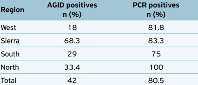
Serology for enzootic bovine leukosis
Through the serology of the 454 blood samples collected from 31 dairy cattle farms distributed in four regions of Santa Catarina state, 191 seropositive animals (42.1%) were found and 21 properties with at least one seropositive animal (67.7%).
In the western region, 72 animals were sampled in four properties, totalizing 13 seropositive animals, the lowest num‑ ber of positive animals observed in the state. In the sierra region, 167 animals from seven properties were evaluated, which 114 animals were seropositive — the highest number of positivity in the state. In the southern region, 176 animals from 12 properties were sampled, totalizing 51 seropositive animals. In the northern region, 39 animals from eight prop‑ erties were analyzed, and 13 were seropositive (Table 1).
Bovine leukemia virus
provirus
detection by polymerase chain
reaction technique
Of the 191 serologically positive samples, PCR reaction was performed on 77 samples, and in 62 samples the fragment of interest of 440 bp of the env gene was amplified (Fig. 1).
Bovine leukemia virus
classification
by restriction fragment length
polymorphism analysis of the env gene
polymerase chain reaction products
One amplicon of one sample from each property that pre‑ sented BLV seropositive animals was digested by five restriction
Table 1. Relationship between collecting regions, performed analysis and results.
Region AGID positives n (%)
PCR positives n (%)
West 18 81.8
Sierra 68.3 83.3
South 29 75
North 33.4 100
Total 42 80.5
endonucleases — BamHI, HaeIII, Tru9I, TaqI and MwoI —, totaling 19 samples.
BamHI enzyme digestion showed two different cleavage patterns, one forming bands of 240 and 200 bp and the other one forming only a band of 440 bp.
Digestion with the enzyme HaeIII cleaved all amplicons into five bands: 200, 90, 65, 35 and 25 bp.
In the Tru9I digestion, two different cleavage patterns were obtained: one forming two bands of 400 and 40 bp, observed in most of the samples; and one sample with cleav‑ age in three bands, 270, 130 and 40 bp.
TaqI also showed two different patterns of cleavage: one formed two bands with size of 280 and 160 bp; and the other one with three bands of 230, 160 and 50 bp.
Three different cleavage patterns — one with three bands of 250, 160 and 30 bp; one with two bands of 280 and 160 bp; and a third one with three bands of 250, 100 and 90 bp — were observed in the samples digested by the MwoI enzyme. These different cleavage patterns are described in Table 2.
Genotypes I and II identified in this study are consistent with previous findings described by INOUE et al. (2011). However, we identified three genotypes not yet described, classified as VIII, IX and X. These results indicate the exis‑ tence of new variants of BLV circulating in Santa Catarina.
Prevalence of viral genotypes
by restriction fragment length
polymorphism analysis
Genotype I presented the lowest prevalence (5%): only one animal displayed it. For genotype II, two animals (11%), genotype VIII, four animals (21%), and genotype IX, three animals were identified (16%), and genotype X showed the highest prevalence (47%), with nine animals among the 19 tested.
Thus, it is worth mentioning that in this study we could observe that there are five circulating genotypes in Santa Catarina state, and the most prevalent genotype of them is X.
DISCUSSION
In the present study, 42.1% (191/454) of the analyzed serum samples were positive for BLV. This result is different from the one found by MORAES et al. (1996), with only 12% of Rio Grande do Sul positive animals, CARVALHO et al. (1996), with 7% in cattle from Londrina, Paraná state, and LUDERS (2001), with 7.6% positive cattle in the northern region of Santa Catarina. Other authors, however, found similar results. BARROS FILHO et al. (2009) found prev‑ alence of 56.3% in Curitiba, Paraná; CORDEIRO et al. (1994), of 35% in Itajaí, Santa Catarina; D’ANGELINO et al. (1998), of 54% in São Paulo; and RAJÃO (2008) in Minas Gerais found the highest prevalence (79.7%), as did MONTI et al. (2005), with prevalence of 70% in the PCR and 90% in the AGID.
As for the prevalence of positive properties, 21/31 (67.7%) were found, a rather high result when compared with the ones found by other authors such as MORAES et al. (1996) (29.1%), CARVALHO et al. (1996) (35.7%) and LUDERS (2001) (10.8%).
In the positive samples, there was amplification through the PCR technique of a 440 bp fragment of the env gene, as well as in the study by INOUE et al. (2011) and MATSUMURA
Figure 1. Amplification of the 440 bp fragment of the env gene by the polymerase chain reaction technique.
Table 2. Products generated by restriction enzyme cleavage of the 440 bp fragment of the bovine leukemia virus env gene
RFLP Type Restriction enzymes
BamHI HaeIII Tru9I TaqI MwoI
I 240, 200 200, 90, 65, 35, 25 270, 130, 40 280, 160 250, 160, 30
II 240, 200 200, 90, 65, 35, 25 400, 40 280, 160 250, 160, 30
VIII 240, 200 200, 90, 65, 35, 25 400, 40 230, 160, 50 280, 160
IX 440 200, 90, 65, 35, 25 400, 40 280, 160 250, 160, 30
X 240, 200 200, 90, 65, 35, 25 400, 40 280, 160 250, 100, 90
et al. (2010) — both also amplified this fragment, correspond‑ ing to position 5029‑5468 of the viral genome.
In this study, PCR products from the env gene were submitted to enzymatic restriction analysis with restric‑ tion endonucleases, similar to the study by INOUE et al. (2011), who also used the same enzymes, except Tru9I. MseI enzyme was used instead, but both have the same cleavage site.
In the present investigation, two different cleavage pat‑ terns were observed for the 440 bp fragment by BamHI enzyme, one forming two sizes of bands, of 240 and 200 bp found in genotypes I, II, VIII, X; and the other one only a band of 440 bp. That is, the fragment was not cleaved, being found in genotype IX. These findings were similar to those established by INOUE et al. (2011), who observed only a cleavage pattern, with bands of 240 and 200 bp, in tumor samples, thus not being the definitive standard for genotyping.
The cleavage of the amplicons with the enzyme HaeIII gave rise to five fragments of 200, 90, 65, 35 and 25 bp. This pat‑ tern was not definitive for genotyping, because it was found in all genotypes, matching the pattern also found by INOUE et al. (2011), which further identified a different cleavage pat‑ tern, forming four bands with sizes of 290, 65, 35, and 25 bp for the 440 bp fragment.
The Tru9I enzyme, obtained two different cleavage pat‑ terns, one forming two bands of 400 and 40 bp, was observed in most of the samples, and one sample with cleavage in three bands, 270, 130 and 40 bp, was seen, in agreement with the findings of INOUE et al. (2011). However, these authors still found a third pattern, with bands of 360 and 40 bp.
The TaqI also presented two different cleavage patterns, one forming two bands with size of 280 and 160 bp and the other one with three bands of 230, 160 and 50 bp. INOUE et al. (2011) also found these two cleavage patterns and a third one, with bands of 240, 160 and 40 bp.
The samples that were digested by the MwoI enzyme presented three different cleavage patterns: one with three bands of 250, 160 and 30 bp, another one with two bands of 280 and 160 bp, and the last one with three bands of 250, 100 and 90 bp, in agreement with the findings of INOUE et al. (2011), but we also found a third pattern with three bands of 250, 100 and 90 bp, not described by the afore‑ mentioned authors.
In this study, we could observe five circulating gen‑ otypes in the analyzed samples of dairy cattle in Santa Catarina. Among them, the genotypes I and II described by INOUE et al. (2011) in Japan, which identified a total of seven BLV genotypes by RFLP analysis of the 440 bp fragment of the env gene, from bovine tumor samples using restriction enzymes with the same restriction site used in this study. Seven BLV genotypes were also identified by
MORATÓRIO et al. (2010) through sequencing and phy‑ logenetic analysis of bovine blood samples from Uruguay, and by BALIC et al. (2012) and MATSUMURA et al. (2010), carrying out a sequential and phylogenetic analy‑ sis of 356 bp fragment of the env gene from bovine blood samples in Croatia and tumors in Japan, also observed a new genotype.
Through sequencing and phylogenetic analysis of the env gene of bovine samples from Argentina, RODRIGUEZ et al. (2009) identified six genotypes, as well as LICURSI et al. (2002), and proposed the existence of a seventh gen‑ otype, similar to previous findings. CAMARGOS et al. (2007) observed in cattle of the Brazilian states of Paraná, Minas Gerais, Mato Grosso do Sul e Goiás and ABADNEH et al. (2012) in Jordanian dairy cattle by the RFLP analy‑ sis of a 444 bp fragment of the env gene with restriction enzymes, the presence of genotype I, also found in this study, and genotype VI, which was not found to be circulating in Santa Catarina. MONTI et al. (2005) through RFLP analysis also found two genotypes, classified as Australian and Argentine.
Among the five genotypes found — I, II, VIII, IX, X —, genotype I has already been described by MORATÓRIO et al. (2010), which detected the genotypes V, VI, and VII in Brazilian samples, similar to those obtained by CAMARGOS et al. (2007) in Paraná, Minas Gerais, Mato Grosso do Sul e Goiás samples, also finding genotypes I and VI.
Genotype X, so named because it did not fit into any of the genotypes described, was the most prevalent among the analyzed samples, followed by genotypes VIII, IX, II and I, disagreeing with results found by MATSUMURA et al. (2010), who identified the highest prevalence of genotype I through phylogenetic analysis of bovine tumors in Japan. However, these authors described the presence of two major viral pop‑ ulations in the country.
As for the predominance of viral genotypes by region of the state, it is worth mentioning that among genotypes the genotype X was found in the north, south and sierra, being absent in the western region. Genotype IX was found in the southern and sierra regions. Genotype VIII was present in the south and west regions, and genotype I was restricted to the southern region, as well as the genotype II to the sierra. These findings corroborate with RODRIGUEZ et al. (2009), who reported that there is a limited group of genotypes, which are distributed irregularly.
area such as foot‑and‑mouth disease, this type of screening is extremely relevant and necessary.
It is known that the development of retrovirus vac‑ cines is extremely difficult because of the constant genetic variation of the agent. However, identifying BLV geno‑ types can be an important step in trying to develop vac‑ cines that cover the most prevalent samples in a particu‑ lar region or herd.
CONCLUSIONS
There are at least five BLV genotypes circulating in Santa Catarina state, and these genotypes are distributed in the analyzed mesoregions.
Among the genotypes identified, three were not previously described, and one of them, classified as genotype X, was the most prevalent among the analyzed samples.
REFERENCES
ABADNEH, M.M.; AL‑RUKIBAT, R.K.; HANANEH, W.M.; NASAR, A.T.; AL‑ZGHOUL, M.B. Detection and molecular characterization of bovine leukemia viruses from Jordan. Archives of Virology,
Austria, v.157, n.12, p.2343‑2348, 2012.
BALIC, D.; LOJKIC, I.; PERISKIC, M.; BEDEKOVIC, T.; JUNGIC, A.; LEMO, N.; ROIC, B.; CAC, Z.; BARBIC, L.; MADIC, J. Identification of a new genotype of bovine leukemia virus.Archives of Virology, Austria, v.157, n.7, p.1281–1290, 2012.
BARROS FILHO, I.R.; GUIMARÃES, A.K.; BIONDO, A.W.; KRÜGER, E.R.; WAMMES, E.V.; OLLHOFF, R.D.; PIEKARZ, C.H.; SPONCHIADO, D. Prevalência da Leucose enzoótica em bovinos leiteiros criados na região metropolitana de Curitiba – Paraná. Ciência Animal Brasileira, Goiânia, p.11‑14, 2009.
CAMARGOS, M.F.; PEREDA, A.; STANCEK, D.; ROCHA, M.A.; DOS REIS, J.K.P.; GREISER‑WILKE, I.; LEITE, R.C. Molecular characterization of the env gene from Brazilian field isolates of Bovine Leukemia Virus. Virus genes, [s.l.], v.34, n.3, p.343‑ 350, 2007.
CANN, A.J. Principles of Molecular Virology. 4 ed. Estados Unidos: Elsevier Academic Press, 2005. 332p.
CARVALHO, L.; BENESI, F. J.; BIRGEL JUNIOR, E.H.; BIRGEL, E.H. Prevalência de anticorpos séricos de anti‑vírus da leucose dos bovinos em animais da raça Holandesa Preta e Branca e zebuínos da raça Nelore, criados no pólo regional de Londrina, Estado do Paraná. Semina: Ciências Agrárias, Londrina, v.17, n.1, p.53‑57, 1996.
CORDEIRO, J.L.F.; DESCHAMPS, F.C.; MARTINS, E.; MARTINS, V.M.V. Identificação e controle da leucose enzoótica bovina (LEB) em um rebanho leiteiro. PesquisaAgropecuária Brasileira, Brasília, v.29, n.8, p.1287‑1292, 1994.
D’ANGELINO, J.L.; GARCIA, M.; BIRGEL, E.H. Epidemiological study of enzootic bovine leukosis in Brazil: Short Comunication.
Tropical AnimalHealth and Production, Amsterdam, v.30, n.1, p.13–15, 1998.
HEMMATZADEH, F. Sequencing and phylogenetic analysis of gp51 gene of bovine leukaemia virus in Iranian isolates. Veterinary Research Communications, Holanda, v.31, n.6, p.783–789, 2007.
INOUE, E.; MATSUMURA, K.; MAEKAWA, K.; NAGATSUKA, K.; NOBUTA, M.; HIRATA, M.; MINAGAWA, A.; OSAWA, Y.; OKAZAKI, K. Genetic heterogeneity among bovine leukemia viruses in Japan and their relationship to leukemogenicity. Archives of Virology, Austria, v.156, n.7, p.1137‑114, 2011.
JULIARENA, M.A.; LENDEZ, P.A.; GUTIERREZ, S.E.; FORLETTI, A.; RENSETTI, D.E.; CERIANI, M.C. Partial molecular characterization of different proviral strains of bovine leukemia virus. Archives of Virology, Austria, v.158, n.1, p.63–70, 2013.
LEUZZI JR, L.A.; ALFIERI, A. F.; ALFIERI, A.A. Leucose enzoótica bovina e vírus da leucemia bovina. Semina: Ciências Agrárias, Londrina, v.22, n.2, p.211‑221, 2001.
LICURSI, M.; INOSHIMA, Y.; WU, D.; YOKOYAMA, T.; GONZÁLEZ, E.T.; SENTSUI, H. Genetic heterogeneity among bovine leukemia virus genotypes and its relation to humoral responses in hosts.
Virus Research, Philadelphia, v. 86, n.1 – 2, p. 101 –110, 2002.
LICURSI, M.; INOSHIMA, Y.; WU, D.; YOKOYAMA, T.; GONZÁLEZ, E.T.; SENTSUI, H. Provirus variants of bovine leukemia virus in naturally infected cattle from Argentina and Japan. Veterinary Microbiology, [s.l.], v.96, n.1, p.17–23, 2003.
LUDERS, M.A. Prevalência de anticorpos contra o vírus da leucose enzoótica bovina em fêmeas com mais de dois anos no Rebanho de bovinos leiteiros no Município de Mafra‑SC. 2001, 30f. Dissertation (Master in Agroveterinary Sciences / Animal Sanity) ‑ Universidade do Estado de Santa Catarina, Lages, 2001.
MATSUMURA, K.; INOUE, E.; OSAWA, Y.; OKAZAKI, K. Molecular epidemiology of bovine leukemia virus associated with enzootic bovine leukosis in Japan. Virus research, Philidelphia, v.155, n.1, p.343–348, 2011.
MONTI, G.; SCHRIJVER, R.; BEIER, D. Genetic diversity and spread of Bovine leukaemia virus isolates in Argentine dairy cattle.
Archives of Virology, Austria, v.150, n.3, p.443–448, 2005.
© 2018 Instituto Biológico This is an open access article distributed under the terms of the Creative Commons license. MORATORIO, G.; OBAL, G.; DUBRA, A.; CORREA, A.; BIANCHI, S.;
BUSCHIAZZO, A.; CRISTINA, J.; PRITSH, O. Phylogenetic analysis of bovine leukemia viruses isolated in South America reveals diversification in seven distinct genotypes. Archives of Virology, Austria, v.155, n.4, p.481–489, 2010.
OIE, Terrestrial Manual. Enzootic Bovine Leukosis, cap. 2.4.11, p. 1‑11. Available from: <http://www.oie.int/fileadmin/Home/ eng/Health_standards/tahm/2008/pdf/2.04.11_EBL.pdf>. Accessed on: Jul. 16 2014.
RAJÃO, D.S.Efeito da infecção pelo vírus da Leucose Enzoótica Bovina na produção de leite e reprodução de rebanhos leiteiros.
2008, 26f. Dissertation (Master of Animal Science / Preventive
Veterinary Medicine) – Escola de Veterinária da Universidade Federal de Minas Gerais, Belo Horizonte, 2008.
RODRIGUEZ, S.M.; GOLEMBA, M.D.; CAMPOS, R.H.; TRONO, K.; JONES, L.R. Bovine leukemia virus can be classified into seven genotypes: evidence for the existence of two novel clades. Journal of General Virology, [s.l.], v.90, p.2788–2797, 2009.
TOSTES, R.A. Situação da Leucose Bovina no Brasil: uma revisão.
Colloquium Agrariae, Presidente Prudente, v.1, n.1, p.42–50, 2005.