A se arch fo r RNA inse rtio ns and NS3
ge ne duplicatio n in the ge no m e o f
cyto pathic iso late s o f bo vine viral
diarrhe a virus
Setor de Virologia, Departamentos de Medicina Veterinária Preventiva,
Microbiologia e Parasitologia, Universidade Federal de Santa Maria, Santa Maria, RS, Brasil
V.L. Q uadros, S.V. Mayer, F.S.F. Vogel, R. Weiblen, M.C.S. Brum,S. Arenhart and E.F. Flores
Abstract
Calves born persistently infected with non-cytopathic bovine viral diarrhea virus (ncpBVDV) frequently develop a fatal gastroenteric illness called mucosal disease. Both the original virus (ncpBVDV) and an antigenically identical but cytopathic virus (cpBVDV) can be isolated from animals affected by mucosal disease. Cytopathic BVDVs originate from their ncp counterparts by diverse genetic mechanisms, all leading to the expression of the non-structural polypeptide NS3 as a discrete protein. In contrast, ncpBVDVs express only the large precursor polypeptide, NS2-3, which contains the NS3 sequence within its carboxy-terminal half. We report here the investigation of the mechanism leading to NS3 expression in 41 cpBVDV isolates. An RT-PCR strategy was employed to detect RNA insertions within the NS2-3 gene and/or duplication of the NS3 gene, two common mechan-isms of NS3 expression. RT-PCR amplification revealed insertions in the NS2-3 gene of three cp isolates, with the inserts being similar in size to that present in the cpBVDV NADL strain. Sequencing of one such insert revealed a 296-nucleotide sequence with a central core of 270 nucleotides coding for an amino acid sequence highly homolo-gous (98%) to the NADL insert, a sequence corresponding to part of the cellular J-Domain gene. One cpBVDV isolate contained a duplica-tion of the NS3 gene downstream from the original locus. In contrast, no detectable NS2-3 insertions or NS3 gene duplications were ob-served in the genome of 37 cp isolates. These results demonstrate that processing of NS2-3 without bulk mRNA insertions or NS3 gene duplications seems to be a frequent mechanism leading to NS3 expression and BVDV cytopathology.
Co rre spo nde nce
E.F. Flores
Departamento de Medicina Veterinária Preventiva, UFSM 97105-900 Santa Maria, RS Brasil
Fax: + 55-55-3220-8034 E-mail: flores@ ccr.ufsm.br Research supported by MCT, CNPq, CAPES, and a FINEP grant (PRO NEX em Virologia Veterinária, No. 215/96). E.F. Flores and R. Weiblen are recipients of CNPq fellowships (Nos. 101666/2004-0 and 301339/2004-0, respectively). V.L. Q uadros and S.V. Mayer are MS students with CNPq fellowships.
Received September 12, 2005 Accepted March 29, 2006
Ke y words
•Bovine viral diarrhea virus •cpBVDV
•NS3 gene •Cytopathology •RNA processing
Intro ductio n
Bovine viral diarrhea virus (BVDV) is an important pathogen of cattle which causes significant economic losses to the livestock
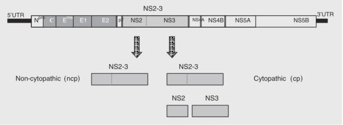
genome is a linear, single-stranded, positive sense RNA molecule of approximately 12.5 kb in length (3). The genome contains a single large open reading frame (ORF) en-coding a polyprotein of about 3900 amino acids. The ORF is flanked by a 5' 381-386-nucleotide long untranslated region (5'UTR) and by a 3' 229-nucleotide long UTR (3). The 5'UTR contains a secondary structure believed to act as an internal ribosomal entry site to direct the translation of the ORF upon internalization of the viral genome into the host cell (4). Translation of the ORF pro-duces a long polyprotein, which is co- and post-translationally processed by viral and cellular proteases giving rise to 11 to 12 mature viral proteins (3,5). The structural proteins (the exception is Npro, a non-struc-tural protein) are encoded by the 5' third of the genome as follows: Npro, C, Erns, E1, E2, and p7. Non-structural proteins are encoded downstream: NS2-3, NS4A, NS4B, NS5A, and NS5B (Figure 1) (3,5,6). The vast ma-jority of BVDV field isolates do not induce cytopathology in cell culture and are called non-cytopathic (ncp); cytopathic (cp) iso-lates are found almost exclusively in cattle suffering from mucosal disease (6,7).
Infection of seronegative cattle with BVDV may result in a variety of clinical manifesta-tions ranging from inapparent infecmanifesta-tions to gastroenteric, respiratory, hemorrhagic syn-drome, severe acute BVDV, and the fatal mucosal disease (7-9). Infection of pregnant cows is often associated with reproductive
losses, including early or late embryonic or fetal deaths, abortion or mummification, con-genital malformations, stillbirths and the birth of weak, non-thriving calves (8,10). Fetuses infected between 40 and 120 days of gestation with ncp isolates may survive the infection and be born as immunotolerant, persistently infected calves (8,10). Most persistently in-fected animals develop and die of mucosal disease within the first 6 to 24 months of life;
cp and ncp BVDV biotypes can usually be isolated from sick animals (7,8,10). Cyto-pathic and noncytoCyto-pathic viruses isolated from cases of mucosal diseases are antigeni-cally identical to each other and constitute what has been called a virus pair (7,11). Molecular analyses of cp-ncp pairs have indicated that the cp virus originates from the original ncp counterpart by diverse ge-netic mechanisms (12-17).
The most important molecular difference between the cp and ncp biotypes of BVDV is the expression of the non-structural poly-peptide NS3 (formerly known as p80). Whereas ncpBVDVs express a single poly-peptide of approximately 125 kDa called NS2-3 (or p125), cpBVDVs express both the entire NS2-3 and NS3, a separate protein which corresponds to the carboxy-terminal two thirds of 3 (Figure 1) (5,11). NS2-3 is a multifunctional protein believed to be involved in several steps of viral replication. It contains a zinc finger motif and a hydro-phobic domain in its amino-terminal third (NS2), and helicase, NTPase and protease
motifs in its carboxy-terminal half (NS3) (5,6). Although the expression of NS3 as a single polypeptide is the molecular marker of cpBVDV isolates, its exact role in the production of cytopathology is still unclear. Several mechanisms of expression of NS3 by cp viruses have been described, including insertions of cellular RNA sequences in NS2-3 near the boundary between NS2 and NSNS2-3 (12,13), downstream duplication of the NS3 gene (16); expression of NS3 from a defec-tive RNA genome (15), point mutations in the NS2-3 gene (14), and insertions of cellu-lar sequences plus viral gene duplications in the N-terminus of the polyprotein (17).
The differential expression of NS3 and its contribution to BVDV cytopathology in vitro and the pathogenesis of mucosal dis-ease in vivo constitute a major issue in pestivirus biology and pathogenesis and have been a matter of intensive investigation in the last two decades. The number of possible mechanisms of NS3 expression - and there-fore, the generation of cp viruses - increases as more cpBVDV isolates are characterized. The molecular mechanism of NS3 ex-pression was investigated here by searching for insertions and/or gene duplications in the genome of 41 cpBVDV isolates. RNA inser-tions were detected in three isolates, NS3 duplication in one, and 37 isolates showed no detectable genomic alterations.
Mate rial and Me thods
Forty-one cytopathic isolates of BVDV were biologically cloned to obtain pure cpBVDV. Individual cp clones were then propagated and their genomic RNA was extracted and submitted to RT-PCR analysis for the presence of insertions in NS2-3, close to the boundary between the NS2 and NS3 genes, and for duplications of the NS3 gene. The viral genomes containing insertions and/ or duplications were partially sequenced to determine the nature of the genomic rear-rangements.
Ce lls and viruse s
All procedures of virus multiplication, quantitation and plaque assays for biologi-cal cloning were performed in pestivirus-free Madin-Darby bovine kidney cells (MDBK, American Type Culture Collec-tion, Rockville, MD, USA). Cells were main-tained in minimal essential medium contain-ing 1.6 mg/L penicillin, 0.4 mg/L streptomy-cin, and 0.0025 mg/L fungizone, supple-mented with 5% horse serum. Sixty cyto-pathic BVDV isolates obtained from cases of mucosal disease were kindly provided by Dr. Fernando A. Osorio (Department of Vet-erinary and Biomedical Sciences, Univer-sity of Nebraska at Lincoln, UNL, Lincoln, NE, USA). After isolation in primary bovine lung cells and identification by fluorescent antibody assay, the viruses were further propagated in MDBK cells. The standard BVDV strains cpSinger, cpNADL, cpOregon, and cpTGAC were provided by Dr. Ruben O. Donis (Department of Veterinary and Biomedical Sciences, UNL, Lincoln, NE, USA).
Biological cloning of cytopathic viruse s
After propagation in MDBK cells, the isolates were biologically cloned in order to obtain pure cp virus clones and further propa-gated. Biological cloning for cp viruses was performed by standard plaque assays and the purified cp viruses were propagated and submitted to RT-PCR analysis.
RNA pre paration
g for 10 min. The cell pellet was submitted to total RNA extraction using Trizol reagent, according to the manufacturer’s protocol (Gibco-BRL, Carlsbad, CA, USA). The RNA pellet was rinsed with 70% ethanol and re-suspended in 50 µL diethylpyrocarbamate (DEPC)-treated water. All procedures of RNA manipulation were performed using disposable pipettes and tubes and DEPC-treated water.
Prim e rs
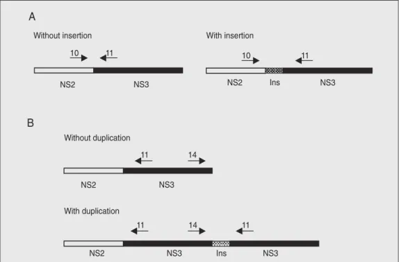
Oligonucleotide primers corresponding to conserved sequences of the published cpNADL and cpOsloss genomes were used (17). The sequences of the primers are listed below: primer 11 (cDNA primer) 5'-CTGTT GTTGCTTTGGCAA-3' (position 5703-5686); primer 10 (PCR primer) 5'-GGACTTT ATGTACTAC-3' (4546-4564); primer 14 (PCR primer) 5'-TCCCAATGATAACAG ACATA-3' (7545-7564). The strategy used for detection of insertions or gene duplica-tion was based on previous reports on the location of insertions of cellular mRNA in the viral genome (17,18). Primers 10 and 11 were used for detection of insertions in the
NS2-NS3 gene boundary and primers 11 and 14 were used to detect downstream du-plication of the NS3 gene (Figure 2) (17).
RT-PCR amplification
RNAs from all cp clones were initially submitted to RT-PCR using primers 10 and 11 (for insertions). RNAs of the standard cpBVDVs Singer and NADL were used as positive controls. Total RNA extracted from mock-infected MDBK cells was used as negative control and subsequently, submit-ted to RT-PCR using primers 11 and 14 (for NS3 gene duplication). RNAs from TGAC, a virus containing an NS3 gene duplication downstream from the original gene (17), were used as control. RT-PCR was performed in a 50-µL reaction using the Ready-to-Go RT-PCR Beads kit (Amersham Biosciences, Piscataway, NJ, USA), according to manu-facturer recommendations. Briefly, 44 µL DEPC-treated water was added to 2 µL of the RNA suspension (approximately 1 µg of total RNA) and 2 µL of each primer (100 ng each) and mixed with the RT-PCR beads preparation. Each 50-µL reaction contained 2 units of Taq DNA polymerase, 10 mM Figure 2.RT-PCR strategy used
Tris-HCl, pH 9.0, 60 mM KCl, 1.5 mM MgCl2, 200 µM of each dNTP, MuLV re-verse transcriptase, RNAse inhibitor and sta-bilizers. The tubes were incubated for 20 min at 42ºC for RT and 5 min at 95ºC and then submitted to 35 cycles of 95ºC for 1 min, 50ºC for 1 min and 72ºC for 1 min, followed by an extension of 5 min at 72ºC. RT-PCR products were visualized under UV light on ethidium bromide-stained 1% aga-rose gel after electrophoresis.
Nucle otide se que ncing and analysis
The 4 amplicons were directly purified by polyethyleneglycol precipitation (19). DNA sequencing was then performed di-rectly from the purified amplicons using a MegaBACE 500 (Amersham Biosciences Corp., Piscataway, NJ, USA) automatic se-quencer. The dideoxi chain-termination re-action was implemented with the use of the DYEnamic ET® kit (Amersham) and the primers 10 and 11. Sequence analysis and sequence alignments were performed with the Clone Manager Professional Suite, Align plus 5, version 5.10, software (Sci Ed. Cen-tral, Cary, NC, USA).
Re sults
Sixty BVDV isolates which produced cytopathology upon virus isolation in bo-vine lung cells were initially propagated in MDBK cells. Since field cpBVDV isolates usually contain a mixture of related cp and
ncp viruses - called a virus pair (11), the isolates were biologically cloned by plaque assay to yield pure populations of each cp
counterpart. For some isolates, plaque puri-fication was performed twice or even three times to ascertain the purity of the cloned cp
viruses. The identification of each isolate was confirmed on the basis of the character-istic cytopathology and by fluorescent anti-body assay (data not shown). After plaque purification, cloned cp viruses were
propa-gated in MDBK cells to produce viral stocks and subsequently used to infect cells for RNA extraction. For some isolates, biologi-cal cloning of the ncp counterparts was per-formed by limiting dilution. Forty-one pure clones of cpBVDV were further used in the study. In the remaining 19 cp isolates, bio-logical cloning of pure cp viruses could not be achieved.
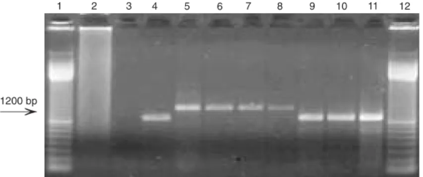
Total RNA extracted from cells infected with each of the 41 cloned cpBVDV was initially submitted to RT-PCR amplification using primers 10 and 11. In viral genomes lacking insertions within the NS2-3 gene, the PCR product obtained by using these primers should be a 900-bp fragment (Fig-ures 2 and 3) (17). RNA extracted from cells infected with the cpBVDV Singer strain, a virus known to lack cellular insertions in the NS2-3 gene (14), and the RNA from the cpBVDV NADL strain, whose genome con-tains a 270-nucleotide insert (12), were used as controls. RT-PCR amplification of the RNA of 38 cpBVDV with primers 10 and 11 produced amplicons indistinguishable in size from that of the Singer strain (Table 1, Fig-ure 3). Further separation of DNA fragments by extending the time of electrophoresis did not permit detection of size differences among these amplicons (data not shown).
Therefore, the genome of the 38 cpBVDV isolates examined does not contain inser-tions within the NS2-3 in the region com-prised between primers 10 and 11, as ascer-tained by agar gel electrophoresis of PCR products. The possible mechanisms of NS3 expression by these viruses will be discussed later.
The genome of cpBVDVs 26, 44, and 63 harbors insertions in the NS2-3 gene which are indistinguishable in size from the inser-tion present in the NADL strain, as ascer-tained by agar gel electrophoresis. The am-plicons obtained by PCR amplification of genomes of cpBVDVs 26, 44, and 63 were then submitted to nucleotide sequencing to determine the nature of the insertion present in these genomes. After several sequencing attempts, only the PCR product of isolate 44 yielded a reliable sequence that could be edited and analyzed. The insert is 296 nucleo-tides long and contains a core sequence (270 nucleotides) which is 98% identical and whose predicted amino acid sequence is 100% identical to the cIns insert present in the genome of the NADL strain. (Figure 4). This insert is highly homologous to a se-quence within the bovine gene coding for the J-Domain and is likely to result from recombination between the viral and cellular RNAs during genome replication (20). In the sequences of the other amplicons (cpBVDVs 26 and 63) stretches of mixed nucleotides were frequently observed, prob-ably reflecting co-amplification of different products.
After the first round of RT-PCR with primers 10 and 11, the RNAs of all 41 cpBVDV clones were submitted to another amplification reaction using primers 11 and 14. These primers were designed to detect possible duplications of the NS3 gene (Fig-ure 2). RNA from the TGAC strain, whose genome contains a downstream insertion of ubiquitin RNA plus a duplication of the NS3 gene (17), was used as positive control. The RNA of the Singer strain, which contains no
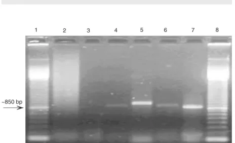
genome rearrangement, was used as nega-tive control. The amplicon produced by prim-ers 11 and 14 in TGAC genomeis about 900 bp (17). The use of primers 11 and 14 re-sulted in RT-PCR amplification in only one cpBVDV isolate (Table 1, Figure 5). The amplicon generated for cpBVDV 14 was smaller than the TGAC amplicon, with an estimated size of about 800-850 bp (Figure 5). No other cpBVDV isolate originated amplicons in the reactions using primers 11
Figure 5. RT-PCR analysis for the detection of NS3 gene duplication in the genome of cytopathic bovine viral diarrhea virus (BVDV) isolates. Lane 1, Molecular weight marker (100-bp ladder); lane 2, negative control; lane 3, no template; lane 4, TGAC strain; lane 5, TGAC strain; lane 6, isolate 14; lane 7, isolate 14; lane 8, molecular weight marker. Lanes 4
and 6 show amplified products of isolates TGAC and 14 using primers 10 and 11. Lanes 5
and 7 show the amplified products of isolates TGAC and 14 using primers 11 and 14. RT-PCR products were submitted to 1% agarose gel electrophoresis, stained with ethidium bromide, and visualized under UV light. The size of the amplified products from the isolate containing duplication is indicated by the arrow.
Table 1. Summary of RT-PCR analysis of the genome of cytopathic bovine viral diarrhea virus isolates for insertions in the NS2-3 gene and duplications of the NS3 gene.
Genetic mechanism Primers Isolates Amplicon Negative Amplicon (N) size (bp) isolates (N) size (bp)
Insertion in the 10 and 11 3 ~1200 38 ~900 NS2-3 gene
Duplication of the 11 and 14 1 ~850 40 None NS3 gene
Undetermined 10 and 11 37 Primers 10-11: ~900 NA NA 11 and 14 40 Primers 11-14: §
and 14. Obtaining amplicons from these RNAs by using primers 10 and 11 elimi-nated any possible problems with RNA qual-ity or with PCR. This result indicates that cpBVDV isolate 14 does possess a duplica-tion of the NS3 gene downstream of the original site that would be amplified by prim-ers 11 and 14. Attempts to determine the complete nucleotide sequence of such ge-nome rearrangement are currently under-way.
D iscussio n
Analysis of the genomes of 41 cpBVDV isolates by RT-PCR revealed that three iso-lates contain RNA insertions in the NS2-3 gene and one virus contains an NS3 gene duplication downstream from the original locus. The sizes of the three insertions are indistinguishable when examined by agar gel electrophoresis, their size corresponding to the NADL insert. In fact, one such insert was shown to be of the cIns type, and was highly homologous to the insert found in the genome of cpBVDV NADL. Expression of NS3 from these rearranged genomes prob-ably occurs either by protease cleavage within the inserted sequences (for the viruses har-boring inserts) or by direct translation of the NS3 polypeptide from the duplicated gene (21). In contrast, the genomes of 37 cp iso-lates do not contain detectable insertions within the region examined or NS3 gene duplication. The most likely mechanism of NS3 generation in these viruses, which lack large genomic rearrangements, appears to be the presence of point mutations within the NS2 gene leading to cleavage of NS2-3 (14). Nonetheless, as the PCR analysis only fo-cused on two sites of genomic rearrange-ments, we cannot rule out other possible rearrangements being responsible for NS3 expression in these viruses.
The identification of BVDV isolates ca-pable of inducing a cytopathic effect in cul-ture cells dates back to the late 50’s (18).
However, data concerning differences of the viral genomes (cp/ncp) were not available for a long time. As a consequence, the origin of cpBVDVs and their role in the pathogen-esis of mucosal disease were only elucidated three decades later (7,22). Once the origin of cpBVDV was determined, molecular mark-ers of cpBVDV at genomic and polypeptide levels were shortly identified (11,12). There-after, expression of NS3 as a discrete poly-peptide, and not as part of NS2-3 as in ncpBVDVs, was universally considered to be the molecular marker of cpBVDV (11-17). As the cytopathic phenotype of BVDV is strictly correlated with the expression of NS3, and no exceptions have been reported to date, we did not investigate the expression of NS3 in all isolates. Only a few isolates were examined by Western blot to confirm the expression of NS3 as a discrete polypep-tide (data not shown).
frequent means of generating cpBVDVs (12,16-18,21,23,26). In addition, other cel-lular insertions have been detected in this genomic region of cpBVDV isolates, namely part of light chain 3 microtubule-associated proteins (LC3) (13), SMT3B (27), riboso-mal S27a-coding sequence (28), NEDD8 cellular sequences (29), ubiquitin-like pro-teins, and part of a chaperon called Jiv (25). The expression of NS3 from genomes con-taining cellular insertions within NS2-3 prob-ably occurs by proteolytic cleavage in the inserted sequence contained within NS2-3. In the present study, three cp viruses had insertions within the NS2-3 gene. One such insert has been sequenced and showed a high nucleotide homology with the NADL cIns insert, which corresponds to sequences of a cellular gene called J-Domain protein (25). The cellular insert present in the NADL genome is essential for generation of NS3 by proteolytic cleavage of NS2-3 and produc-tion of cytopathology (24). In the present study, the nucleotide sequence of the insert in the genome of two isolates could not be determined. The size of the amplified prod-ucts, taken together with their frequent oc-currence, suggests that they might also be of the cIns type. Although insertions leading to proteolytic cleavage of NS2-3 seem to be the most common mechanism of generation of NS3 in cpBVDV isolates (21), other mechan-isms responsible for expression of NS3 have been identified. NS3 can be expressed from a duplicated gene, associated or not with other sequence rearrangements, including duplication of Npro right upstream of the duplicated NS3 gene (30), or from a defec-tive genome with deletion of all structural genes (15). Recently, a novel mechanism of NS3 generation was described. BVDV CP8 contains cellular insertions and viral sequence duplications in the N-terminal region of the polyprotein - and not in the NS2-3 boundary as most isolates characterized thus far. Thus, it is tempting to speculate that the number of mechanisms of generation of NS3 will
in-crease as more cpBVDV isolates are charac-terized. In the present study, one isolate presented an NS3 gene duplication down-stream of the original locus, yielding an amplicon of approximately 800-850 nucleo-tides. Attempts to determine the nucleotide sequence of this region are currently ongo-ing. In genomes containing a duplicated NS3 gene, the expression of NS3 polypeptide is believed to occur by direct translation of the duplicated gene (17,21).
Despite the initial emphasis on cellular insertions and gene duplications as a molec-ular mechanism of generation of NS3, ex-amination of a large number of isolates failed to detect such genomic alterations (17,18,30). Analysis of the genome of cpBVDV Or-egon, a cp strain whose genome does not contain NS2-3 insertions or NS3 duplica-tion, revealed a novel mechanism of NS3 generation (14). In the genome of BVDV Oregon and other cp isolates including the Singer strain, the information necessary for NS2-3 processing resides within the NS2 gene and probably involves a set of point mutations that somehow affect the cleava-bility of NS2-3 (14). Regardless of the mo-lecular mechanism leading to NS3 genera-tion, these findings demonstrated that, in addition to RNA recombination, a different genetic mechanism, point mutations within the NS2 gene, may be responsible for the production of cpBVDV. Indeed, examina-tion of a large number of cpBVDV isolates has indicated that this mechanism seems to be more frequent than previously expected (17,18,30).
of primers restricted the possible detectable genomic changes. Likewise, the limitation of resolution of agar gel electrophoresis anal-ysis of RT-PCR products would not allow the detection of small insertions.
The results presented here demonstrate that expression of NS3 in cytopathic BVDV isolates, although frequently associated with
NS2-3 insertions and/or NS3 gene duplica-tion, appears to be more frequently associ-ated with cleavage of the NS2-3 polypeptide without the presence of bulk genome rear-rangements. It is tempting to speculate that additional mechanisms of generation of NS3 may arise as more cpBVDV isolates are examined.
Re fe re nce s
1. Horzinek MC. Pestiviruses - taxonomic perspectives. Arch Virol
1991; 3 (Suppl): 1-5.
2. Francki RIB, Fauquet CM, Knudson DL, Brown F. Classification and nomenclature of viruses. Fifth report of the International Committee on the Taxonomy of Viruses. Arch Virol 1991; 2: 223-233. 3. Collett MS, Larson RE, Gold C. Molecular cloning and nucleotide
sequence of the pestivirus bovine virus diarrhea virus. Virology
1988; 70: 253-266.
4. Poole TL, Wang C, Popp RA, Potgieter LN, Siddiqui A, Collett MS. Pestivirus translation initiation occurs by internal ribosome entry.
Virology 1995; 206: 750-754.
5. Collett MS, Larson R, Belzer SK, Retzel E. Proteins encoded by bovine viral diarrhea virus: the genomic organization of a pestivirus.
Virology 1988; 165: 200-208.
6. Donis RO. Molecular biology of bovine viral diarrhea virus and its interactions with the host. Vet Clin North Am Food Anim Pract 1995; 11: 393-423.
7. Brownlie J. The pathways for bovine virus diarrhoea virus biotypes in the pathogenesis of disease. Arch Virol Suppl 1991; 3: 79-96. 8. Baker JC. The clinical manifestations of bovine viral diarrhea
infec-tion. Vet Clin North Am Food Anim Pract 1995; 11: 425-445. 9. Corapi WV, French TW, Dubovi EJ. Severe thrombocytopenia in
young calves experimentally infected with noncytopathic bovine viral diarrhea virus. J Virol 1989; 63: 3934-3943.
10. Moennig V, Liess B. Pathogenesis of intrauterine infections with bovine viral diarrhea virus. Vet Clin North Am Food Anim Pract
1995; 11: 477-487.
11. Donis RO, Dubovi EJ. Differences in virus-induced polypeptides in cells infected by cytopathic and noncytopathic biotypes of bovine virus diarrhea-mucosal disease virus. Virology 1987; 158: 168-173. 12. Meyers G, Tautz N, Dubovi EJ, Thiel HJ. Viral cytopathogenicity correlated with integration of ubiquitin-coding sequences. Virology
1991; 180: 602-616.
13. Meyers G, Stoll D, Gunn M. Insertion of a sequence encoding light chain 3 of microtubule-associated proteins 1A and 1B in a pestivirus genome: connection with virus cytopathogenicity and induction of lethal disease in cattle. J Virol 1998; 72: 4139-4148.
14. Kummerer BM, Stoll D, Meyers G. Bovine viral diarrhea virus strain Oregon: a novel mechanism for processing of NS2-3 based on point mutations. J Virol 1998; 72: 4127-4138.
15. Tautz N, Thiel HJ, Dubovi EJ, Meyers G. Pathogenesis of mucosal disease: a cytopathogenic pestivirus generated by an internal dele-tion. J Virol 1994; 68: 3289-3297.
16. Meyers G, Tautz N, Stark R, Brownlie J, Dubovi EJ, Collett MS, et al.
Rearrangement of viral sequences in cytopathogenic pestiviruses.
Virology 1992; 191: 368-386.
17. Qi F, Ridpath JF, Lewis T, Bolin SR, Berry ES. Analysis of the bovine viral diarrhea virus genome for possible cellular insertions.
Virology 1992; 189: 285-292.
18. Greiser-Wilke I, Haas L, Dittmar K, Liess B, Moennig V. RNA inser-tions and gene duplicainser-tions in the nonstructural protein p125 region of pestivirus strains and isolates in vitro and in vivo. Virology 1993; 193: 977-980.
19. Sambrook J, Russel D. Molecular cloning: A laboratory manual. Cold Spring Harbor: Cold Spring Harbor Laboratory Press; 2001. 20. Muller A, Rinck G, Thiel HJ, Tautz N. Cell-derived sequences in the
N-terminal region of the polyprotein of a cytopathogenic pestivirus. J Virol 2003; 77: 10663-10669.
21. Vilcek S, Greiser-Wilke I, Nettleton P, Paton DJ. Cellular insertions in the NS2-3 genome region of cytopathic bovine viral diarrhoea virus (BVDV) isolates. Vet Microbiol 2000; 77: 129-136.
22. Lee KM, Gillespie JH. Propagation of viral diarrhea virus of cattle in tissue culture. Am J Vet Res 1957; 18: 952-953.
23. Bolin SR, McClurkin AW, Cutlip RC, Coria MF. Severe clinical disease induced in cattle persistently infected with noncytopathic bovine viral diarrhea virus by superinfection with cytopathic bovine viral diarrhea virus. Am J Vet Res 1985; 46: 573-576.
24. Mendez E, Ruggli N, Collett MS, Rice CM. Infectious bovine viral diarrhea virus (strain NADL) RNA from stable cDNA clones: a cellu-lar insert determines NS3 production and viral cytopathogenicity. J Virol 1998; 72: 4737-4745.
25. Rinck G, Birghan C, Harada T, Meyers G, Thiel HJ, Tautz N. A cellular J-domain protein modulates polyprotein processing and cytopathogenicity of a pestivirus. J Virol 2001; 75: 9470-9482. 26. Tautz N, Meyers G, Thiel HJ. Processing of poly-ubiquitin in the
polyprotein of an RNA virus. Virology 1993; 197: 74-85.
27. Qi F, Ridpath JF, Berry ES. Insertion of a bovine SMT3B gene in NS4B and duplication of NS3 in a bovine viral diarrhea virus ge-nome correlate with the cytopathogenicity of the virus. Virus Res
1998; 57: 1-9.
28. Becher P, Orlich M, Thiel HJ. Ribosomal S27a coding sequences upstream of ubiquitin coding sequences in the genome of a pestivirus. J Virol 1998; 72: 8697-8704.
29. Baroth M, Orlich M, Thiel HJ, Becher P. Insertion of cellular NEDD8 coding sequences in a pestivirus. Virology 2000; 278: 456-466. 30. Kummerer BM, Tautz N, Becher P, Thiel H, Meyers G. The genetic


