SY
STEMA
TIC REVIEW
1Physical therapist, professor of the course of Physical Therapy of the Instituto Federal de Educação, Ciência e Tecnologia do Rio de Janeiro – Rio de Janeiro (RJ), Brazil.
2Physical therapist, professor of the course of Physiotherapy of the Centro Universitário Barão de Mauá – Ribeirão Preto (SP), Brazil. 3Centro de Reabilitação of the Clinical Hospital of the Faculdade de Medicina de Ribeirão Preto of the Universidade de São Paulo – Ribeirão Preto (SP), Brazil.
4Physical therapist, resident physiotherapist of the Universidade Federal de São Paulo – São Paulo (SP), Brazil.
5PhD of the Department of Gynecology and Obstetrics of the Faculdade de Medicina de Ribeirão Preto of the Universidade de São Paulo – Ribeirão Preto (SP), Brazil.
6PhD of the Department of Internal Medicine of the Faculdade de Medicina de Ribeirão Preto of the Universidade de São Paulo – Ribeirão Preto (SP), Brazil.
88
Functional assessment of the pelvic floor muscles by
electromyography: is there a normalization in data
analysis? A systematic review
Avaliação funcional dos músculos do assoalho pélvico pela eletromiografia: existe a
normalização na análise de dados? Uma revisão sistemática
Evaluación funcional de los músculos del suelo pélvico por la electromiografía: ¿la
normalización existe en el análisis de datos? Una revisión sistemática
Aline Moreira Ribeiro
1, Elaine Cristine Lemes Mateus-Vasconcelos
2,3, Thaís Daniel da Silva
4,
Luiz Gustavo de Oliveira Brito
5, Harley Francisco de Oliveira
6Corresponding address: Aline Moreira Ribeiro – Professora Lady Alvarenga Neves Street, 30, Jd. Europa – Campo Belo (MG), Brazil – Zip Code: 37270-000 – E-mail: [email protected] – Finance source: Nothing to declare – Conflict of interests: Nothing to declare – Presentation: June 21st, 2016 – Accepted for publication: Dec. 18th, 2017.
ABSTRACT | This study aims to evaluate the method of analysis of electromyographic data considering the functional assessment of pelvic floor muscles (PFM). We have included in our search strategy the following databases: Medline, PubMed, Cochrane Central Register of Controlled Trials and Cochrane Database of Systematic Reviews, PEDro, and IBECS, considering articles published in the last ten years (2004-2014). The identified articles were independently examined by two evaluators, according to these inclusion criteria: (1) population: female adults; (2) PFM assessment by electromyography (EMG) with vaginal/ anal probe; and (3) description of how electromyographic data analysis is performed. The Newcastle-Ottawa Scale (NOS) was used to assess the risk of bias. We identified 508 articles, of which 23 were included in the review. The data showed differences between the collection protocols, and a significant number of studies did not normalize the electromyographic data. Physiotherapists are among the clinicians who most frequently use EMG to evaluate the function and dysfunction of the neuromuscular system. Although some previous studies have provided an overview to guide the evaluator in the assessment, few succeeding studies followed their recommendations.
Keywords | Pelvic Floor; Electromyography; Review.
RESUMO | Este estudo objetiva analisar o método de análise dos dados eletromiográficos na avaliação funcional dos músculos do assoalho pélvico (MAP). As bases de dados consultadas foram: Medline, PubMed, Cochrane Central Register of Controlled Trials e Cochrane Database of Systematic Reviews, PEDro e IBECS, considerando-se artigos publicados nos últimos dez anos (2004-2014). Os artigos identificados foram avaliados independentemente por dois avaliadores, de acordo com os critérios de inclusão: (1) população: mulheres adultas; (2) avaliação dos MAP utilizando a eletromiografia (EMG) com sonda vaginal ou anal; (3) descrição de como é feita a análise dos dados eletromiográficos. O instrumento Newcastle-Ottawa Scale (NOS) foi usado para avaliação da qualidade metodológica dos estudos. Foram identificados 508 artigos, dos quais 23 foram incluídos na revisão. Os dados mostraram diferenças entre os protocolos de coleta, e um número significativo de estudos não utilizaram a normalização na análise dos dados eletromiográficos. Os fisioterapeutas estão entre os profissionais que mais utilizam a EMG para avaliar a função e disfunção do sistema neuromuscular. Apesar de alguns estudos anteriores fornecerem uma visão geral para orientar o avaliador no uso deste instrumento, poucos estudos subsequentes seguiram suas recomendações.
RESUMEN |Se evaluó, en este estudio, el método de análisis de los datos electromiográficos en la evaluación funcional de los músculos del suelo pélvico. Las bases de datos consultadas fueron: Medline, PubMed, Cochrane Central Register of Controlled Trials, Cochrane Database of Systematic Reviews, PEDro y IBECS. La investigación fue refinada considerándose artículos publicados en los últimos diez años (2004 a 2014). Se evaluaron de forma independiente los artículos identificados por dos evaluadores, según los siguientes criterios de inclusión: (1) población: mujeres adultas; (2) evaluación de los músculos del suelo pélvico utilizándose la electromiografía (EMG) con sonda vaginal o anal; (3) descripción de cómo es el análisis de los datos electromiográficos. Se utilizó
el instrumento Newcastle-Ottawa Scale (NOS) para evaluar la calidad metodológica de los estudios. Se identificaron 508 artículos y 23 de estos fueron incluidos en la revisión. Los datos mostraron diferencias entre los protocolos de recolección y un número significativo de estudios no utilizaron la normalización en el análisis de los datos electromiográficos. Los fisioterapeutas están entre los profesionales que más utilizan la EMG para evaluar la función y disfunción del sistema neuromuscular. Aunque algunos estudios anteriores proporcionen una visión general para orientar el evaluador en el uso de este instrumento, pocos estudios posteriores han seguido sus recomendaciones.
Palabras clave | Diafragma de la Pelvis; Electromiografía; Revisión.
INTRODUCTION
The correlation between pelvic floor muscles (PFM)
strength and dysfunctions in this region is still an open field
of research, with few published data. Several functional
PFM assessment methods are used in clinical practice
and research, such as digital palpation, perineometry,
surface electromyography (sEMG), dynamometry, and
imaging modalities, such as ultrasonography and magnetic
resonance imaging
1,2.
Among these assessment methods, electromyography
(EMG) outstands as an alternative method to monitor
muscle tone at rest, muscle strength, and muscular
endurance, and to obtain data on the normal and abnormal
physical functions of the PFM. EMG is indicated as a
reliable and objective muscle evaluation method that
causes no harm to patients
3,4.
EMG monitors the electrical activity generated by
the depolarization of muscle fibers, as a function of
voltage effect over time. It is the algebraic sum of all the
signals detected in a certain area and can be considered
as an indirect measurement of muscle strength. The
electrical activity can be collected either with surface or
intramuscular electrodes
1,5. Because PFMs lie deep to
the skin surface and the superficial and deep layers of
PFMs have distinct functions, to use surface electrodes
adhered to the perineum is not ideal to study the deep
layer of the PFMs, as unwanted activity (crosstalk)
would inevitably be recorded from the superficial PFMs
and potentially from other nearby muscles including
the anal sphincter, the gluteus, and the obturator hip
musculature. Considering that deep PFMs lie adjacent
to the vaginal walls, electrodes positioned against the
lateral vaginal walls are a convenient mean to record
their EMG activity
6.
Thus, in clinical practice, surface electrodes (vaginal
and anal probes) are more widely used due to the high
sensitivity of the perineal region and the skills required
in using the needle or fine-wire electrodes
2,7.
The characteristics of the amplitude and frequency of
EMG signal are sensible to intrinsic (type of muscle fiber,
depth, diameter and amount of tissue between the muscle
and electrode) and extrinsic factors (location, orientation
and shape of the area of the electrodes). Therefore, the
signal amplitude cannot be analyzed directly
3,5,8.
The “normalization” is required to analyze and
compare EMG signals from different individuals,
muscles and acquisition. It is a form of transforming
the absolute amplitude values into relative values relating
to an amplitude value characterized as 100%
8. There are
several forms of normalization of EMG signal. Among
them, the most used is the normalization by maximum
voluntary contraction (MVC), in which the reference to
the standards is the highest value found among certain
muscle contractions in question
9.
Normalization prevents interference on the intensity
of contraction, since they remove the effect of other
factors that influence the signal capture. Thus, we can
compare different muscles and individuals to the amount
of energy produced during a certain contraction only
after the process of normalization
5.
Thus, the aim of this study was to evaluate the methods
of analysis of EMG data in studies that encompass the
PFM functional evaluation.
METHODOLOGY
Eligibility criteria
Inclusion and exclusion criteria are:
• Inclusion criteria:
- Scientific articles published in the last ten years
(2004–2014), which aimed to perform pelvic
floor muscles (PFM) functional assessment
using EMG;
- Articles published in English and in full.
Design
- Full-text articles of cross-sectional studies.
Participants
- Female adult participants.
PFM functional assessment
- Performed using sEMG with a vaginal/anal
probe;
- With a description of how the analysis of EMG
data is performed.
• Exclusion criteria:
- Studies including children and adolescents;
- Studies including men;
- Animal experiments;
- Treatment with EMG biofeedback, without
EMG rating;
- Other exams that included EMG (urodynamic,
for example);
- Modified custom probes;
- Review articles, guidelines and other studies
with different design.
Information sources and search
To select studies related to the topic investigated,
we have performed a computer-aided and manual
search between September 2013 and February 2014.
The consulted databases included: Medline PubMed,
Cochrane Central Register of Controlled Trials
(CENTRAL), Cochrane Database of Systematic
Reviews, PEDro, and IBECS. We have limited the
search to English-language manuscripts published
between 2004 and 2014. Articles published in full
and available in the databases were included in
this study. Our search strategy was broad, aiming
to have as many manuscripts as possible to make a
good triage. The following keyword was used: “pelvic
floor electromyography”. Articles identified in the
initial search strategy were selected according to the
inclusion criteria.
This review followed the methodological quality
parameters of systematic reviews and the Preferred
Reporting Items for Systematic Reviews and
Meta-Analyses (PRISMA) criteria were observed.
Data collection process
A standardized data extraction form was used to
collect the following data: authors, year, publication,
country of origin, study design, sample, age (years),
probe used, data collection protocol and whether the
normalization of electromyographic data was performed.
This process was performed using two independent
raters (AMR and ECLMV).
Risk of bias assessment
The risk of bias was assessed using the
Newcastle-Ottawa Scale (NOS). The original NOS was developed
to assess the quality of the observational studies; it
contains eight items that analyze three dimensions:
selection, comparability and outcome (in cohort studies)
or exposure (case-control studies). There is a series of
options for each item, which reflects the quality of the
studies and is scored by a star: the higher the number
of the star, the higher is the study quality
10.
A study may receive a maximum of one star for each
numbered item within the “selection” and “outcome”
categories. A maximum of two stars may be given for
“comparability”. The total score is nine: four stars in
“selection”, two stars in “comparability” and three stars
in “outcome”
10.
Summary measures
RESULTS
Study selection
A total of 213 publications were identified from
MEDLINE, 221 from PubMed, 51 from CENTRAL
and the Cochrane Database of Systematic Reviews, 18
from PEDro, and five from IBECS, resulting in 508 related
publications. Of the articles identified, 107 were selected after
applying the established inclusion and exclusion criteria.
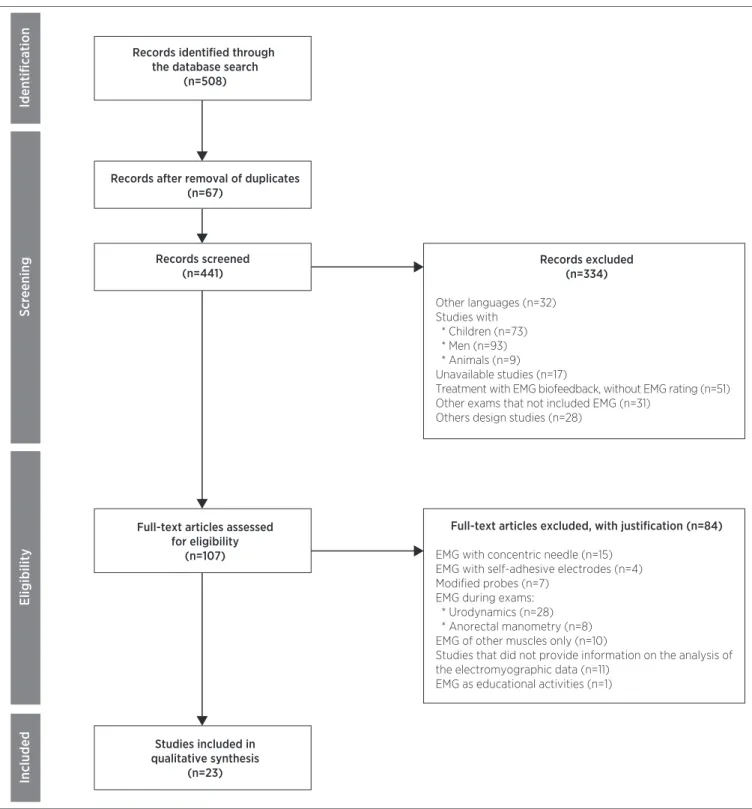
Only 23 articles were included for this review (Figure 1).
Assessment of quality of manuscripts
Table 1 summarizes the results of the methodological
quality assessment.
Figure 1. Flowchart with the numbers of articles identified, excluded, and included in the systematic review
Records identified through the database search
(n=508)
Iden
tifica
tion
Studies included in qualitative synthesis
(n=23)
Included
Full-text articles assessed for eligibility
(n=107)
Eligibilit
y
Full-text articles excluded, with justification (n=84)
EMG with concentric needle (n=15) EMG with self-adhesive electrodes (n=4) Modified probes (n=7)
EMG during exams: * Urodynamics (n=28) * Anorectal manometry (n=8) EMG of other muscles only (n=10)
Studies that did not provide information on the analysis of the electromyographic data (n=11)
EMG as educational activities (n=1)
Records after removal of duplicates (n=67)
Records screened (n=441)
Records excluded (n=334)
Other languages (n=32) Studies with
* Children (n=73) * Men (n=93) * Animals (n=9) Unavailable studies (n=17)
Treatment with EMG biofeedback, without EMG rating (n=51) Other exams that not included EMG (n=31)
Others design studies (n=28)
Scr
Table 1. Evaluation of the methodological quality of selected studies.
References Selection Comparability Outcome
Auchincloss and McLean11
-Botelho et al.3
-Chen et al.12
-Devreese et al.13
Frederice, Amaral and Ferreira14
-Grape, Dedering and Jonasson15
-Halski et al.16
-Halski, Ptaszkowski, Słupska and Dymarek17
-Junginger, Baessler, Sapsford and Hodges18
-Lauper, Kuhn, Gerber, Luginbühl and Radlinger19 Luginbuehl et al.20
Pereira et al.21
Petricelli et al.22
-Resende et al.23
Resende et al.24
-Sapsford, Richardson, Maher and Hodges25 Smith, Coppieters and Hodges26
Smith, Coppieters and Hodges27
Soljanik et al.28
-Stüpp et al.29
-Thompson, O’Sullivan, Briffa and Neumann30
-Thompson. O’Sullivan, Briffa and Neumann31
-Zhang, Wang and Zheng32
to 307 women (mean 50.0; median 30.0). The mean age
of the participants was 35.2 years (median 30.4; range
22-63). Seven studies were performed in Brazil and six
in Australia.
General characteristics of the studies
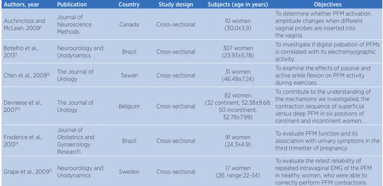
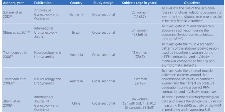
The characteristics of the articles included in this study
are listed in Table 2. The sample sizes ranged from nine
Table 2. General characteristics of the 23 selected studies.
Authors, year Publication Country Study design Subjects (age in years) Objectives
Auchincloss and McLean, 200911
Journal of Neuroscience Methods
Canada Cross-sectional 10 women
(30.0±3.9)
To determine whether PFM activation amplitude changes when different vaginal probes are inserted into the vagina.
Botelho et al., 20133
Neurourology and
Urodynamics Brazil Cross-sectional
307 women (23.93±5.78)
To investigate if digital palpation of PFMs is correlated with its electromyographic activity.
Chen et al., 200912 The Journal of
Urology Taiwan Cross-sectional
31 women (46.48±7.24)
To examine the effects of passive and active ankle flexion on PFM activity during exercises.
Devreese et al., 200713
The Journal of
Urology Belgium Cross-sectional
82 women (32 continent, 52.38±9.68;
50 incontinent, 52.78±7.99)
To contribute to the understanding of the mechanisms we investigated, the contraction sequence of superficial versus deep PFM in six positions of continent and incontinent women.
Frederice et al., 201314
Journal of Obstetrics and Gynaecology Research
Brazil Cross-sectional 91 women
(24.3±4.9)
To evaluate PFM function and its association with urinary symptoms in the third trimester of pregnancy.
Grape et al., 200915 Neurourology and
Urodynamics Sweden Cross-sectional
17 women (26, range 22-34)
To evaluate the retest reliability of repeated intravaginal EMG of the PFM in healthy women, who were able to correctly perform PFM contractions.
Authors, year Publication Country Study design Subjects (age in years) Objectives
Halski et al., 201316 BioMed Research
International Poland Cross-sectional
20 women (22.3±1.28)
To determine how the depth of probe placement affects functional and resting bioelectrical activity of the PFM and whether the recorded signal might be dependent on the direction in which the probe is rotated.
Halski et al., 201417 BioMed Research
International Poland Cross-sectional
16 women (63.1±4,1, range 55-70)
To evaluate resting and functional bioelectrical activity of the PFM and synergistic muscles, depending on the orientation of the pelvis, in anterior (P1) and posterior (P2) pelvic tilt.
Junginger et al., 201018
International Urogynecology Journal
Australia Cross-sectional 9 women (42, range 32-59)
To investigate the relationship between bladder neck displacement, EMG activity of the PFM and abdominal muscles and intra-abdominal pressure during different pelvic floor and abdominal contractions.
Lauper et al. 200919
Neurourology and
Urodynamics Switzerland Cross-sectional
38 women (21 control, 30.0±4.7; 17 post-partum, 31.7±3.4)
To determine if two different whole-body vibrations, sinusoidal vibration and SR-WBV, lead to a reactive activation of PFM when using various intensities.
Luginbuehl et al., 201220
Neurourology and
Urodynamics Switzerland Cross-sectional
50 women (28 np/pp group, 47.0±9.6; 22 pp group
33.1±4.8)
To determine the SR-WBV load modality regarding PFM activity to complete the SR-WBV training methodology for future PFM training with SR-WBV.
Pereira et al., 201321 Neurourology and
Urodynamics Brazil Cross-sectional
81 women (20 nulliparous, 24.4±3.6;
25 pregnant, 24.6±5.6; 19 vaginal delivery, 21.8±3.1; 17 cesarean section, 23.1±5.9)
To simultaneously evaluate both transversus abdominis/internal oblique muscles and PFM during isometric exercises in nulliparous, pregnant, and postpartum women.
Petricelli et al., 201422
BioMed Research
International Brazil Cross-sectional
60 women (26.06±5.58)
To compare the role of the PFM between nulliparous and multiparous women in the third trimester of pregnancy, analyzing the relationship between EMG, vaginal palpation (modified Oxford Grading Scale), and perineal distensibility (EPI-NO).
Resende et al., 201223 International Urogynecology Journal Brazil Cross-sectional 30 women (Group 1, 28.2±6.5; Group 2, 26.4±3.2)
To compare the MVC and strength of PFM of pregnant and non-pregnant women using sEMG.
Resende et al., 201124
International Urogynecology Journal
Brazil Cross-sectional 34 women
(28±5.9)
To determine whether Paula method of circular muscles contraction (those surrounding the eyes, mouth and fingers) can increase PFM activity.
Sapsford et al., 200825 Archives of Physical Medicine and Rehabilitation Australia Cross-sectional 17 women (9 asymptomatic, 45, range 32–66; 8 symptomatic, 41.8, range
33–55)
To determine whether resting activity of the PFM and abdominal muscles varied in different sitting postures in parous women with and without SUI.
Smith et al., 200726 Neurourology and
Urodynamics Australia Cross-sectional
30 women (14 continent, 52.5±12.5; 16 incontinent, 49.8±12.0;
7 mild-incontinence, 48.1±12.2; 9 more severe incontinence, 51.0±12.4)
To determine whether activity of the PFM and abdominal muscles differs between continent and incontinent women in response to a postural perturbation with a moderately full or empty bladder.
Smith et al., 200827 Neurourology and
Urodynamics Australia Cross-sectional
29 women (16 with SUI, 49.8± 12.0;
13 controls, 53.1±12.7)
To investigate whether there are differences in center of pressure displacement, trunk motion, and trunk muscle activity in women with and without SUI during static balance tasks when the bladder is empty and moderately full.
Authors, year Publication Country Study design Subjects (age in years) Objectives
Soljanik et al., 201228
Archives of Gynecology and Obstetrics
Germany Cross-sectional 23 women
(22±3.7)
To evaluate the role of the ischioanal fossa in functional relations between the levator ani and gluteus maximus muscles in healthy female volunteers.
Stüpp et al., 201129
International Urogynecology Journal
Brazil Cross-sectional 34 women
(28.1±6.0)
To investigate PFM and transversus abdominis activation during the abdominal hypopressive technique through sEMG.
Thompson et al., 200630
Neurourology and
Urodynamics Australia Cross-sectional
13 women (38±7)
To investigate the muscle activation patterns of the abdominopelvic region used by incontinent women during a PFM contraction and a Valsalva maneuver compared to healthy and asymptomatic subjects.
Thompson et al., 2006b31
Neurourology and
Urodynamics Australia Cross-sectional
13 women (37±9)
To investigate the different muscle activation patterns around the abdominopelvic cavity in continent women and their effect on pressure generation during a correct PFM contraction and a Valsalva maneuver.
Zhang et al., 200632
International Journal of Gynecology and Obstetrics
China Cross-sectional
114 women (57 with SUI, 41.3±10.2;
57 controls, 38.8±11)
To obtain perineal electrophysiological data and assess the clinical usefulness of measuring the sEMG activity of the PFM in predicting and diagnosing SUI.
EMG: electromyography; MVC: maximal voluntary contraction; PFM: pelvic floor muscles; SR-WBV: stochastic resonance whole-body vibration; SUI: stress urinary incontinence; np: nulliparous; pp: primiparous
Characteristics of data collection and analysis of
surface electromyography
The type of vaginal probe used varied between the
studies. The most commonly used were FemiScan (n=4)
and Periform (n=9). One study did not mention the
type of vaginal probe used. The EMG data collection
protocols also varied. Only one study examined the
evaluation of the two separate types of muscle fibers
(types I and II). Six studies evaluated the PFM in
other positions besides the supine position; and three,
during effort (cough) or Valsalva maneuver. Only four
studies reported a normalization of the sEMG data
for analysis.
Table 3. Characteristics of the probe used, collection protocol and other data on EMG
Authors, year Probe used Data collection protocol Frequencies and approaches to filter the signal Normalization?
Auchincloss and McLean, 200911
Vaginal: Periform and FemiScan
One MVC and maximal effort cough.
The signals were preamplified using a Delsys™ DE-2.1 Sensor for active electrode amplifiers (200×) before final amplification using Delsys™ AMT-8 amplifiers [common mode rejection ratio (CMRR)=90 dB at 60 Hz, bandwidth of 20-450 Hz, input impedance>1015 Ω] to produce an overall gain of 1000.
No
Botelho et al., 20133
Vaginal: PhysioMed Services
Three 5-s MVCs with 10-s rest.
Band-pass filter with cutoff frequencies at 20-500 Hz, an amplifier gain of 1,000 times and a CMRR>120 dB. A 12-bit A/D signal converter plate was used, to convert analog signals into digital ones with a 2.0 kHz anti-aliasing filter sampling frequency, with an input range of 5 mN.
No
Chen et al., 200912
Vaginal: FemiScan
One contraction standing and one contraction in eight different positions of the ankle joint.
No description. No
Devreese et al., 200713
Vaginal: No description
Supine + knees flexed, supine + knees straight, sit leaning forward, sit upright, stand leaning forward and stand upright: 2-min rest preceded by a MVC for 6 s. The onset of increased activity was defined as any muscle activity with a minimum amplitude of a sliding window of 25 ms exceeding resting EMG by 2 SD.
Band-pass filter of 10 to 500 Hz. Data were sampled by a 12- bit analog-to-digital converter at a rate of 1,000 samples per second, stored on a computer, full-wave rectified and low-pass filtered at 50 Hz to smooth the data.
No
Authors, year Probe used Data collection protocol Frequencies and approaches to filter the signal Normalization?
Frederice et al., 201314
Vaginal: Miotec
Basal tone at rest, one contraction with 1-min rest, MVC (highest of 3 contractions), and one 10-s contraction.
No description. No
Grape et al., 200915
Vaginal: FemiScan
Three series of contractions with 10-s sustenance and 10-s rest.
A band-pass filter of 8 to 500 Hz (3 dB points) and a low-pass filter of 500 Hz was used. The signal was sampled at 10 Hz after full-wave rectification.
No
Halski et al., 201316
Vaginal: Periprobe Optima 3
Standing position: five contractions with 5-s rest between each contraction (resting activity).
This device is characterized by a continuous amplitude of 0.2-2000 µVRMS in the frequency band of 2-100 Hz and pulse-width modulation from 50-450 µS for signals recorded generated by muscles. Device sensitivity is established at a level of 0.1 µV (4% accuracy; readings ±0.3 mV at 200 Hz), with selectable band-pass filter (3 dB bandwidth) and 50 Hz notch filter (33 dB; 0.1% accuracy).
No
Halski et al., 201417
Vaginal: Lifecare PR-02 Vaginal Probe
EMG activity was recorded with the pelvis rotated forward (P1) and backward (P2) around the transverse axis. In P1 and P2, the participants made five 5-s MVCs (functional sEMG activity) with a 5-s rest (resting sEMG activity). They took 60 s of rest between trials.
Analog output gain: x1000 standard (5000 selected units); minimum CMRR: 100 dB at 50-60 Hz; input impedance>100 MΩ on sEMG channels (isolated to>3000 V; sEMG amplifier performance: 1 µV sensitivity and <1
µVRMS baseline noise; data acquisition: 8 channels of 12-bit resolution, and USB update to PC every millisecond; cutoff of high-pass filter: 10 Hz first-order filter on sEMG channels; selectable low-pass cutoff of 500 or 1000 Hz on sEMG channels.
No
Junginger et al., 201018
Vaginal:
Periform One 5-s MVC with 30- to 120-s rest.
EMG data were amplified 2,000 times, bandpass-filtered
between 10 Hz to 1 kHz and sampled at 2 kHz. Yes
Lauper et al., 200919
Vaginal:
Periform PFM activity at rest and in 5-s MVC.
The EMG was sampled at a rate of 1 kHz, the cutoff frequency of the low-pass filter (Butterworth, 24dB/ Oct.) was set at 500 Hz. Expecting vibration artifacts in the EMG, no high-pass filter was applied to detect the fundamental frequency of vibration as well as the harmonic content in the EMG signal.
Yes
Luginbuehl et al., 201220
Vaginal: Periform
Orthostatism: PFM activity at rest and two 5-s MVCs with 60-s rest.
The EMG signal was transmitted to a measurement amplifier (UMVE, uk-labs). The cutoff frequency of the low-pass filter was set at 500 Hz to avoid aliasing in accordance with the Nyquist-Shannon sampling theorem.
Yes
Pereira et al., 201321
Vaginal: PHYSIOMED Services
Three 5-s MVCs with 10-s rest
Band-pass filter with cutoff frequencies at 20-500 Hz, an amplifier gain of 1,000 and a CMRR>120 dB. A 12-bit A/D signal converting plate was used to convert analog signals to digital ones with a 2.0 kHz anti-aliasing filter sampling frequency, with an input range of 5 mn.
No
Petricelli et al., 201422 Vaginal: Cone-shaped intravaginal sensor (Chattanooga Group)
Three MVCs followed by relaxation (10 s). The best of the three contractions was selected for the study.
This is a signal processer with band-pass filters, cutoff frequencies of 20-500 Hz, instrumentation preamplifier (20-fold gain), differential amplifier with bipolar input, and CMRR>100 dB.
No
Resende et al., 201223
Vaginal: Chattanooga Group
Two contractions sustained for 5 s with 30-s rest. The best of the contractions was considered as the MVC.
Band-pass filter with cutoff frequencies at 20-500 Hz, an amplifier gain of 1,000, and a CMRR>120 dB and a 12-bit analog-to-digital signal converting plate with a 2.0 kHz anti-aliasing filter sampling frequency for each channel was used.
No
Resende et al., 201124
Vaginal: Chattanooga Group
Three 30-s MVCs. The best of the 3 contractions was used for the analysis.
Band-pass filter with cutoff frequencies at 20-500 Hz, an amplifier gain of 1,000 and a CMRR>120 dB and an A/D 12-bit analog-to-digital signal converting plate with a 2.0 kHz anti-aliasing filter for each channel and an input range of 5 mV was used.
No
Sapsford et al., 200825
Vaginal: Periform
PFM activity was conducted for 10 s in 3 different sitting postures: slump supported, upright unsupported, and very tall unsupported.
EMG data were bandpass-filtered between 20 and 1000 Hz and sampled at 2 kHz using an AMLAB-based data acquisition system.
No
Authors, year Probe used Data collection protocol Frequencies and approaches to filter the signal Normalization?
Smith et al., 200726
Vaginal: Periform
EMG activity was recorded prior and subsequent to a postural perturbation in which a 1-kg weight was dropped 30 cm into a bucket held. Subjects were instructed to remain relaxed before loading and to catch the load when it contacted the bucket. In the first ten repetitions the subject held the switch and controlled the timing of the drop of the weight (expected condition). In the subsequent ten trials, subjects wore a blindfold and earplugs and the weight release was controlled by the researcher (unexpected condition).
EMG data were amplified 2,000 times, band-pass filtered
between 30 and 1,000 Hz, and sampled at 2,000 Hz. Yes
Smith et al., 200827
Vaginal: Periform
Subjects stood on a force plate during six static balance conditions: eyes open, eyes closed, standing on foam with eyes open, standing on foam with eyes closed, tandem stance, and standing on a short base.
EMG data were amplified 2,000 times, band-pass filtered
between 30 and 1,000 Hz and sampled at 2,000 Hz. Yes
Soljanik et al., 201228
Vaginal: VS0 2000, Haynl-Elektronik Corp.
Six body positions: MVC sustained for 5 s and rest for 10 s, with 10-min rest between positions.
The sEMG signal-processing unit consists of one portable analog-to-digital channel of an EMG unit operating at voltage between 0 and 100 V, band-pass filtered at 50 Hz.
No
Stüpp et al., 201129
Vaginal: Chattanooga Group
Three 30-s MVCs. The best of the three contractions was used for the analysis.
Band-pass filter with cutoff frequencies at 20-500 Hz, an amplifier gain of 1,000 and a CMRR>120 dB. An A/D 12-bit analog-to-digital signal converting plate with a 2.0 kHz anti-aliasing filter sampling frequency for each channel was used. The plate had an input range of 5 mV.
No
Thompson et al., 200630
Vaginal: Periform
One contraction for 3 s and a maximal straining Valsalva maneuver. The EMG activity was recorded for 3 s and repeated three times with 1-min rest.
The gain setting was 2,000, and the signal was sampled at 1,000 Hz. The EMG raw data was reduced, rectified, band-pass filtered at 4-400 Hz using a fourth-order zero-lag Butterworth filter (National Instruments).
Yes
Thompson et al., 200631
Vaginal: Periform
One contraction for 3 s and a maximal straining Valsalva maneuver. The EMG activity was recorded for 3 s and repeated three times with 1-min rest.
The amplifier gain was 2,000, and the signal was sampled at 1,000 Hz. The EMG raw data was reduced, rectified, band-pass filtered at 4-400 Hz using a fourth-order zero-lag Butterworth filter (National Instruments).
Yes
Zhang, 200632 Vaginal: FemiScan
Four 5-s contractions preceded by 10-s
relaxation. No description. No
EMG: electromyography; MVC: maximal voluntary contraction; PFM: pelvic floor muscles; sEMG: surface electromyography Table 3. Continuation
DISCUSSION
This systematic review aimed to determine how
EMG data are analyzed in the functional assessment
of PFM. Only seven studies normalized the data,
according to the recommendations proposed in
the
Guide for Use and Interpretation of Kinesiologic
Electromyographic Data
33.
The characteristics of the amplitude and frequency
of the electromyography signal are sensitive to intrinsic
(muscle fiber type, depth, diameter, and amount of
tissue between the muscle and electrode) and extrinsic
factors (location, orientation, and shape of the area
of the electrodes). Thus, the signal amplitude cannot
be analyzed directly
3,5,8. To analyze and compare
electromyography signals from different individuals,
muscles, and acquisition modes, it is necessary to
“normalize” them, which is a form of transforming the
absolute values of the amplitude into relative values
related to an amplitude value characterized at 100%
8.
Normalization methods impede any interference on
the intensity of the contraction, as they remove the effect
of other factors that influence on signal capture. Thus,
it is only after the standardization process that we can
compare different muscles and individuals considering the
We have several ways to normalize the electromyographic
signal. Usually, it is performed by dividing the obtained
values by a reference point. The most referenced point
in the literature is the normalization by the maximum
voluntary contraction (MVC); in this point, a reference
is attributed to the highest value found among certain
contractions from that muscle. In general, patients are
oriented to perform three MVCs and the highest value
is recorded. The other contractions of the collected
protocol will be percentages of the MVC
9. Some authors
use the mean between two or three MVC as reference
value
8. Another possibility of normalization is to use the
maximum peak of the electromyographic signal. Similarly,
the value of 100% is attributed to the maximum peak,
and all the electromyographic signal is normalized using
this value
8.
In this systematic review, only seven studies performed
the normalization of the electromyographic signal using
the MVC
18-20,26,27,30,31.
As aforementioned, the most common method
for the normalization of the amplitude of the EMG
signal is to use the MVC. This method quantifies more
precisely the relative effort of muscular groups, allowing
the comparison between patients with and without
neuromuscular dysfunction. It may also be defined as
the Muscular Utilization Ratio (%), characterized by the
ratio between the mechanic demand imposed during the
motor activity and the maximum capacity of the muscular
group to perform the activity. This ratio is multiplied by
one hundred to obtain a percentage (%) based on the
MVC to develop a specific motor task
33.
According to Soderberg and Knutson
34, the decision
to normalize or not is based on the type of description
and whether one of the research objectives is to compare
data. If comparisons are made between subjects, days,
muscle, or studies, the normalization is required
35.
Meanwhile, if the subjects act independently and the
collection is held on the same day, assessing the same
muscle without electrode removal, the normalization
is not considered necessary. However, we recommend
normalization of data, because this step is required in
case the results are compared with similar data from
other studies in the future.
Besides depending on physiological properties,
sEMG is also influenced by non-physiological properties,
such as probe configuration (size, shape, how it is applied,
and type of filter used for signal detection)
8. Some studies
have assessed the reliability of the comparison between
different probes; however, few studies have evaluated
this aspect in Brazil.
Ten different probes were used in the studies included
in this review, except for those that did not mention the
type of vaginal probe used. Data collection protocols
also varied, and patients were evaluated in different
positions. These are limiting factors for a systematic
review of literature that seeks to evaluate the contribution
of sEMG in the functional assessment of PFM in
a given population. The heterogeneity of the studies
hampered their comparison and systematization of
data. However, positively, our study opens space for
reflection and discussions on the subject to move toward
a standardization of the technique used in the PFM
functional assessment.
Another important factor worth mentioning is
the sample heterogeneity between studies. Functional
assessment of PFM of nulliparous, primiparous, and
multiparous women; patients with PFM disorders, such
as urinary incontinence and pelvic organ prolapse; and
women in menopause and postmenopause contribute to
the differences in results due to the effects of delivery,
mode of delivery, and hormone changes related to
aging, for example, on the PFM function. Variations in
sample size and study design are also relevant factors
that limit the systematization of data due to the distinct
methodologies between studies.
The strength of this review is the originality and
the analysis of the risk of bias with a specific tool for
cohort studies
10.
As weaknesses of the study we can mention the
low methodological quality of the included studies.
A considerable amount of the NOS scoring was lost
when we analyzed the “comparability” item due to the
lack of a control group for comparisons. “Outcome”
was another item with a reduced score due to the lack
of information on blinding and/or a follow-up long
enough so that results may occur. Unfortunately, this
was not found for most of the selected results.
The research question within this systematic review
is very important. EMG studies are central to evaluate
studies on pelvic floor training efficacy, and, as such,
comparison across studies with heterogeneity in the
methods used to capture sEMG activity is important.
REFERENCES
1. Matheus-Vasconcelos ECL, Ribeiro AM. Força e função muscular do assoalho pélvico: como avaliar? Fisioter Bras. 2013;14(6):465-9.
2. Bø K, Sherburn M. Evaluation of female pelvic-floor muscle function and strength. Phys Ther. 2005;85(3):269-82. doi: 10.1093/ptj/85.3.269
3. Botelho S, Pereira LC, Marques J, Lanza AH, Amorim CF, Palma P, et al. Is there correlation between electromyography and digital palpation as means of measuring pelvic floor muscle contractility in nulliparous, pregnant, and postpartum women? Neurourol Urodyn. 2013;32(5):420-3. doi: 10.1002/nau.22321 4. Marques J, Botelho S, Pereira LC, Lanza AH, Amorim CF,
Palma P, et al. Pelvic floor muscle training program increases muscular contractility during first pregnancy and postpartum: electromyographic study. Neurourol Urodyn. 2012;32(7):998-1003. doi: 10.1002/nau.22346
5. Ocarino JM, Silva PLP, Vaz DV, Aquino CF, Brício RS, Fonseca ST. Eletromiografia: interpretação e aplicações nas ciências da reabilitação. Fisioter Bras. 2005;6(4):305-10.
6. Keshwani N, McLean L. State of the art review: intravaginal probes for recording electromyography from the pelvic floor muscles. Neurourol Urodyn. 2015;34(2):104-12. doi: 10.1002/ nau.22529
7. Turker KS. Electromyography: some methodological problems and issues. Phys Ther. 1993;73(10):698-710.
8. de Luca CJ. The use of surface electromyography in biomechanics. J Appl Biomech. 1997;13(2):135-63. doi: 10.1123/ jab.13.2.135
9. Burden A, Bartlett R. Normalisation of EMG amplitude: an evaluation and comparison of old and new methods. Med Eng Phys. 1999;21(4):247-57. doi: 10.1016/S1350-4533(99)00054-5 10. Stang A. Critical evaluation of the Newcastle-Ottawa scale
for the assessment of the quality of nonrandomized studies in meta-analyses. Eur J Epidemiol. 2010;25(9):603-5. doi: 10.1007/s10654-010-9491-z
11. Auchincloss C, McLean L. The reliability of surface EMG recorded from the pelvic floor muscles. J Neurosci Methods. 2009;182(1):85-96. doi: 10.1016/j.jneumeth.2009.05.027 12. Chen HL, Lin YC, Chien WJ, Huang WC, Lin HY, Chen PL. The
effect of ankle position on pelvic floor muscle contraction activity in women. J Urol. 2009;181(3):1217-23. doi: 10.1016/j. juro.2008.10.151
13. Devreese A, Staes F, Janssens L, Penninckx F, Vereecken R, de Weerdt W. Incontinent women have altered pelvic floor muscle contraction patterns. J Urol. 2007;178(2):558-62. doi: 10.1016/j.juro.2007.03.097
14. Frederice CP, Amaral E, Ferreira NO. Urinary symptoms and pelvic floor muscle function during the third trimester of pregnancy in nulliparous women. J Obstet Gynaecol Res. 2013;39(1):188-94. doi: 10.1111/j.1447-0756.2012.01962.x 15. Grape HH, Dedering A, Jonasson AF. Retest reliability of
surface electromyography on the pelvic floor muscles. Neurourol Urodyn. 2009;28(5):395-9. doi: 10.1002/nau.20648 16. Halski T, Słupska L, Dymarek R, Bartnicki J, Halska U, Król
A, et al. Evaluation of bioelectrical activity of pelvic floor
muscles and synergistic muscles depending on orientation of pelvis in menopausal women with symptoms of stress urinary incontinence: a preliminary observational study. Biomed Res Int. 2014;2014:[8 p.]. doi: 10.1155/2014/274938
17. Halski T, Ptaszkowski K, Słupska L, Dymarek R. The evaluation of bioelectrical activity of pelvic floor muscles depending on probe location: a pilot study. Biomed Res Int. 2013;2013:[7 p.]. doi: 10.1155/2013/238312
18. Junginger B, Baessler K, Sapsford R, Hodges PW. Effect of abdominal and pelvic floor tasks on muscle activity, abdominal pressure and bladder neck. Int Urogynecol J. 2010;21(1):69-77. doi: 10.1007/s00192-009-0981-z
19. Lauper M, Kuhn A, Gerber R, Luginbühl H, Radlinger L. Pelvic floor stimulation: what are the good vibrations? Neurourol Urodyn. 2009;28(5):405-10. doi: 10.1002/nau.20669 20. Luginbuehl H, Lehmann C, Gerber R, Kuhn A, Hilfiker R,
Baeyens JP, et al. Continuous versus intermittent stochastic resonancewhole body vibration and its effect on pelvic floor muscle activity. Neurourol Urodyn. 2012;31(5):683-7. doi: 10.1002/nau.21251
21. Pereira LC, Botelho S, Marques J, Amorim CF, Lanza AH, Palma P, et al. Are transversus abdominis/oblique internal and pelvic floor muscles coactivated during pregnancy and postpartum? Neurourol Urodyn. 2013;32(5):416-9. doi: 10.1002/nau.22315 22. Petricelli CD, Resende AP, Elito Júnior J, Araujo Júnior E,
Alexandre SM, Zanetti MR, et al. Distensibility and strength of the pelvic floor muscles of women in the third trimester of pregnancy. Biomed Res Int. 2014;2014:[6 p.]. doi: 10.1155/2014/437867
23. Resende AP, Petricelli CD, Bernardes BT, Alexandre SM, Nakamura MU, Zanetti MR. Electromyographic evaluation of pelvic floor muscles in pregnant and nonpregnant women. Int Urogynecol J. 2012;23(8):1041-5. doi: 10.1007/ s00192-012-1702-6
24. Resende AP, Zanetti MR, Petricelli CD, Castro RA, Alexandre SM, Nakamura MU. Effects of the Paula method in electromyographic activation of the pelvic floor: a comparative study. Int Urogynecol J. 2011;22(6):677-80. doi: 10.1007/ s00192-010-1331-x
25. Sapsford RR, Richardson CA, Maher CF, Hodges PW. Pelvic floor muscle activity in different sitting postures in continent and incontinent women. Arch Phys Med Rehabil. 2008;89(9):1741-7. doi: 10.1016/j.apmr.2008.01.029
26. Smith MD, Coppieters MW, Hodges PW. Is balance different in women with and without stress urinary incontinence? Neurourol Urodyn. 2008;27(1):71-8. doi: 10.1002/nau.20476 27. Smith MD, Coppieters MW, Hodges PW. Postural response of
the pelvic floor and abdominal muscles in women with and without incontinence. Neurourol Urodyn. 2007;26(3):377-85. doi: 10.1002/nau.20336
28. Soljanik I, Janssen U, May F, Fritsch H, Stief CG, Weissenbacher ER, et al. Functional interactions between the fossa ischioanalis, levatorani and gluteus maximus muscles of the female pelvic floor: a prospective study in nulliparous women. Arch Gynecol Obstet. 2012;286(4):931-8. doi: 10.1007/s00404-012-2377-4 29. Stüpp L, Resende AP, Petricelli CD, Nakamura MU, Alexandre
surface electromyography. Neurourol Urodyn. 2011;30(8):1518-21. doi: 10.1002/nau.21151
30. Thompson JA, O’Sullivan PB, Briffa NK, Neumann P. Altered muscle activation patterns in symptomatic women during pelvic floor muscle contraction and Valsalva manouevre. Neurourol Urodyn. 2006;25(3):268-76. doi: 10.1002/nau.20183 31. Thompson JA, O’Sullivan PB, Briffa NK, Neumann P.
Differences in muscle activation patterns during pelvic floor muscle contraction and Valsalva maneuver. Neurourol Urodyn. 2006;25(2):148-55. doi: 10.1002/nau.20203
32. Zhang Q, Wang L, Zheng W. Surface electromyography of pelvic floor muscles in stress urinary incontinence.
Int J Gynaecol Obstet. 2006;95(2):177-8. doi: 10.1016/j. ijgo.2006.07.006
33. Silva Junior RA. Normalização EMG: considerações da literatura para avaliação da função muscular. Conscientiae Saúde, 2013;12(3):470-9.
34. Soderberg GL, Knutson LM. A guide for use and interpretation of kinesiologic electromyographic data. Phys Ther. 2000;80(5):485-98.