Frozen Section in the Management of Ovarian and
Uterine Tumors: The Past 5 Years in a Tertiary Centre
Exame de congelação no tratamento de tumores uterinos e
do ovário: experiência de 5 anos em um centro terciário
Joana Aidos
1Renata Veríssimo
1Joana Almeida
1Teresa Carvalho
2Nuno Nogueira Martins
1Francisco Nogueira Martins
11Department of Obstetrics and Gynecology, Centro Hospitalar Tondela Viseu, Viseu, Portugal
2Department of Pathological Anatomy, Centro Hospitalar Tondela Viseu, Viseu, Portugal
Rev Bras Ginecol Obstet 2018;40:458–464.
Address for correspondence Joana Aidos, MD, Departamento de Obstetrícia e Ginecologia, Centro Hospitalar Tondela Viseu, Av. Rei D. Duarte, Viseu, 3504-509, Viseu, Portugal
(e-mail: joanaaidos@hotmail.com).
Keywords
►
intraoperative frozen
section
►
ovarian tumors
►
uterine tumors
Abstract
Objective
Intraoperative frozen section (IFS) is a valuable resource, and its use in
gynecological pathology has not been suf
fi
ciently emphasized. The main goal of the
present study is to evaluate the reliability and agreement rates between IFS and the
fi
nal paraf
fi
n section (PS) and determine how reliable IFS is.
Methods
A retrospective study of all IFSs performed on uterine tumors and suspicious
adnexal masses between January 2012 and December 2016 (excluding metastases) at
the department of obstetrics and gynecology of the Centro Hospitalar Tondela Viseu.
Frozen versus permanent section diagnosis were compared regarding the histologic
type of the tumor, and the depth of myometrial invasion.
Results
A total of 286 cases were eligible for the present study, including 102 (35.7%)
IFSs of uterine tumors, and 184 (64.3%) IFSs of ovarian tumors. The overall rate of
deferred cases was 5.2% (15/286). The accuracy of the diagnosis in cases of
endome-trial carcinoma was 96.25% (77/80). Among the ovarian tumors, misdiagnoses
occurred in 2 cases (1.1%), corresponding to a borderline tumor (serous type) and a
clear cell intracystic adenocarcinoma.
Conclusion
The IFS analysis plays an important role in selected situations and is
associated to a high sensitivity and speci
fi
city in cases of ovarian and endometrial
tumors. Its high accuracy is almost universally associated with the possibility of
obtaining an optimal surgical treatment at the time of the
fi
rst surgical approach.
Resumo
Objetivo
O diagnóstico intraoperatório por congelação é um recurso importante cujo
uso em patologia ginecológica não tem sido su
fi
cientemente enfatizado. O objetivo do
presente estudo foi avaliar as taxas de concordância entre o diagnóstico
intraopera-tório por congelação e o estudo anatomopatológico de
fi
nitivo e determinar o quanto o
diagnóstico intraoperatório por congelação é um método con
fi
ável.
received
February 14, 2018
accepted
June 13, 2018
DOI https://doi.org/ 10.1055/s-0038-1668526.
ISSN 0100-7203.
Copyright © 2018 by Thieme Revinter Publicações Ltda, Rio de Janeiro, Brazil Original Article
Introduction
Intraoperative frozen section (IFS) can prove to be a valuable resource, and its use in gynecological pathology has not been emphasized in the literature to the same degree as in other surgicalfields. It is important for pathologists and surgeons to understand the role and limitations of IFS in gynecological oncology.1
Intraoperative frozen section plays a critical role in guid-ing gynecological tumor surgical procedures, determinguid-ing whether the sample tissues are benign or malignant.2
The use of IFS is one of the most important steps in the operative management of suspicious adnexal masses. The IFS is usually requested to define the adequate surgical plan either by obtaining histological confirmation of suspected malignant or borderline primary ovarian tumors, or by ruling out malignancy in a suspicious adnexal mass.3
Regarding endometrial cancer (EC), IFS can potentially define the grade of the tumor, the depth of myometrial invasion, the histological type, and the existence or absence of cervical extension. Intraoperative frozen section is the only way to identify during surgery the subgroup of patients who are at a higher risk of extrauterine disease and therefore provide adequate guidance toward optimal surgical staging.4,5 Relevant clinical information should include not only the previous history of malignancy, pathology reports, imaging studies such as ultrasonography or computed tomography (CT) scan and serum markers, but also the impression of the surgeon during the operation. Ovarian tumors represent the most common request site for intraoperative diagnosis, followed by endometrium, cervix, and vulva tumors.2
Intraoperative frozen section requires pathologists to possess excellent gross and microscopic diagnostic skills. The overdiagnosis can lead to unnecessary surgical interven-tion and increased morbidity and mortality. On the other
hand, underdiagnosis is associated with tumor spread and need for additional surgeries.3
Surgeons should be aware that IFS diagnosis is based on the assessment of a few sections from the grossly most suspicious or representative portion of the tumor, while thefinal diagnosis is made after the evaluation of an inter-nationally agreed standard of a minimum of one section per centimeter of maximal tumor diameter.4
An ideal IFS would have 100% accuracy for the evaluation of ovarian tumors. Nevertheless, the sensitivity and specificity for IFS in ovarian tumors range from 65 to 97% and from 97 to 100%, respectively.6Large diameter, mucinous type and borderline tumors were shown to increase the discrepancy between IFS and the definitive pathological result.6,7
Regarding endometrial tumors, the accuracy of IFS has been extensively discussed in the literature and concordance rates between IFS and paraffin section (PS) range from 68 to 95% for tumor grade and from 72 to 95% for depth of myometrial invasion.4,5
Several investigators have suggested that IFS is an accu-rate and useful tool to guide intraoperative decision making for surgical staging in EC.2,6In contrast, several others have presented data that question the reliability of IFS.7,8Simple total hysterectomy (TH) plus bilateral salpingo-oophorecto-my (BSO) remains a cornerstone for the management of EC, whereas the value of systematic lymphadenectomy is a matter of great debate.9 Many gynecologic oncologists have turned to IFS analysis as a means of determining which women should undergo lymph node sampling.10
At our institution, lymphadenectomy in the context of IFS is usually performed on patients with grade 1 or 2 endome-trioid adenocarcinoma presenting with cervical involvement and/or more than 50% myometrial invasion in the IFS. Regarding ovarian tumors, the same is true for those with an IFS diagnosis of malignancy.
Métodos
Um estudo retrospectivo de todos os diagnósticos intraoperatórios por
congelação realizados em tumores uterinos e massas anexiais suspeitas entre janeiro e
2012 e dezembro de 2016 (excluindo metástases) no serviço de ginecologia e
obstetrícia do Centro Hospitalar Tondela Viseu. Comparação do diagnóstico
intraope-ratório por congelação com o resultado do estudo de
fi
nitivo em relação ao tipo
histológico do tumor e profundidade de invasão miometrial.
Resultados
Um total de 286 casos foram elegíveis para o estudo, incluindo 102
(35.7%) tumores uterinos e 184 (64.3%) tumores ovarianos. A taxa global de casos
deferidos foi de 5.2% (15/286). Entre os tumores uterinos, a acuidade de diagnóstico
nos casos de carcinoma endometrial foi de 96.25% (77/80). Entre os tumores
ovarianos, não se veri
fi
cou concordância em 2 casos (1.1%), correspondendo a um
tumor borderline do tipo seroso e a um adenocarcinoma de células claras intracístico.
Conclusão
O diagnóstico intraoperatório por congelação apresenta-se com um
importante papel em situações selecionadas, sendo acompanhado de elevada taxa
de sensibilidade e especi
fi
cidade para tumores endometriais e ovarianos. A sua elevada
acuidade diagnóstica encontra-se associada à possibilidade de obter um tratamento
cirúrgico adequado na primeira abordagem cirúrgica
Palavras-chave
►
diagnóstico
intraoperatório por
congelação
The aim of the present study is to evaluate the reliability and agreement rates between IFS and thefinal PS and to determine whether IFS is a reliable method for guiding the intraoperative decision-making or not, namely regarding the need for lymphadenectomy.
Methods
In the present retrospective study, all IFSs performed on uterine and suspected ovarian tumors between January 2012 and December 2016 (excluding metastases) at the department of obstetrics and gynecology of the Centro Hospitalar Tondela Viseu were included. Frozen versus permanent section diagno-ses were compared regarding the histologic type of the tumor, and the depth of myometrial invasion. During surgery, the uterus or/and adnexa were given to the pathologist for IFS examination. The pathologist determined the number of sec-tions to be examined; assessed tumor size, grade, histologic subtype and, regarding endometrial tumors, the invasion of the cervix or of the external half of the myometrium. The results were reported to the operating surgeon while the patient was still anesthetized. The surgeon then determined the surgical conduct accordingly. In cases in which no definitive conclusions can be achieved by IFS, the pathology report is deferred. In most of these cases, a strong suspicion or possible exclusion of a particular diagnosis is given orally to the surgeon.
After reporting the results of the frozen section, the specimens were processed routinely for final PS analysis. Regarding ovarian tumors, the results of IFS and PS were divided in benign, borderline, and malignant. Regarding uterine tumors, the results were divided in benign (excluded) and malignant, including invasion of the cervix and of the external half of the myometrium. After that, the sensitivity, specificity, positive predictive value (PPV) and negative predictive value (NPV) were determined separately for be-nign, borderline, and malignant cases by considering thefinal PS diagnosis as the gold standard (►Table 1).
A total of 307 patients underwent hysterectomy and/or adnexectomy including IFS, 123 of which had a preoperative diagnosis of endometrioid adenocarcinoma of the uterus, complex atypical hyperplasia (CAH) or suspected cancer in hysteroscopy combined with inconclusive sampling.
Twenty-one patients were excluded from the study group: six cases of polypoid endometrium, three cases of adenomyosis, three cases of tubal benign tumors, two cases of endometrial hyperplasia without atypia, two cases of uterinefibroids, two cases of ovarian metastasis (one from the colon and another from the appendix), one case of cervical carcinoma, one case of parametrialfibroid and one case of ovarian varicocele.
Hence, 286 patients were eligible for analysis. These patients were analyzed with IFS to determine the need for complete surgical staging, including pelvic and para-aortic lymphadenectomy.
The approval of the institutional review board was obtained for the reviews of the medical records and pathol-ogies of all patients. The ethical approval protocol was based on the World Medical Association’s Declaration of Helsinki. No patient consent was required because the data were analyzed anonymously.
Results
There were 102 (35.7%) uterine tumors and 184 (64.3%) ovarian tumors. The overall rate of deferred cases was 5.2% (15/286).
Ovarian Tumors
A total of 184 patients were identified. The mean age was 54.715.9 years old (range 16–86 years old). The majority of patients (84.7%) were>40 years old and 47.8% (88) of them were postmenopausal.
The median serum concentration of the tumor marker CA125 was 20.6 (range 2.4–12548.0) IU/L (n¼133). The risk of ovarian malignancy algorithm (ROMA) was calculated in 100 cases with a high-risk result in 57 (57.0%) cases (n¼100).
The intraoperative frozen section of the 184 ovarian specimens revealed 75% (138) benign tumors, 1.6% (3) borderline tumors, 16.8% (31) malignant tumors, and 6.5% (12) deferred diagnoses. The final PS diagnoses revealed 77.1% (142) benign tumors, 4.9% (9) borderline tumors, and 17.9% (33) malignant tumors.
Of the 142 benign cases, 62 were non-neoplastic cystic lesions of the ovary, including endometriotic, follicular, and corpus luteal cysts. The most common benign neoplastic tumor was serous cystadenoma (28 cases), followed by mucinous cystadenoma (24 cases), mature cystic teratoma (21 cases), fibromas (6 cases) and Brenner tumor (1 case). Among the borderline neoplasms, 7 cases were of borderline serous and 2 cases of borderline mucinous neoplasms. The most common malignant tumors were serous carcinoma (17 cases) followed by endometrioid carcinoma (6 cases), clear cell carcinoma (5 cases), mucinous carcinoma (2 cases), malignant granulosa cells tumor (1 case), malignant epithelioid mesothelioma tumor (1 case) and Brenner malignant tumor (1 case).
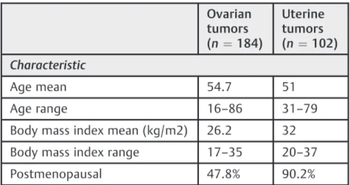
Among the ovarian tumors, misdiagnosis occurred in 2 cases (1.1%), corresponding to a borderline tumor (serous type) and a clear cell intracystic adenocarcinoma, which were underdiagnosed as benign mucinous proliferation and borderline serous tumor, respectively, on the IFS. Table 1 Characteristics of the patients
Ovarian tumors (n¼184)
Uterine tumors (n¼102)
Characteristic
Age mean 54.7 51
Age range 16–86 31–79
Body mass index mean (kg/m2) 26.2 32
Body mass index range 17–35 20–37
Postmenopausal 47.8% 90.2%
In the 12 cases in which the IFS conclusions were deferred, thefinal diagnoses were: borderline mucinous tumor in 6 (5 serous and 1 mucinous type) cases, serous cystadenoma in 3 cases, mucinous cystadenoma in 1 case, teratoma in 1 case, and bilateral serous adenocarcinoma in 1 case (►Table 2).
The sensitivity and specificity for benign, borderline, and malignant tumors were 100%, 66.7%, 96.9%, and 97.1%, 99.4%, 100%, respectively. The PPV and NPV for benign, borderline, malignant tumors were 99.3%, 66.7%, 100%, and 100%, 99.4% and 100%, respectively (►Table 3).
Uterine Tumors
A total of 102 patients were included. The mean age was 51.0 years old (range 31–79 years old). In the study group, 92 (90.2%) women were postmenopausal and 59 (64.1%) pre-sented with postmenopausal bleeding. Ten patients were premenopausal (9.8%) and 6 of them (60%) had complaints of heavy and/or irregular menstrual bleeding.
As shown in ►Table 2, there were 64 patients with a preoperative diagnosis of endometrial carcinoma, 31 patients with CAH, 1 patient with complex hyperplasia without atypia, and 6 patients with suspected carcinoma at hysteroscopy with inconclusive biopsy. All of the patients were evaluated initially by IFS, and then by a definitive PS to determine the degree of concordance between IFS and PS.
A total of 26 patients underwent lymphadenectomy in the same operative process (18 bilateral pelvic and para-aortic, 6 bilateral pelvic and 2 unilateral pelvic). Infive cases, there was metastatic disease in the pelvic lymph nodes, and in
three cases both in the pelvic and para-aortic lymph nodes. One patient presented with metastatic disease in the peri-ganglionar adipose tissue. Infive cases, lymphadenectomy was not performed due to technical difficulties, such as lack of access, obesity or other significant comorbidities.
Concerning the tumor staging according to the Internation-al Federation of Gynecology and Obstetrics (FIGO, in the French acronym) classification system, 47 patients (58.7%) were included in stage IA, 19 patients (23.75%) in IB, 6 patients (7.5%) in IIA, 2 patients (2.5%) in IIIA and 6 patients (7.5%) in IIIC. From the study group, 75 (93.75%) women had type I and the remaining patients had type II endometrial carcinomas, including 2 with mixed adenocarcinoma (clear cell and se-rous); 1 with carcinosarcoma with cervical invasion; 1 with a mixed adenocarcinoma with neuroendocrine elements and 1 with a large cells neuroendocrine carcinoma (►Table 4). The Accuracy of the Diagnosis of Endometrial Carcinoma was 96.25% (77/80).
The IFS correctly diagnosed the histologic type in 75 of 80 patients (93.75%). The depth of myometrial invasion was accurately diagnosed in 94.8% of the patients (73/77).
In thefinal PS, 75/80 tumors remained endometrioid adeno-carcinomas, whereas 5 that were originally diagnosed as endometrioid adenocarcinoma in the IFS were changed to mixed histology in the PS. One was read as a grade 3 carcino-sarcoma with cervical invasion, 1 as a grade 3 mixed papillary Table 2 Comparison between intraoperative frozen section
andfinal histological diagnoses of ovarian masses
Frozen Diagnosis Final histological diagnosis
Malignant Borderline Benign
Malignant 31 0 0
Borderline 1 2 0
Benign 0 1 137
Total 32 3 137
Table 3 Sensitivity, specificity, positive predictive value and negative predictive value of the intraoperative frozen section for ovarian neoplasms
Benign Borderline Malignant
Sensitivity 100% 66.7% 96.9%
Specificity 97.1% 99.4% 100%
Positive predictive value
99.3% 66.7% 100%
Negative predictive value
100% 99.4% 100%
Six patients (3.3%) had bilateral disease, and 21 patients (11.4%) had tumor spread beyond the ovaries.
All the patients with malignant intraoperative frozen section diagnosis underwent radical surgery, except four cases in which the tumor was unresectable.
Table 4 Correlation results between intraoperative frozen section andfinal histological diagnoses in endometrial tumors
Number Percentage
Preoperative diagnosis
Cancer 64 62.7
Complex atypical hyperplasia
31 30.4
Complex hyperplasia without atypia
1 1.0
Suspected carcinoma 6 5.9
IFS
Cancer 77 75.5
▪Inner half 50
▪Outer half 27
Without invasion 22 21.6
Deferred 3 2.9
Number Percentage
Postoperative diagnosis (PS)
Cancer 80 78.4
▪Inner half 50
▪Outer half 30
Complex atypical hyperplasia
11 10.8
Benign 11 10.8
serous adenocarcinoma, 1 as a grade 3 mixed adenocarcinoma with neuroendocrine elements, 1 as large cells neuroendocrine tumor carcinoma, and 1 as a grade 3 mixed adenocarcinoma (clear cells and serous). Our correlation rate between IFS and PS for histological subtype was 93.75% (75/80). It is important to note that the intraoperative management was not affected in any of thefive cases mentioned above (►Table 5).
Complex Atypical Hyperplasia
A total of 31 patients had a preoperative diagnosis of CAH. In the IFS study, 11 cases were read as malignant, 17 as negative for malignant lesions and 3 with deferred diagnosis. In the postoperative PS, 11 cases were read as CAH, 7 cases were read as no residual disease, and 13 as endometrial cancer. It is important to note that 2 cases thought to be non-cancerous in the IFS were later determined to be cancerous in thefinal PS. These two cases had received a diagnosis of“no residual disease”in the IFS diagnosis. They were non-invasive grade 1 cancers, in which the discrepancy was not relevant to the surgical management, and they were staged according to the FIGO classification system as IA.
Discussion
Intraoperative Frozen Section in Ovarian Tumors Ovarian cancer is the third more common malignant tumor, and its incidence has increased, especially in younger women.11 The clinical diagnosis of ovarian malignancy is challenging due the difficulty in obtaining a histological diagnosis before the definitive treatment.12
The optimal surgical management of ovarian tumors depends very much on their correct categorization as benign, borderline or malignant. The need for an additional surgical procedure may arise from an incomplete preoperative eval-uation of a complex adnexal mass, followed by an inadequate surgery. It is to avoid this unwanted sequence that the IFS represents a potentially powerful tool for the gynecologic oncology surgeons in the right setting.
The IFS analysis of ovarian masses allows gynecological oncologists to perform the optimal surgery to a given patient, therefore preventing the unnecessary morbidity of excessive surgical staging in benign cases and the need for restaging procedures in early-stage malignant tumors.12The IFS anal-ysis is only valuable if it may alter the procedure that the surgeon performs at the time of the operation, which is the case of ovarian cancer surgery.
The use of IFS offers a very good diagnostic accuracy in distinguishing women with malignant and benign ovarian tumors. In contrast, for borderline ovarian tumors, IFS results in more diagnostic discrepancies.
In a study of 274 patients, the sensitivity and specificity of IFS for benign, borderline and malignant tumors were 97% and 81%, 62% and 96% and 88% and 99%, respectively. The histologic type (mucinous), tumor size (<10 cm), the borderline com-ponent (<10%) and the pathologist experience predicted the misdiagnosis of borderline tumors.13In another study, the IFS diagnosis agreed with the PS diagnosis in 94% of all cases (98.5% for malignant tumors, 94% for benign tumors, and 78.6% for borderline tumors). The sensitivity and specificity values for malignant tumors were 93 and 99%; for borderline tumors, 61 and 99%; and for benign tumors, 98 and 93%, respectively.14 In a majority of studies, the sensitivity, specificity and predictive values of IFS diagnoses for benign and malignant tumors were found to be relatively high.5–7,15,16On the other hand, the sensitivity of IFS for borderline ovarian tumors was 60% in previous reports.14,17
In our experience, the sensitivity and specificity for benign, borderline, and malignant tumors were 100%, 66.7%, 96.9%, and 97.1%, 99.4%, 100%, respectively.
Out of the 184 IFSs reported in the present study, 98.9% of the women were submitted to the correct operative proce-dure at the initial surgical operation, only 0.54% of the women were under-staged at thefirst surgery, and 0.54% of the women were over-staged based on the IFS report.
The present study shows that IFS can contribute signifi -cantly to determine the malignant or benign nature of epithelial ovarian tumors, whereas for borderline tumors, its accuracy appears to be more dependent on the experience of the pathologist and on the characteristics of the tumor.
The main limitation of the IFS is the difficulty to obtain an accurate diagnosis of borderline ovarian tumors, mainly of the mucinous type.
Various reasons have been proposed for the relative inaccuracy of IFS in the diagnosis of borderline tumors. In a large borderline tumor, there may be only occasional foci of atypia amounting to the borderline category. On the other hand, severe atypia and/or invasion may be focal, but amounting to frank malignancy in thefinal reporting. Ovari-an mucinous borderline tumors may contain benign, border-line and malignant areas in the same tumor. Therefore, the final reporting may require a large number of sections to be processed, an option not usually available during IFS, as it is very labor-intensive and time-consuming. It has also been suggested that it may be more difficult to diagnose border-line mucinous tumors compared with borderborder-line serous tumors because of their larger average size.18
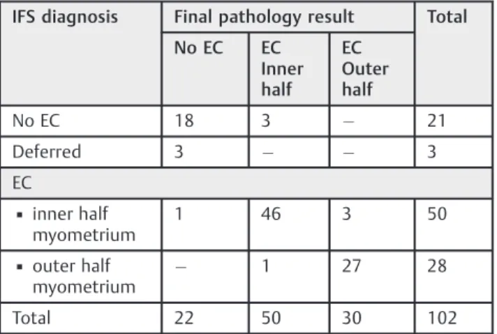
Table 5 Comparison between intraoperative frozen section andfinal histological diagnoses in terms of the presence or absence of endometrial cancer
IFS diagnosis Final pathology result Total
No EC EC Inner half
EC Outer half
No EC 18 3 21
Deferred 3 3
EC
▪inner half myometrium
1 46 3 50
▪outer half myometrium
1 27 28
Total 22 50 30 102
In a univariate analysis, underdiagnosis was shown to be more likely in non-serous epithelial tumors.15Another study showed a 9% inaccuracy rate in serous tumors compared with 36.6% in mucinous tumors.19Most false negatives occur in mucinous neoplasms and borderline tumors of various types.12
In summary, IFS can be of clinical use and the surgeons may feel confident enough to base appropriate surgical action upon a given result in experienced centers. Proximity between surgeons and pathologists seems advisable.
Intraoperative Frozen Section in Uterine Tumors Endometrial cancer is the most common malignant tumor of the genital tract worldwide.7
In endometrial carcinoma, the incidence of pelvic/para-aortic lymph node metastasis is related to the grade of the tumor, the depth of myometrial invasion and the presence of cervical involvement. These factors determine the type of initial surgery and the extent of the surgical staging.20The IFS analysis has been used for this purpose to identify patients requiring pelvic/para-aortic lymphadenectomy.
The keyfinding from the study was that, in experienced hands, the IFS analysis is accurate in identifying the subgroup of patients with high-risk EC who will benefit from full surgical staging at the time of their primary surgery.
In the present study, IFS for both histologic grade and depth of myometrial invasion correlated strongly with the final pathology analysis, supporting the use of IFS as a means to guide intraoperative decisions regarding lymphadenectomy.
The depth of myometrial invasion was accurately diagnosed in 94.8% (73/77) of the cases. The inaccuracy resulted in sub-optimal intraoperative surgical care in only 3.9% (3/77) of the patients, with 2.6% and 1.3% being under and over-treated, respectively, at the time of the primary surgery.
The correlation between IFS and paraffin histology in patients with endometrial carcinoma was 96.25% (77/80). However, the three cases not detected in IFS were not surgically undertreated in the end.
Lymphadenectomy was performed in 32.5% (26/80) of the patients with malignant disease. In 5 cases (6.25%), lymphadenectomy was not performed due to technical diffi -culties, while 61.25% (49/80) were considered low-risk and were spared of being submitted to lymphadenectomy. Two patients (2.5%) were incorrectly diagnosed as low-risk and were not submitted to lymphadenectomy.
There is controversy about the use of IFS in the evaluation of endometrial tumors. In some studies, IFS accurately identified 90% of the patients requiring pelvic/para-aortic lymphadenectomy. Histologic grade on IFS correlated with thefinal diagnosis in 91.4 to 94% of the cases.6,16The depth of myometrial invasion was accurately reported in 80 to 95% of the cases.2,5,7,16However, other studies showed that IFS for histological grade and depth of myometrial invasion in EC correlates poorly (58%) with thefinal pathology analysis.7In some studies, the evaluation of the depth of myometrial invasion with IFS has a sensitivity and a specificity of 74 and 95%, respectively, and this is not significantly higher than the radiological assessment.21,22
It is important to say that, in IFS, we cannot expect a high accuracy concerning the histopathological classification of the tumor and its grade because both depend on:
1. Sampling criteria (for example, is it necessary to count the percentage of solid areas in microscopy); 2. evaluation with complementary techniques (for example, diverse his-tological types, mixed tumors, heterologous elements, etc.); 3. we can rarely make a diagnosis of a neuroendocrine tumor in the IFS; it may be suspected in extreme cases of very good differentiation or extremely small differentiation, but only with paraffin processing and immunophenotype study can we make thefinal diagnosis.
Criteria 1 and 2 are not subject to definitive observation in IFS; they imply total tumor inclusion. Neither is it allowed to freeze tissue more than necessary, because freezing alters the processing and complementary immune techniques, such as irreversibly preventing a correct histopathological diagnosis.
There are several reasons for inaccuracies related to all IFS samplings, including inadequate sampling and potential artifacts. If the tumor is macroscopically confined to the endometrium, it is difficult to reveal myometrial invasion in IFS, even with multiple cuts, leading to a sampling error.
For the group of patients with a preoperative diagnosis of CAH, endometrial carcinoma was diagnosed at the IFS study in 35.5% (11/31) of the cases, and all of them were confirmed in the final PS. However, the IFS failed to identify 2 cases of endome-trial carcinoma. According to several studies, a considerable number of patients with EC can be missed on IFS. In one study, the diagnosis of EC was missed in 7/20 patients with perioper-ative CAH.23In another study, the diagnosis was missed in 14/ 125 patients.24It is important to reinforce that in the present study, as well as in other reports, these cases were early stage ECs without myometrial invasion, which can provide an expla-nation for the discrepancies in these cases, findings which, again, were not relevant to the surgical management.
Conclusion
The limitations of the present study are inherent to its retro-spective nature, as well as to the relatively limited sample. Regular audits, including specific analysis of cases in which IFS and PS are different, should be conducted by both surgeons and pathologists as part of a quality assurance process for the intraoperative management of patients with suspected or proven ovarian or uterine cancer.
Conflicts of Interest
The authors have no conflicts of interest to declare.
References
1 Baker P, Oliva E. A practical approach to intraoperative consulta-tion in gynecological pathology. Int J Gynecol Pathol 2008;27(03): 353–365 Doi: 10.1097/PGP.0b013e31815c24fe
2 Quinlivan JA, Petersen RW, Nicklin JL. Accuracy of frozen section for the operative management of endometrial cancer. BJOG 2001; 108(08):798–803 Doi: 10.1016/S0306-5456(00)00196-0 3 Bige O, Demir A, Saygili U, Gode F, Uslu T, Koyuncuoglu M. Frozen
section diagnoses of 578 ovarian tumors made by pathologists with and without expertise on gynecologic pathology. Gynecol Oncol 2011;123(01):43–46 Doi: 10.1016/j.ygyno.2011.06.030 4 Malviya VK, Deppe G, Malone JM Jr, Sundareson AS, Lawrence
WD. Reliability of frozen section examination in identifying poor prognostic indicators in stage I endometrial adenocarcinoma. Gynecol Oncol 1989;34(03):299–304
5 Kucera E, Kainz C, Reinthaller A, et al. Accuracy of intraoperative frozen-section diagnosis in stage I endometrial adenocarcinoma. Gynecol Obstet Invest 2000;49(01):62–66 Doi: 10.1159/000010215 6 Shim JU, Rose PG, Reale FR, Soto H, Tak WK, Hunter RE. Accuracy of frozen-section diagnosis at surgery in clinical stage I and II endo-metrial carcinoma. Am J Obstet Gynecol 1992;166(05):1335–1338 Doi: 10.1016/0002-9378(92)91600-F
7 Case AS, Rocconi RP, Straughn JM Jr, et al. A prospective blinded evaluation of the accuracy of frozen section for the surgical management of endometrial cancer. Obstet Gynecol 2006;108 (06):1375–1379 Doi: 10.1097/01.AOG.0000245444.14015.00 8 Frumovitz M, Slomovitz BM, Singh DK, et al. Frozen section analyses
as predictors of lymphatic spread in patients with early-stage uterine cancer. J Am Coll Surg 2004;199(03):388–393 10.1016/j. jamcollsurg.2004.05.258
9 Creasman WT, Mutch DE, Herzog TJ. ASTEC lymphadenectomy and radiation therapy studies: are conclusions valid? Gynecol Oncol 2010;116(03):293–294 Doi: 10.1016/j.ygyno.2009.10.065 10 Fanning J, Tsukada Y, Piver MS. Intraoperative frozen section diagnosis of depth of myometrial invasion in endometrial adeno-carcinoma. Gynecol Oncol 1990;37(01):47–50 Doi: 10.1016/0090-8258(90)90306-6
11 Hashmi AA, Naz S, Edhi MM, et al. Accuracy of intraoperative frozen section for the evaluation of ovarian neoplasms: an institutional experience. World J Surg Oncol 2016;14:91 Doi: 10.1186/s12957-016-0849-x
12 Cross PA, Naik R, Patel A, et al. Intra-operative frozen section analysis for suspected early-stage ovarian cancer: 11 years of Gateshead Cancer Centre experience. BJOG 2012;119(02): 194–201 Doi: 10.1111/j.1471-0528.2011.03129.x
13 Brun JL, Cortez A, Rouzier R, et al. Factors influencing the use and accuracy of frozen section diagnosis of epithelial ovarian tumors. Am J Obstet Gynecol 2008;199(03):244.e1–244.e7 Doi: 10.1016/j. ajog.2008.04.002
14 Pinto PB, Andrade LA, Derchain SF. Accuracy of intraoperative frozen section diagnosis of ovarian tumors. Gynecol Oncol 2001; 81(02):230–232 Doi: 10.1006/gyno.2001.6133
15 Houck K, Nikrui N, Duska L, et al. Borderline tumors of the ovary: correlation of frozen and permanent histopathologic diagnosis. Obstet Gynecol 2000;95(6 Pt 1):839–843
16 Zorlu CG, Kuscu E, Ergun Y, Aydogdu T, Cobanoglu O, Erdas O. Intraoperative evaluation of prognostic factors in stage I endome-trial cancer by frozen section: how reliable? Acta Obstet Gynecol Scand 1993;72(05):382–385 Doi: 10.3109/00016349309021118 17 Medeiros LR, Rosa DD, Edelweiss MI, et al. Accuracy of
frozen-section analysis in the diagnosis of ovarian tumors: a systematic quantitative review. Int J Gynecol Cancer 2005;15(02):192–202 Doi: 10.1111/j.1525-1438.2005.15203.x
18 Bhurgri Y, Shaheen Y, Kayani N, et al. Incidence, trends and morphology of ovarian cancer in Karachi (1995-2002). Asian Pac J Cancer Prev 2011;12(06):1567–1571
19 Kayikçioglu F, Pata O, Cengiz S, et al. Accuracy of frozen section diagnosis in borderline ovarian malignancy. Gynecol Obstet Invest 2000;49(03):187–189 Doi: 10.1159/000010244
20 Kumar S, Bandyopadhyay S, Semaan A, et al. The role of frozen section in surgical staging of low risk endometrial cancer. PLoS One 2011;6(09):e21912 Doi: 10.1371/journal.pone.0021912 21 Altintas A, Cosar E, Vardar MA, Demir C, Tuncer I. Intraoperative
assessment of depth of myometrial invasion in endometrial carcinoma. Eur J Gynaecol Oncol 1999;20(04):329–331 22 Fishman A, Altaras M, Bernheim J, Cohen I, Beyth Y, Tepper R. The
value of transvaginal sonography in the preoperative assessment of myometrial invasion in high and low grade endometrial cancer and in comparison to frozen section in grade 1 disease. Eur J Gynaecol Oncol 2000;21(02):128–130
23 Stephan JM, Hansen J, Samuelson M, et al. Intra-operative frozen section results reliably predictfinal pathology in endometrial cancer. Gynecol Oncol 2014;133(03):499–505 Doi: 10.1016/j. ygyno.2014.03.569