Abstract published online: February 12, 2010 Full paper published online: May 30, 2010
Original paper. ISSN 1678-9199.
Antiophidian properties of plant extracts against Lachesis muta venom
De Paula RC (1), Sanchez EF (2), Costa TR (3), Martins CHG (4), Pereira PS (5), Lourenço MV (5), Soares AM (3), Fuly AL (1)
(1) Department of Cell Biology, Institute of Biology, Fluminense Federal University, Niterói, Rio de Janeiro State, Brazil; (2) Center for Research and Development, Ezequiel Dias Foundation, Belo Horizonte, Minas Gerais State, Brazil; (3) Department of Clinical, Toxicological and Bromatological Analysis, School of Pharmaceutical Sciences, University of São Paulo, Ribeirão Preto, São Paulo State, Brazil; (4) Research Group on Natural Products, Laboratory of Applied Microbiology, University of Franca, Franca, São Paulo State, Brazil; (5) Biotechnology Unit, University of Ribeirão Preto, Ribeirão Preto, São Paulo State, Brazil.
ABSTRACT: Snakebites comprise a serious health problem in several countries due to their global incidence, which exceeds 2.5 million per year, and the elevated number of victim fatalities. To counteract envenomations, antivenoms have been used regularly for more than a century. Apart from side effects including anaphylactic reactions, antivenoms are not able to efficiently neutralize local tissue damage, which contributes to increasing the severity and morbidity observed in patients. This fact, in turn, may be responsible for economic hardship, particularly in rural populations of developing countries. In the present work, we evaluated the antiophidian properties of 12 Brazilian plant extracts against the hemolytic, coagulant, hemorrhagic and proteolytic effects of Lachesis muta venom. Taken together, our data revealed that
most of these aqueous products were capable of inhibiting those activities at different levels, except for Sapindus saponaria extract. In contrast, Stryphnodendron barbatiman extract completely neutralized all the analyzed biological activities. Thus,
we may conclude that Brazilian flora may also be useful against L. muta accidents.
KEY WORDS: snake venom, Lachesis muta, plant extract, antivenom, biological
activities.
CONFLICTS OF INTEREST: There is no conflict.
FINANCIAL SOURCE: FAPERJ, FAPESP, CNPq, CAPES and IFS (grant F/4571-1).
CORRESPONDENCE TO:
INTRODUCTION
Snake venoms are a complex mixture of components that affect a wide range of tissues and organs. We may consider the main active ones to be phospholipases A2
enzymes, metallo- and serine proteases, phosphodiesterases, myotoxins, lectins, L-amino acid oxidases, disintegrins and neurotoxins, which are responsible for hemorrhage, necrosis, edema, blood clotting, hypotension and neurotoxicity effects in victims (1). In Brazil, Bothrops, Crotalus, Lachesis and Micrurus genus are
responsible for, respectively, 90, 7, 2 and 0.5% of the accidents. But, although the accidents caused by Lachesis are rare, the incidence of death (also denominated
lethality or fatality index) should be considered (2).
Envenomation by snakes is often treated by parenteral administration of antiophidian serum (antivenoms) obtained from hyperimmunized equine serum. This serum therapy efficiently neutralizes the systemic toxic effects, thus preventing victim fatalities. However, antivenoms present some disadvantages. They can induce adverse reactions ranging from mild symptoms to serious ones and they do not neutralize the local tissue damage (3). Thus, it is important to search for new venom inhibitors, whether synthetic or from a natural source, to complement the traditional serum therapy. Hence, the aim of this work was to evaluate the antiophidian properties of twelve aqueous plant extracts from Brazilian flora against biological effects caused by L. muta snake venom.
MATERIALS AND METHODS Venom and Animals
Lachesis muta snake venom was kindly supplied by the Ezequiel Dias Foundation,
Belo Horizonte, Minas Gerais state, Brazil, vacuum dried and stored at –20oC until experiments. Male Swiss Wistar mice (18-20 g) were obtained from the Center of Laboratory Animals (NAL) of the Fluminense Federal University (UFF).
Plant Extracts
The number of columns, which appears in parenthesis, represents the plant extracts in the graphs displayed in the Results section. Leaves, stems and roots of Eclipta
alba (column 1), Mandevilla velutina (column 2), Mikania glomerata (column 3),
Jatropha ellyptica (leaves, column 4), Jatropha ellyptica (stems, column 5), Miconia
Stryphnodendron barbatiman (column 9), Tibouchina stenocarpa (column 10),
Casearia sylvestris (column 11) and Sapindus saponaria (column 12) were collected
from the Biotechnology Unit of the University of Ribeirão Preto (UNAERP), São Paulo state, Brazil. A voucher specimen of each plant has been deposited at UNAERP and authenticated by Professor Hermógenes de Freitas Leitão Filho of the Institute of Biology, State University of Campinas (UNICAMP). Plant extracts were prepared according to Maiorano et al. (4) and further dissolved in physiological saline
solution yielding aqueous extracts.
Inhibition of the Venom
For inhibition experiments, a fixed amount of L. muta snake venom was mixed with
varying amounts of plant extracts to obtain two venom:plant ratios (w/w), 1:10 and 1:20. Then, the mixture was incubated for 30 minutes at room temperature (r.t.), after which the biological assays were performed.
Phospholipase A2 (PLA2) Activity
PLA2 activity was determined by the indirect hemolytic test using human erythrocytes
and hen’s egg yolk emulsion as substrate (5). One minimum indirect hemolytic dose (MIHD) was defined as the amount of L. muta venom (µg/mL) able to produce
70-80% hemolysis. To verify the inhibitory action of plant extracts, they were preincubated with one MIHD and the hemolytic assay was performed. The maximal effect (100% hemolysis) was obtained after lysing erythrocytes with distilled water to constitute the control.
Clotting Activity
A normal pool of human plasma from healthy volunteers was mixed with L. muta
crude venom and clotting experiments were performed in an Organon Teknika BE Coagulator 1® (Germany) (6). The amount of venom (µg/mL) that clots plasma in 60
Hemorrhagic Activity
Hemorrhagic lesions produced by L. muta venom were quantified by intradermally
injecting samples into abdominal skin of mice two hours before the animals were euthanized according to the UFF Institutional Committee for Ethics in Animal Experimentation (7). Then, abdominal skin was removed, stretched and the site of injection analyzed. One minimum hemorrhagic dose (MHD) was defined as the amount of venom (µg/g) that produced a hemorrhagic halo of 10 mm. Hemorrhagic activity was expressed by the means (in mm) of the hemorrhagic halos induced by L.
muta venom in the absence or presence of plant extracts. Negative control
experiments were performed by injecting saline, in parallel.
Proteolytic Activity
Proteolytic activity was determined according to the method of Garcia et al. (8) using
azocasein as substrate (0.2% w/v, in 20 M Tris-HCl, 8 mM CaCl2, pH 8.8) in the
presence or absence of plant extracts. One minimum proteolytic dose (MPD) was defined as the amount of venom (µg/mL) able to produce a variation of 0.2 at A405
nm.
Ethics Committee
The experiments were approved by the UFF Institutional Committee for Ethics in Animal Experimentation and are in accordance with the guidelines of the Brazilian College for Animal Experimentation (COBEA).
Statistical Analysis
Results are expressed as means ± SD obtained with the indicated number of animals or experiments performed. The statistical significance of differences among experimental groups was evaluated using the ANOVA test and p values less than
0.05 were considered statistically significant.
RESULTS
A previous study found that L. muta venom presented phospholipase A2 (indirect
Antihemolytic and Anticlotting Effects
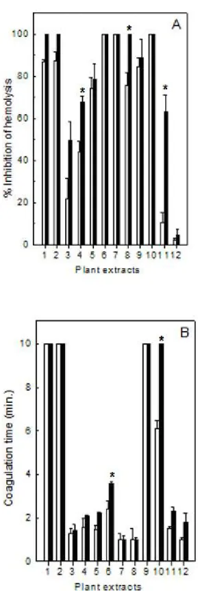
As shown in Figure 1, plant extracts were able to inhibit the indirect hemolytic (Panel A) and clotting (Panel B) activities caused by L. muta venom (12 µg/mL and 15
µg/mL), but with different potencies. The venom concentrations used above represent the MIHD and MCD, respectively. However, the extracts of E. alba (column
1), M. velutina (column 2), M. fallax (column 6), M. albicans (column 7), S.
barbatiman (column 9) and T. stenocarpa (column10) inhibited the indirect hemolytic
activity (100%), in contrast to S. saponaria (column 12) which did not neutralize this
effect. Meanwhile, E. alba (column 1), M. velutina (column 2), S. barbatiman (column
9) and T. stenocarpa (column 10) fully inhibited coagulation of L. muta (100%), since
no clotting was observed throughout ten minutes of observation. However, M.
glomerata (column 3), J. ellyptica (column 4), J. ellyptica (column 5), M. albicans
(column 7), M. sellowiana (column 8), S. barbatiman (column 9), T. stenocarpa
(column 10), C. sylvestris (column 11) and again S. saponaria (column 12) did not
counteract the clotting effect of L. muta venom. The M. fallax extract (column 6) was
Figure 1. Inhibition of hemolytic (Panel A) or blood clotting (Panel B) activities of L. muta crude venom by plant extracts at 1:10 (white column) or 1:20 (black column)
venom:plant ratio (w/w). Data are expressed as means ± SD of three individual experiments (n = 3).
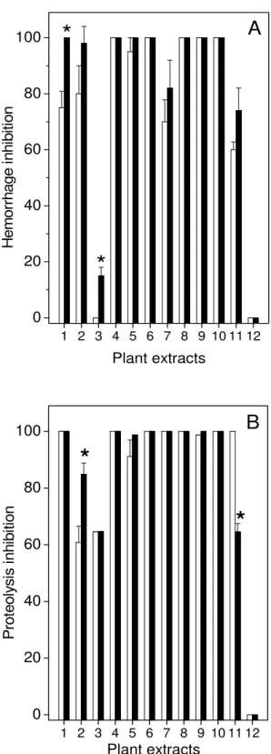
Antihemorrhagic and Antiproteolytic Effects
The effects of aqueous extracts on hemorrhagic (Figure 2A) and proteolytic (Figure 2B) activities induced by L. muta venom were also analyzed. As observed in Figure
2A, the extracts of M. glomerata (column 3) and S. saponaria (column 12) did not
counteract L. muta venom-induced hemorrhage (12.5 µg/g). In contrast, all of the
other plant extracts (J. ellyptica, column 4; J. ellyptica, column 5; M. fallax, column 6;
M. sellowiana,column 8; S. barbatiman, column 9 and T. stenocarpa, column 10) did
neutralize such lesions induced by L. muta venom and produced inhibition of around
100 % (Figure 2A).
It is worth noting that S. barbatiman extract (column 9) produced 100% inhibition of a
three-fold MHD (30 µg/g) of L. muta (data not shown). In parallel, plant extracts also
inhibited one MPD of L. muta venom (10 µg/mL) with different potencies (Figure 2B).
As shown, the extracts of M. velutina (column 2) and M. glomerata (column 3)
presented the lowest inhibitory effect (60 and 65 % for 1:10 venom:plant ratio; 85% and 65% for 1:20) while S. sapindus (column 12) was not capable of inhibiting this
phenomenon. The other plant extracts – J. ellyptica (column 4), J. ellyptica (column
5), M. fallax (column 6), M. albicans (column 7), M. sellowiana (column 8), S. barbatiman (column 9) and T. stenocarpa (column 10) – inhibited 100% of L. muta
1 2 3 4 5 6 7 8 9 10 11 12 0 20 40 60 80 100
*
**
A
H em or rh age i nhi bi tion Plant extracts1 2 3 4 5 6 7 8 9 10 11 12 0 20 40 60 80 100
*
*
B
Pr ot eol ys is inh ibi tion Plant extractsFigure 2. Inhibition of L. muta venom-induced hemorrhagic (Panel A) or proteolytic
(Panel B) activities by plant extracts at 1:10 (white column) or 1:20 (black column) venom:plant ratio (w/w). Data are expressed as means ± SD of four individual experiments (n = 4).
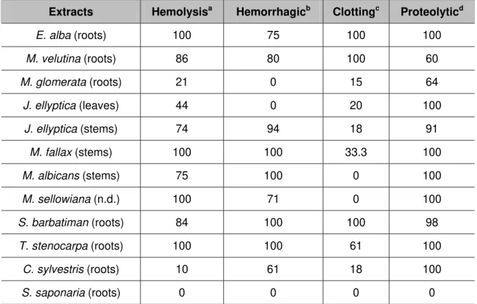
Inhibitory Profile of Plant Extracts
Furthermore, we compared the inhibitory profile of plant extracts according to their indirect hemolytic (hemolysis), clotting, hemorrhagic and proteolytic biological activities (Table 1). As shown in Table 1, none of these activities was affected by S.
saponaria. However, M. glomerata and J. ellyptica (stems) extracts were not able to
completely inhibit any activity; but M. velutina, J.ellyptica (leaves) or C. sylvestris
extracts fully inhibited at least one activity (clotting, proteolytic and proteolytic, respectively). On the other hand, the extracts of M. fallax (hemolysis and
hemorrhagic), M. albicans (hemorrhagic and proteolytic), M. sellowiana (hemolysis
and proteolytic), S. barbatiman (hemolysis, hemorrhagic and proteolytic) and T.
stenocarpa (hemolysis, hemorrhagic and proteolytic), inhibited 100% of at least two
of those activities. Also, it is worth highlighting that only the S. barbatiman extract
fully abolished all of the activities assessed in this work (Table 1).
Table 1. Inhibitory percentage of plant extracts against L. muta snake venom’s
biological activities
Extracts Hemolysisa Hemorrhagicb Clottingc Proteolyticd
E. alba (roots) 100 75 100 100
M. velutina (roots) 86 80 100 60
M. glomerata (roots) 21 0 15 64
J. ellyptica (leaves) 44 0 20 100
J. ellyptica (stems) 74 94 18 91
M. fallax (stems) 100 100 33.3 100
M. albicans (stems) 75 100 0 100
M. sellowiana (n.d.) 100 71 0 100
S. barbatiman (roots) 84 100 100 98
T. stenocarpa (roots) 100 100 61 100
C. sylvestris (roots) 10 61 18 100
S. saponaria (roots) 0 0 0 0
Plant extracts were preincubated with L. muta crude venom at 1:10 ratio (venom:plant, w/w) and then assayed for these activities. The values above are expressed as inhibition percentage (%) of the venom amount able to produce: 70-80% hemolysis (MIHD)a; hemorrhagic halo of 10 mm (MHD)b; clot
plasma in 60 seconds (MCD)c; produce a variation of 0.2 nm at 420 nm (MPD)d.
DISCUSSION
Given the high incidence of accidents caused by Bothrops and Crotalus species,
data from the literature have focused much attention on them (10). But, although the accidents caused by L. muta snake venom are rare, they also represent a medical
problem in Brazil, especially because of the high lethality index (1.0%) when compared to approximately 0.3% for the Bothrops genus (2).
Envenomation by L. muta promotes a wide range of symptoms including nociception,
edema and muscle necrosis, as well as neurotoxic, hemorrhagic, hemolytic and blood clotting disorders. These effects are related to the presence of phospholipase A2 enzymes, serino- and metalloproteases, L-amino acid oxidases, nucleotidases
and others. The pathophysiological alterations may cause victims to die. To counteract those systemic and local effects of venoms, an antiserum is used as treatment.
According to the Brazilian Health Ministry and World Health Organization (11) guidelines, the antiophidic serum is produced by the following three public institutes in Brazil: the Butantan Institute in São Paulo city, São Paulo state; Ezequiel Dias Foundation in Belo Horizonte city, Minas Gerais state; and the Vital Brasil Institute, in Niterói city, Rio de Janeiro state; and then distributed free of charge to hospitals. As far as we know, this treatment is not efficient enough to neutralize local effects, thus contributing to high morbidity indexes observed in Brazil (2, 12-15).
Indeed, antisera possess some disadvantages such as side effects in victims, poor availability in distant areas, delays in administration, need for refrigeration, high cost and low efficacy (3, 10). Furthermore, snake venoms have both inter- and intraspecific variation in composition that, in turn, may affect antivenom neutralization ability (16). Because of these factors, alternative treatments for snakebite are under investigation and plant extracts constitute a rich source of molecules with several pharmacological properties (4, 17, 18).
pterocarpans and terpenoids and crude extracts are able to neutralize toxic snake venom effects (17, 19-23).
In the present experiment we demonstrated for the first time the ability of different aqueous plant extracts to neutralize biological effects of L. muta snake venom, due
to the scarcity of data on the use of plants to neutralize its venom. All of the plant extracts tested in this work inhibited at least one biological activity, except for S.
saponaria that failed to neutralize any activity. The biological activities evaluated
herein are relevant to envenomation symptoms such as necrosis, hemolysis, hemorrhage, inflammation, edema and pain, which obviously may contribute to victim fatalities. Therefore, vegetal extracts appears as an alternative treatment against local damage to complement the serum therapy or may be useful as prototypes for designing new antiophidian molecules to improve the current treatment used for L.
muta bites.
ACKNOWLEDGMENTS
This work was supported by the State of Rio de Janeiro Research Foundation (FAPERJ), the State of São Paulo Research Foundation (FAPESP), the National Council for Scientific and Technological Development (CNPq), the Coordination for the Improvement of Higher Education Personnel (CAPES) and the International Foundation for Science (IFS grant F/4571-1).
REFERENCES
1. de Paula RC, Castro HC, Rodrigues CR, Melo PA, Fuly AL. Structural and pharmacological features of phospholipases A2 from snake venoms. Protein Pept
Lett. 2009;16(8):899-07.
2. Ministério da Saúde do Brasil. Fundação Nacional de Saúde. Manual de diagnóstico e tratamento de acidentes por animais peçonhentos. 2nd ed. Brasília: FUNASA; 2001. 120 p.
3. Gutiérrez JM, Fan HW, Silvera CL, Angulo Y. Stability, distribution and use of antivenoms for snakebite envenomation in Latin America: report of a workshop. Toxicon. 2009;53(6):625-30.
4. Maiorano VA, Marcussi S, Daher MA, Oliveira CZ, Couto LB, Gomes OA, et al. Antiophidian properties of the aqueous extract of Mikania glomerata. J
5. Fuly AL, Machado OL, Alves EW, Carlini CR. Mechanism of inhibitory action on platelet activation of a phospholipase A2 isolated from Lachesis muta (Bushmaster)
snake venom. Thromb Haemost. 1997;78(5):1372-80.
6. Quick AJ. Hemostasis and blood coagulation. Science. 1973;182(4107):87.
7. Melo PA, do Nascimento MC, Mors WB, Suarez-Kurtz G. Inhibition of the myotoxic and hemorrhagic activities of crotalid venoms by Eclipta prostrata (Asteraceae)
extracts and constituents.Toxicon. 1994;32(5):595-03.
8. Garcia ES, Guimarães JA, Prado JL. Purification and characterization of a sulfhydryl-dependent protease from Rhodnius prolixus midgut. Arch Biochem
Biophys. 1978;188(2):315-22.
9. Fuly AL, Francischetti IM, Zingali RB, Carlini CR. Partial purification and some physicochemical properties of phospholipases A2 from the venom of the bushmaster
snake (Lachesis muta). Braz J Med Biol Res. 1993;26(5):459-63.
10. Cruz LS, Vargas R, Lopes AA. Snakebite envenomation and death in the developing world. Ethn Dis. 2009;19(1 Suppl 1):42-6.
11. WHO. Normes relatives aux immunsèrums d'origine animale. WHO Sér Rapp Tech. 1969;413(1)47-61.
12. da Silva JO, Fernandes RS, Ticli FK, Oliveira CZ, Mazzi MV, Franco JJ, et al. Triterpenoid saponins, new metalloprotease snake venom inhibitors isolated from
Pentaclethra macroloba. Toxicon. 2007;50(2):283-91.
13. Gutiérrez JM, Rojas G, Lomonte B, Gene JA, Chaves F, Alvarado J, et al. Standardization of assays for testing the neutralizing ability of antivenoms. Toxicon. 1990;28(10):1127-29.
14. Zamunér SR, da Cruz-Hofling MA, Corrado AP, Hyslop S, Rodrigues-Simioni L. Comparison of the neurotoxic and myotoxic effects of Brazilian Bothrops venoms and
their neutralization by commercial antivenom. Toxicon. 2004;44(3):259-71.
15. Gutiérrez JM, Lomonte B. Local tissue damage induced by Bothrops snake. Mem
Inst But. 1989;51(1):211-23.
16. Queiroz GP, Pessoa LA, Portaro FC, Furtado M de F, Tambourgi DV. Interspecific variation in venom composition and toxicity of Brazilian snakes from
Bothrops genus. Toxicon. 2008;52(8):842-51.
18. Mors WB, Nascimento MC, Pereira BM, Pereira NA. Plant natural products active against snake bite: the molecular approach. Phytochemistry. 2000;55(6):627-42. 19. Ticli FK, Hage LIS, Cambraia RS, Pereira OS, Magro AJ, Fontes MRM, et al. Rosmarinic acid, a new snake venom phospholipase A2 inhibitor from Cordia
verbenacea (Boraginaceae): antiserum action potentiation and molecular interaction.
Toxicon. 2005;46(3):318-27.
20. Nishijima CM, Rodrigues CM, Silva MA, Lopes-Ferreira M, Vilegas W, Hiruma-Lima CA. Anti-hemorrhagic activity of four Brazilian vegetable species against
Bothrops jararaca venom.Molecules. 2009;14(3):1072-80.
21. Lindahl M, Tagesson C. Selective inhibition of group II phospholipase A2 by
quercetin. Inflammation. 1993;17(5):573-82.
22. Girish KS, Kemparaju K. Inhibition of Naja naja venom hyaluronidase by
plant-derived bioactive components and polysaccharides. Biochemistry (Mosc). 2005;70(8):948-52.