Department of Conservation and Restoration
COCHINEAL, A PRECIOUS SOURCE OF RED
Cochineal Dyes Characterization by High Performance Liquid Chromatography
with Diode Array Detection and Principal Component Analysis
By
Ana Filipa Albano Serrano
Presented at Faculdade de Ciências e Tecnologias, Universidade de Lisboa, to obtain the Master Degree in Conservation and Restoration of Textiles
Supervision:
Dr. Micaela Sousa (FCT-UNL)
Co-supervision:
Dr. Jessica Hallett (CHAM-FCSH), Maria Passos Leite (MCG)
Lisbon
Abstract
The identification of precise cochineal species used to dye historical textiles can provide important information about the provenance and date of these objects. The most widely used method to identify cochineal species in textiles involves quantification of specific minor compounds, after High-performance Liquid Chromatography with Diode Array Detection (HPLC/DAD) analysis. However, there are several factors which are not presently taken in account when characterizing cochineal species on historical textiles. Not only all the species of cochineal are not well studied, but also the current studies, based on a limited number of species, frequently face difficulties with the identification of these on historical textiles, especially due to the analysis conditions and the results treatment.
Therefore, a new approach on the study of cochineal species present in historical textiles was developed. Different parameters for the analysis conditions were undertaken to optimize the results for both insect species and textiles samples. Afterwards, with Principal Components Analysis (PCA), results from textiles samples exhibited a satisfactory correlation when compared with a cochineal reference database. Moreover, High-performance Liquid Chromatography with Diode Array Detector coupled with Mass Spectrometry (HPLC/DAD/MSn) analysis could offer accurate information on cochineal species and textiles samples. The characterization of six species of cochineal allowed, through PCA and HPLC/DAD/MSn analysis, the identification of unidentified cochineal insect samples and a group of Islamic and Italian historical dyed-cochineal textiles, dated from 15th to 17th centuries.
This identification contributes to connect the textiles’ history, and the trade and dyeing technologies on possible different species of cochineal. This fact regards especially textiles produced in the main textile centres, where, after the 16th century, the traded American cochineal was swiftly adopted, as many historical publications assert. Although this study identified American cochineal in a 17th-century Indian textile for the first time, the results for the other analyzed textiles did not reveal the presence of this species. In this way, the possibility of the prompt spread of the American specie in European and Asian textiles dyeing seems to be more complex than what is emphasized by present publications.
Communications and Publications
Ana Serrano, Micaela Sousa, Jessica Hallett, Maria Fernanda Passos Leite, “Cochineal, A Precious Source of Red” – accepted for oral communication in “Colours” conference, Victoria & Albert Museum, London, November 20, 2010.
Sumário
A identificação precisa de espécies de cochinilha usadas no tingimento de têxteis históricos pode fornecer informação importante para a proveniência e a datação dos mesmos. O método mais usado para a identificação dessas espécies nos têxteis caracteriza-se pela quantificação de compostos minoritários específicos, depois de análises por Cromatografia Líquida de Alta Eficiência por Vector de Díodos (HPLC/DAD). Contudo, existem determinados factores que não são tidos em conta quando as espécies de cochinilha são caracterizadas em têxteis históricos. Para além de nem todas as espécies de cochinilha estarem estudadas, estudos recentes, baseados num limitado número de espécies, encontram dificuldades na identificação das mesmas em têxteis, devido às condições de análise e ao tratamento dos resultados.
Assim, desenvolveu-se um novo método de estudo para caracterizar espécies de cochinilha em têxteis históricos. Diferentes parâmetros para as condições de análise foram realizados, de modo a optimizar os resultados para as amostras de insectos e de têxteis. Posteriormente, com Análises por Componente Principal (PCA), os resultados das amostras têxteis demonstraram uma correlação satisfatória quando comparados com a biblioteca de referência de espécies de cochinilha. Adicionalmente, análises por Cromatografia Líquida de Alta Eficiência por Vector de Díodos acoplado a Espectrometria de Massa (HPLC/DAD/MSn) permitiram obter informação mais precisa sobre as espécies de cochinilha e as amostras têxteis. A caracterização de seis espécies de cochinilha, através de análises por PCA e HPLC/DAD/MSn, permitiu identificar insectos de cochinilha de espécie desconhecida e um grupo de têxteis históricos islâmicos e italianos, datados de entre os sécs. XV e XVII.
Esta identificação contribuiu para interligar a história dos têxteis, o comércio e as tecnologias têxteis, com as diferentes espécies de cochinilha. Esta interligação é mais comum em têxteis produzidos nos principais centros têxteis, onde, após o séc. XVI, a cochinilha americana foi rapidamente adoptada, tal como as publicações históricas defendem. Apesar de, neste estudo, se ter identificado pela primeira vez, cochinilha americana num têxtil indiano do séc. XVII, os resultados para os outros têxteis não revelaram a presença desta espécie. Assim, a possibilidade da rápida assimilação da espécie americana nos tingimentos têxteis europeus e asiáticos parece ser mais complexa do que é enfatizado pelas recentes publicações.
Comunicações e Publicações
Ana Serrano, Micaela Sousa, Jessica Hallett, Maria Fernanda Passos Leite, “Cochineal, A Precious Source of Red” – aceite para comunicação oral na conferência “Colours”, Victoria & Albert Museum, Londres, a 20 de Novembro de 2010.
Subjects Index
Abstract 2
Sumário 3
Introduction
1. Cochineal, a Precious Source of Red 5
2. Previous studies on cochineal species identification with HPLC/DAD 6
3. Proposed study on cochineal characterization 7
Experimental
1. Materials and Solvents
1.1 Chemicals 8
1.2 Cochineal samples 8
1.3 Red silk fibres 8
2. Samples preparation
2.1 Insect dye-extraction 9
2.2 Dyed-fibre extraction methods 9
3. HPLC/DAD/UV 9
4. MS (Mass Spectrometry) 10
5. PCA (Principal Components Analysis) 10
Results and Discussion
1. Characterization of cochineal 11
2. Samples Preparation
2.1 Insect dye extracts 13
2.2 Dyed-fibre extraction methods 14
3. Insect samples
3.1 Reference cochineal specimens identified by entomologists 15
3.2 Unidentified cochineal insect species 17
4. Historical textile dye identification 20
Conclusion 25
Acknowledgements 26
References 27
Appendixes
Appendix 1: Cochineal, the dye and its textile history 30
Appendix 2: Textile catalogue 42
Appendix 3: Cochineal markers database 46
Appendix 4: Reference cochineal specimens identified by entomologists 47
Appendix 5: Unidentified cochineal insect species 50
Appendix 6: Historical textile samples 54
Appendix 7: Cochineal species database 58
Appendix 8:Non-cochineal samples database 59
Introduction
1. Cochineal, a Precious Source of Red1
Cochineal was one of the most precious natural red dyes, appreciated by Europeans and Asians alike. Cochineal, like kermes and lac, belongs to the group of coccid dyes, which have been appreciated for yielding brilliant and enduring crimson hues. These costly dyestuffs were applied almost exclusively to luxury textiles owing to the large quantities of insects necessary to make sufficient dye, as well as the special skills involved in gathering and preparing the insects and the complex dyeing process [1,2].
Cochineal species belong to two families from the Coccoidea superfamily, Margarodidae and Dactylopiidae [3,4]. Currently, Margarodidae family includes, among others, Porphyrophora genus, with 47 species, spread all over the Palearctic region [5]. On the other hand, Dactylopiidae family comprises Dactylopius genus, which has 10 species, original from America [6]. Information on dyeing with Porphyrophora species is rare, and other species apart from the well-documented P. polonica, P. hamelii, and the domesticated D. coccus, were probably also used for dyeing textiles in the past, especially in regions remote from the main centres of textile manufacturing and international trade routes [4].
From a taxonomical point of view, many insects from the same family are relatively similar to each other, with very slight differences, and so, accuracy should be taken when analyzing species with the same geographical origin. Especially because different species could be gathered in the past and named under the same designation, due to their high similarity, or even due to a dishonestly meaning, like mixture of high-quality D. coccus specie with other inferior species of wild Dactylopius [4,7]. Consequently, when identifying red cochineal-dyes in historical textiles, great care must be taken when affirming that a dye might belong to P.
polonica,P. hamelii or D. coccus species, since not all the species are studied.
Until 16th century, Porphyrophora insects, as well as kermes, were often used by the wealthiest Europeans and Asians to dye textiles. However, soon after the conquest of Mexico by the Spaniards, in 1521, the first shipments of Mexican cochineal (D. coccus) began arriving in Europe and spread from there to Asia[2,7]. It provided more vivid crimson colours, as well as the possibility of achieving a wider range of mixed hues, which were not possible with other coccid dyes. But the greatest advantage of this dyestuff was its high dye content, which had important economic implications [4]. As demand for it increased dramatically, American cochineal became a great source of income for the Spanish economy, and became, after silver, the most valuable item traded in the Hispanic empire during the 17th century [7,8].
The identification of cochineal in European and Asian textiles dating from immediately after the Spanish conquest of Mexico is commonly associated with the adoption of D. coccus [9, 10]. However, there were many different sources and species of cochineal available for preparing dyes in these regions which have not been well studied, and previous publications have had difficulty distinguishing them [4, 10]. Hence, in this study a diverse group of red European and
1
Islamic textiles produced after the documented arrival of Mexican cochineal are analysed, with the intention of examining the penetration of this insect in dyeing practices.
In Ottoman Turkey, Safavid Iran and Mughal India, red coccid dyes were widely employed in
the production of luxurious silk textiles, which were conceived as expressions of wealth, status
and prestige. Red was often used as a background colour, providing a vivid contrast to designs
woven in blue, green and yellow, and embellished with luxurious precious metal thread [9,11].
Five exquisite velvets and a spectacular Persian silk carpet from these regions and dated from
16th to 17th centuries, were analyzed and compared with a database of cochineal species. In
addition, two Italian velvets, one dating from prior to 1521 (MNAA 1616Tec) and the other from
later in the 16th century (GCM 245) were also analyzed, Appendix 2. These analyses
pretended to identify the possible presence of different cochineal species, and hence contribute to the textiles’ history and the trade on the species of cochineal.
2. Previous studies on cochineal species identification with HPLC/DAD
So far, only four species of cochineal have been characterized [12-16] and simply three are
being characterized in historical textiles through High-performance Liquid Chromatography with
Diode Array Detection (HPLC/DAD) [10,12,16-25]. In the pioneering work on cochineal by
Wouters and Verhecken [13-14], it was determined that chromatograms representative of
cochineal species are characterized by several minor markers in addition to carminic acid (CA), table 1, which are extracted easily from the insect with aqueous and acidic solutions2. These markers vary according to each species as well according to the developmental stage of the insect, figure 1 [13, 14]. For instance, in P. Polonica it was found that insect cysts contains a higher proportion of fk+ka than mature insects, and that some females displayed a very low
content of fk+ka, comparable to D. coccus [13], which could complicate cochineal species
identification. Moreover, in all of the species analyzed, the recovery of dye from insect
specimens using strong acidic solutions resulted in a higher content of fk+ka in the final dye
extract [13]. Nevertheless Wouters et. al developed a graphical system to distinguish the
Dactylopius coccus from Porphyrophora hamelii and Porphyrophora Polonica, based in the
relative percentages of dcII and fk+ka [14].
Table 1 – Representative abbreviations from the compounds present in cochineal species [13,14].
Abreviations Extended designations
dcII Dactylopius coccus II
ppI Porphyrophora polonica I
ppII Porphyrophora polonica II
CA Carminic acid
dcIV Dactylopius coccus IV
dcVII Dactylopius coccus VII
fk+ka Flavokermesic + Kermesic acids
2
Figure 1 – Graphical representation from markers’ relative percentages and respective standard
deviations of cochineal insects aqueous extracts, calculated at 275 nm [13,14].
The dye extraction with HCl [12, 20, 23, 25] can be problematic for anthraquinone red dyes
as shown in previous work [26]. With the application of mild reagents, like formic acid, oxalic
acid or TFA, more information about the dye source can be obtained, mainly for the yellow dyes
[27, 28]. Furthermore, cochineal chromatograms of dyed textiles display poor resolution, and, as a consequence, the quantification of the minor markers is difficult [15]. For instance, in recent work, the dcII compound was co-eluted together with carminic acid, compromising the identification of American cochineal [10,17,29].
3. Proposed study on cochineal characterization
High-resolution chromatograms are fundamental for markers identification, as well as Principal Component Analysis (PCA). Multivariate analysis is required in order to analyze and obtain a more accurate distinction of the cochineal species than the markers method quantification. Previous work has used PCA analysis to distinguish D. coccus of different geographic origins [30]. In this work, PCA analysis is used for the first time to distinguish six cochineal species (D. coccus, D. opuntiae, D. confusus, D. ceylonicus, P. polonica and P. hamelii), and identify the red dye source used in a group of eight historical textiles. In a first
phase, different solvent preparation tests and soft extraction methods were performed on D.
coccus dye-samples and cochineal-dyed textile reproductions, respectively, in order to optimize
conjunction with historical documentation, to establish possible centers and dates of production, as well as the trade routes of these important dyestuffs [18].
Experimental
1. Materials and Solvents
1.1 Chemicals
Water from Millipore Simplicity Simpak 2, R = 18.2MΩ cm, USA, methanol, 99,9%, from Panreac (Barcelona, Spain) and perchloric acid from Riedel-de-Haën (Seelze, Germany) were used in all extractions and mobile phase preparations, in dyes analyses with HPLC/DAD. Acetone, C3H6O, from Aga (Prior Velho, Portugal), formic acid from Riedel-de-Haën (Seelze, Germany), hydrochloric acid from Panreac (Barcelona, Spain); oxalic acid from BDH (Poole, England) and TFA from Riedel-de-Haën (Seelze, Germany) were applied to the red fibres’ extraction solutions.
1.2 Cochineal samples
Analyses were conducted on identified cochineal insect species, Appendix 4: (a) 33 samples of Dactylopius species (D. coccus, D. ceylonicus, D. confusus, D. opuntiae and D. tomentosus), from the 17th and early 20th centuries, from different sources (Canary Islands, Madeira, USA, Ceylon, Argentina, and Mexico) provided by Douglas Miller; (b) 30 samples of D. coccus and 3 samples of D. opuntiae from Mexico, Peru, Chile, and the Canary Islands, given by Liberato Portillo [4] and Mónica González [30]; (c) 9 samples of D. coccus purchased from Dott. Alessandro Bizarri (Florence), Zecchi (Florence, Italy) and Kremer (Aichstetten, Germany); (d)
9 samples of Porphyrophora hamelii and 3 samples of P. medicaginis species, collected in Iran, and obtained from Hassan-Ali Vahedi [5]; (e) 20 samples of P. polonica from Hungary and Poland, obtained from Ferenc Kozár, Katarzyna Golan and Ewa Simon.
Further analyses were carried out on unidentified insect species, Appendix 5: (a) 70 samples from different places and mainly collected through the 19th century, were provided by the Royal Botanic Garden at Kew (London, UK); (b) 12 samples, supplied by Dominique Cardon [4]; (c) and 12 samples offered by Piero Tiano (ICVBC-CNR, Florence), and Jenny Balfour (Exeter University).
1.3 Red silk fibres
HPLC-DAD analyses were conducted on 25 red silk fibres, circa 0,2 mg each, from six historical velvets in the Calouste Gulbenkian Museum (MCG) distinguished by different styles of decoration, but sharing a similar strong crimson colour. Five velvets are from Turkey (MCG 1388A, probably Bursa, 16th/17th century), Iran (MCG 1446 probably Isfahan, 17th century, and MCG 1513, 16th century) and India (MCG 1449 and MCG 1422, probably Mughal India, both 17th century), while the sixth one, MCG 245, is European, and attributed to 16th-century Italy, probably Genoa. In addition, a mid-16th-century “Small Silk Kashan” carpet, MCG T100, from Iran was analyzed, along with an important Chasuble, MNAA 1616Tec, in the Museu Nacional de Arte Antiga (Lisbon,late 15th-century Italian velvet), Appendix 6.
2. Samples preparation
2.1 Insect dye-extraction
Extraction of dye from the cochineal insect specimens was adapted from [32]: after being finely powdered, three samples from the same insect with circa 0,2-0,3 mg were extracted using 200µL water in 1,5mL eppendorfs for 10min in a 60ºC water bath, with constant mechanical agitation. Following the procedure of [14], a dilapidation with methanol/chloroform (2/1, v/v) was performed for one Porphyrophora polonica sample, prior to its dye extraction. The resulting dye extract solutions were filtered and diluted in water, when necessary (1:5, v/v). Prior to HPLC/DAD analysis of the cochineal extract solution, several solvent proportions in the final dye extract were tested: (a) Aqueous dye extract solution 100%; (b) Aqueous dye extract solution: CH4O (50:50, v/v); (c) H2O: CH4O: H2O/HClO4 (50:20:30, v/v/v). The best result acquiredwas applied to all the insect and textile samples.
2.2 Dyed-fibre extraction methods
Four different extraction solutions were undertaken on the cochineal-dyed silk fibre references, to optimize the best extraction method for the cochineal dyestuff: (a) Formic acid method – CO2H2: CH4O (5: 95, v/v) [33]; (b) HCl method - HCl 37%: CH4O: H2O (2:1:1, v/v/v) [34]; (c) Oxalic acid method - C2O4H2 (0,2M): C3H6O: CH4O: H2O (0,1: 3: 3: 4, v/v/v/v) [34]; and
(d) TFA method - TFA 2M [28]. The analyses were performed in six replicates, for each extraction method. Fibre’ samples, with circa 0,2-0,3 mg, collected from the red cochineal-dyed silk fibre references were extracted in 200 µL extraction solution, at 60ºC for 30 min, with constant mechanical agitation. After extraction, each extract was dried in a vacuum system, and the resulting dry residues were reconstituted with H2O: CH3OH: H2O/HClO4 (50:20:30, v/v/v).
3.HPLC/DAD/UV
The dye analyses were carried out in a Thermofinnigan Surveyor HPLC-DAD system with a
Thermofinnigan Surveyor PDA 5 diode-array detector (Thermofinnigan, USA), an autosampler
of 0,5 mL/min at 35ºCconstant temperature, and were injected onto the column by a Rheodyne
injector with 25L loop. A solvent gradient of A-pure methanol and B-0,3% (v/v) aqueous
perchloric acid (v/v) adapted from [32] was applied to the insect extracts and textiles: 0-2 min 7A:93B isocratic, 8 min 15A:85B linear, 25 min 75A:25B linear, 27 min 80A:20B linear, 29min 95A:5B linear, and 33-40min 7A:93B isocratic.
4. MS (Mass Spectrometry)
The characterization of the cochineal minor markers was based in the retention time and mass spectrometry. Optimal ESI-MSn conditions were established for a standard solution of carminic acid prepared in aqueous methanol (H2O: MeOH, 20:80 (v/v)) analyzed by direct injection. The LC-ESI-MS analysis were performed with a ProStar 410 autosampler, two 210-LC chromatograph pumps, a ProStar 335 diode array detector and a 500-MS ion trap mass
spectrometer with an electrospray ionisation (ESI) ion source (Varian Inc., Palo Alto, CA, USA).
Data acquisition and processing were performed using Varian MS Control 6.9 software.
Separations were carried out using a Polaris (Varian) C18-A (150 mm × 2 mm I.D., 5 m of
particle size), with controlled temperature (35 ◦C). The samples were injected onto the column
via a Rheodyne injector with a 20 µL loop. The mobile phase was delivered at a flow rate of 200
µL/min, using a 2-min isocratic elution, with 5% acetonitrile in 0,1% aqueous formic acid, followed by a 30-min linear gradient from 5-60% acetonitrile, a 5-min linear gradient to 100% acetonitrile. The mass spectrometer was operated in negative ESI mode; the optimized operating parameters were: ion spray voltage, -5.2 kV; capillary voltage, 60 V; and RF loading, 80%. Nitrogen was used as nebulising and drying gas, at pressures of 50 and 30 psi, respectively; the drying gas temperature was 350°C. The multistage MS (MSn) spectra were obtained with an isolation window of 2.0 Da, excitation energy values between 1.2 and 1.7 V
and an excitation time of 10ms (collision induced dissociation (CID) experiments up to MS3).
5. PCA (Principal Components Analysis)
Similarity between the cochineal samples was assessed with the chromatogram data. A preliminary analysis was made for samples belonging to the same species and it was found that retention times were consistent (slight retention time shifts were observed). Therefore, no retention time correction was adopted as a pre-processing step.
The PCA models were estimated using the chromatographic data (absorbance at 275 nm) obtained from 15 to 25 min (retention time) since all peaks were found to be within this region. For each chromatogram, 600 points were available for the selected retention time region (1 second intervals). Prior to PCA modelling, all chromatograms were pre-processed using the standard normal variate method (SNV) and mean centering. The consistence between replicates and adjustment of analyzed samples was assessed and guaranteed through the analysis of scores and Hotelling T2/residuals statistics [36].
The major peak at 19 min (CA) was excluded from the analysis since it provides no differentiation between samples. Depending on the analysis purpose different chromatographic regions were selected. The differentiation between Dactylopius and Porphyrophora species was optimally observed considering the chromatogram regions 17,5 - 18.8 min. Distinction between different species of Porphyrophora was performed using the regions 15 - 18,8 min and 19,7 – 25,0 min. Distinction between different species of Dactylopius was performed considering the regions 19,6 - 21,3 min and 23,3 - 25 min.
The analysis of unidentified historical specimens and red textiles samples was performed by projecting the correspondent chromatograms onto PCA models developed using known origin samples of Dactylopius and Porphyrophora species. Therefore, textile samples on score plots were never used to calibrate the model. The matching of these samples to the calibration samples (known origin) was assessed by evaluating the Hotellin T2/residuals statistics. These statistics for the textile samples must be below the confidence level obtained for the calibration samples, in order to validate the projection. The extraction methods produced additional peaks on the chromatograms. This was circumvented by restricting the PCA analysis to elution time regions where chromatograms are consistent.
Results and Discussion
1. Characterization of cochineal
As reported in the literature [14] all the cochineal species are composed by carminic acid (m/z=491), the major red chromophore, and several minor markers, which diverge as well as their concentration according with the cochineal species, figure 23. The results obtained were in agreement with the literature [13, 14], however, the markers dcIII, reported in D. coccus, and ppII and ppIII from P. polonica [12, 14] were not found in the analyzed insect specimens.
3 The structure of these markers’ chromophores and respective UV spectra can be found in Appendix 3.
The representative chromatograms for the other five characterized cochineal species are found in
A
Figure 2 – Representative chromatograms (monitored at 275 nm) of cochineal species A - D. coccus,
with CA structure; B -P. Polonica female (solid line) and P. polonica male (traced line)and C -P. hamelii,
with respective markers. The peaks of ppI and fk+ka in P. polonica insects can vary significantly: in the
female insect (solid line) the ppI and fk+ka are very intense, while in the male insect these peaks are
practically absent and with a concentration comparable to P. hamelii insect (for more details see next
sections).
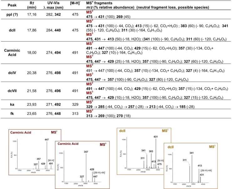
Table 2 – Retention times and MS data of the main anthraquinone components identified in extracts of
several cochineal insects by HPLC/DAD/ESI/MSn. The product ion spectra were acquired in negative ionisation mode [16,37].
Peak Rt (min)
UV-Vis max (nm)
[M-H]- MSn fragments
m/z (% relative abundance) (neutral fragment loss, possible species)
ppI (?) 17,16 282, 342 475 MS
2
475431 (100); 269 (45)
dcII 17,86 284, 440 475
MS2
475431 (100) (- 44, CO2); 413 (15) (- 62, CO2+H2O) ; 383 (60) (- 90, C3H6O3); 341
(55) (- 120, C4H8O4); 311 (30) (-164, C6H12O5)
MS3
475, 431 413 (50) (-18, H2O); (341 (100) (- 90, C3H6O3); 311 (60) (- 120, C4H8O4) Carminic
Acid 18,00 274, 494 491
MS2
491447 (100) (-44, CO2); 429 (15) (- 62, CO2+H2O); 357 (30) (-134, CO2+
C3H6O3); 327 (10) (-164, C6H12O5)
MS3
475, 447 429 (25) (-18, H2O); 357 (100) (-90, C3H6O3); 327 (60) (-120, C4H8O4)
dcIV 20,38 276, 498 491
MS2
491 447 (100) (-44, CO2); 357 (10) (-134, CO2+ C3H6O3); 327 (4) (-164, C6H12O5)
MS3
475, 447 357 (100) (-90, C3H6O3); 327 (80) (-120, C4H8O4)
dcVII 21,58 276, 496 491
MS2
491447 (100) (-44, CO2); 429 (15) (- 62, CO2+H2O); 357 (15) (-134, CO2+ C3H6O3)
MS3
475, 447 429 (10) (-18, H2O); 357 (100) (-90, C3H6O3); 327 (15) (-120, C4H8O4) ka 23,93 271, 492 329 MS
2
329285 (-44, CO2) 257 (-28) 213 (-44, CO2) 185 (-28) fk 23,65 276, 448 313 MS
2
313269 (100); 270 (18)
Figure 3 – MSN spectrums from carminic acid and dcII obtained in negative mode.
2. Samples preparation
2.1 Insect dye extracts
All the cochineal insect species were extracted in 100% H2O, simulating old dyeing recipes [4], in order to avoid the higher formation of fk+ka, produced by strong acidic conditions, and allowing the comparison with soft extraction methods applied in textiles cochineal recovery.The dilapidation of Porphyrophora specimens prior to dyes extraction [14] did not improve significantly the amount of dyestuff extracted and the Porphyrophora specimens were extracted with the same procedure as Dactylopius species.
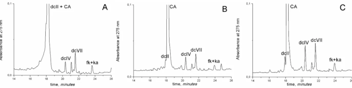
when the final extract is reconstituted with H2O: CH4O: H2O/ HClO4 (50:20:30, v/v/v). When the reconstituted extract, from dyed textiles samples, was not acidified, the chromatograms obtained displayed very poor resolution, comparable to chromatograms reported in literature [17,29] and the dcII peak was co-eluted with carminic acid peak, figure 4a. Better results were obtained by acidifying the extracts, and so the dcII and the carminic acid eluted at distinct retention times, figures 4b and 4c. Therefore, the elution profiles in figure 3 shows that much more information can be obtained with the slight acidification of the reconstituted extract, being this result applied in all the insect and textile specimens’ preparations.
Figure 4 – HPLC/DAD chromatograms (monitored at 275 nm) of dyed cochineal textiles with different
solvent proportions in the final dye extract: A – Dye-sample from textile fibre extracted with TFAmethod,
not acidified, and comparable to chromatograms in [17,29]; B - Dye-sample from textile fibre extracted
with TFAmethod, and reconstituted with H2O: CH3OH: H2O/HClO4 (50:20:30, v/v/v); and C - Dye-sample
from textile fibre extracted with oxalic acid method, and reconstituted with H2O: CH3OH: H2O/HClO4
(50:20:30, v/v/v). All the tests were performed in three replicates.
2.2 Dyed-fibre extraction methods
As reported in the literature, the maximum amount of carminic acid extracted from American cochineal dyed fibres was obtained with oxalic acid [28]. In the performed tests, oxalic acid extracted almost the same amount of carminic acid as TFA solution in agreement with [28], figure 5a. The oxalic acid method showed better chromatograms’ resolution than extraction with other methods, figure 4c, and, consequently, it was selected for all the fibres’ extractions.
Figure 5 – Results obtained with the extraction methods: A – HPLC peak areas for cochineal
chromophores extracted from dyed fibres with formic acid, HCL, oxalic acid and TFA method. B - PCA
scores obtained from mean centered cochineal chromatograms (17,5 – 18,8 min.), acquired at 275nm:
cochineal extracts from D. coccus dyed textiles (solid black symbols) are comparable with D. coccus
insect’ extracts (solid red symbols), as well as with the dyeing solution (open symbols). The TFA method
(solid blue symbols) deviates the extracts from dyed fibres from the D. coccus cluster to the
Porphyrophora insect’ extracts (solid green symbols).
3. Insect samples
3.1 Reference cochineal specimens identified by entomologists
As reported in literature [12,14] D. coccus is distinguished from Porphyrophora species, due to the higher amount of dcII compound and minor amount of fk+ka in D. coccus than in
Porphyrophora species, figures 1 and 2, and table 3. However, it is difficult to distinguish P.
hamelli from P. polonica due to the similar content of fk+ka. Also, it should be noticed that the
standard deviations presented by table 3, either for literature or for the results obtained in this work, are very high, and careful attention should be given when using the marker’s relative percentage for cochineal species recognition. For instance, in P. polonica, the relative percentage of fk+ka has an error of circa 90%. This high value can be explained by the variations in the insect composition according to its development stage [12,14].
Table 3 – Markers’ relative percentages from cochineal insects aqueous extracts calculated at 275nm.
Literature1 Obtained Results2
D. coccus 2,3±1,0% dcII, 95,2±1,3% CA, 1,4±0,4 % dcIV, 0,4±0,2% dcVII, 0,5±0,3% fk + ka
(8 specimens analyzed)
2,4±0,9% dcII, 95,3±1,3% CA, 1,0±0,4% dcIV, 1,0±0,4% dcVII, 0,2±0,1% fk + ka
(39 specimens analyzed) P. polonica 4,5±0,1% ppI, 87,8±5,5% CA, 1,2±0,4% dcIV,
0,8±0,1% dcVII, 5,7±5,8% fk+ka (6 specimens analyzed)
2,2±1,7% ppI (?),~0% dcII, 92,4±3,6% CA, 0,6±0,4% dcIV, 1,4±0,8% dcVII, 3,5±3,1% fk+ka
(20 specimens analyzed) P. hamelli 2,1±1,0% ppI, 0,4±0,1% dcII,92,6±0,6% CA,
3,1±0,4% dcIV, 0,9±0,2% dcVII, 1,2±0,5% fk + ka (4 specimens analyzed)
1,4±1,3% ppI, 0,5±0,3% dcII,96,3±1,3% CA, 0,6±0,2% dcIV,0,6±0,1% dcVII, 1,4±0,8% fk + ka
(9 specimens analyzed)
1 The average values and standard deviation of the cochineal insects relative peak areas were calculated from data
2 The relative peak areas were calculated with the chromatographic program ChromQuest 4.1 at the maximum
wavelength absorption of 275 nm (Appendix 4).
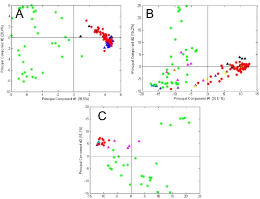
The differentiation of the species can be more reliable if obtained with PCA, considering the chromatographic data between 15 and 25 min.,and not only the minor markers relative peak areas. In figure 6a, Dactylopius family is grouped in a distinctive cluster, from Porphyrophora family. Therefore, PCA is able to distinguish between both cochineal families. With PCA it is also possible to distinguish P. hamelii and P.polonica, inside the Porphyrophora family, figure 6b. P. polonica samples gather in two distinct regions, due to the differences shown by the stage development of the insect, already mentioned before. Thus, samples joined in the upper left quadrant of figure 6a and in the upper right quadrant of figure 6b correspond to females with eggs, which chromatogram have shown a higher content of fk+ka [14]. Also, with PCA analysis it is possible to distinguish D. coccus (solid red symbols) and the wild Dactylopius species, figure 6c. The wild species groups showed some similarity between each other, and with the PLS model it was possible to distinguish the species with circa 80% success.
Due to the few number of D. tomentosus and P. medicaginis specimens analyzed, PCA analyzes could not be accomplished. However, its representative chromatograms can be observed in Appendix 7, along with the other representative chromatograms for each identified species.
A B
C
Figure 6 – PCA scores obtained from mean centered chromatograms acquired at 275 nm, where it is
possible to distinguish Dactylopius and Porphyrophora and the species inside the both families: A -
Dactylopius (solid red symbols)and Porphyrophora (solid green symbols) families, obtained for 17.5-18.8 min.; B –P. polonica (solid blue symbols) and P. hamelii (solid pink symbols) species, acquired in the
region between 15.0-18.8 and 19.7-25.0 min; and C – D. coccus (solid red symbols) and the wild
Dactylopius species, D. confusus (solid green symbols), D. ceylonicus (solid blue symbols) and D.
3.2 Unidentified cochineal insect species
From the 94 specimens of unidentified insect species (historical collection from Royal Botanic Garden, at Kew, and insects supplied by Cardon, Balfour and Tiano, table 4 and Appendix 5), the presence of carminic acid and several minor cochineal markers were detected in 87 specimens. In the remaining seven specimens from Kew (circa 4% of the collection), yellow unidentified compounds were detected in six dye-samples (55338 and 55340, Appendix 8), and one insect sample produced a colorless extraction (73057), excluding these insects as cochineal, table 4.
Table 4 –Unidentified specimens’ labelled as cochineal with respective species attribution, identified by HPLC/DAD, marker’s relative percentages and PCA analysis.
Classification1 Collection
Date Source, donor Observations
Species attribution (markers’ relative percentages) 2 Royal Botanic Garden, at Kew
54387 1856 Índia, Madras, from James A. Mann
Dry, brown, medium dimensions, 10,4±3,0 mg
D. coccus (3,3±0,5% dcII, 94,6±0,6% CA, 1,0±0,1% dcIV, 0,9±0,1% dcVII, 0,3±0,1% fk + ka) 54388 1918 London Drug Market Peru, Callas, from dimensions, 14,2±2,8 mg Dry, black, medium
D. coccus(3,4±0,9% dcII, 94,7±1,2% CA, 0,8±0,1% dcIV, 0,8±0,2% dcVII, 0,3±0,1% fk + ka) 54389 1899
Ecuador, Chimborazo Province, Guana, from Edward Whimper
Dark dry cake mixture of cochineal and other ingredients, 1,0±0,2 mg
Dactylopius sp. (1,2±0,3% dcII, 96,7±0,2% CA, 0,6±0,1% dcIV, 0,2±0,1% dcVII, 1,3±0,6% fk + ka) 54390 Before 1879
Indonesia, Java, Buitenzorg, Tyikoppo, donated by India Museum
Dry, dark brown, medium dimensions, 14±2 mg
D. coccus (2,7±0,3% dcII, 95,4±0,8% CA, 0,9±0,3% dcIV, 0,8±0,1% dcVII, 0,3±0,1% fk + ka) 54391 Before 1879
Indonesia, Java, Buitenzorg, Bandok, donated by India Museum
Dry, dark brown, medium dimensions, 16,2±1,9 mg
D. coccus (2,3±0,3% dcII, 96,0±0,5% CA, 0,8±0,3% dcIV, 0,6±0,0% dcVII, 0,3±0,1% fk + ka) 54392 Late 19
th
century
4
, A.S. Hill & Son, London Dry, dark brown, medium dimensions, 21,7±5,2 mg
D. coccus (2,6±0,2% dcII, 95,1±0,5% CA, 0,8±0,1% dcIV, 1,2±0,5% dcVII, 0,4±0,1% fk + ka) 54393 Before 1879
Índia, Andhra Pradesh, Scinde, Hyderabad, donated by India Museum
Dry, shiny red, medium dimensions, 2,8±1,1 mg
D. coccus (2,5±0,9% dcII, 95,6±1,8% CA, 0,8±0,3% dcIV, 0,9±0,4% dcVII, 0,2±0,3% fk + ka) 54394 1867
India, Calcutta, from International exhibition,
Paris
Dry, dark brown, medium dimensions, 11±2,8 mg
D. coccus (2,6±0,2% dcII, 95,6±0,7% CA, 0,6±0,4% dcIV, 0,8±0,3% dcVII, 0,4±0,1% fk + ka) 54402 Late 19
th
century Honduras and Vera Cruz
Dry, light brown, little dimensions, 1,6±0,4 mg
D. coccus (2,7±0,5% dcII, 94,4±1,2% CA, 1,4±0,2% dcIV, 1,3±0,4% dcVII, 0,3±0,2% fk + ka) 54403 Before 1879 India, Punjab, donated by
India Museum
Dry, dark brown, medium dimensions, 11±4,7 mg
D. coccus (2,9±0,7% dcII, 95,1±0,4% CA, 0,9±0,3% dcIV, 0,8±0,2% dcVII, 0,3±0,1% fk + ka) 54404 Probably 1851
Mexico, Oaxaca, from J. Sadler, probably International exhibition,
London
Dry, dark brown, medium dimensions, 10,8±1,7 mg
D. coccus (4,0±2,1% dcII, 93,9±1,7% CA, 0,9±0,1% dcIV, 0,9±0,3% dcVII, 0,3±0,2% fk + ka) 54410 1977 Madeira, from Jane Stubbs
Dry, greyish brown, medium dimensions,
7,8±1,4 mg
D. coccus (3,0±0,8% dcII, 95,1±1,2% CA, 0,8±0,2% dcIV, 0,9±0,2% dcVII, 0,3±0,1% fk + ka) 55338 1855
Australia (NSW), from International exhibition,
Paris
Dried naturally, rusted red colour, little dimensions, 2,1±0,3 mg
Not cochineal insect, unidentified yellow compounds (Main peak: rt=
19,86 min, λmax= 421 nm)
55340 1862
Australia (Victoria), from International exhibition,
London
Dried naturally, rusted yellow colour, little dimensions, 1,8±0,7 mg
Similar to 55338 58236.1 Late 19
th
century
4
, Ripley, Roberts & Co. 3. Mincing lane
Dry, dark shiny red, medium dimensions,
20,7±1,7 mg
D. coccus (2,5±0,4% dcII, 95,9±0,4% CA, 0,6±0,1% dcIV, 0,7±0,2% dcVII, 0,2±0,1% fk + ka) 58236.2 Late 19
th
century
4, Ripley, Roberts & Co. 3.
Mincing lane
Dry, dark shiny red, medium dimensions,
15,9±2,0 mg
D. coccus (3,0±0,9% dcII, 94,5±0,3% CA, 0,7±0,1% dcIV, 1,2±0,8% dcVII, 0,6±0,3% fk + ka) 58236.3 Late 19
th
century
4, Beazley & Co. Dunster
House, Mincing Lane
Dry, dark shiny red, medium dimensions,
18,7±2,5 mg 0,9±0,2% dcVII, 0,3±0,1% fk + ka) 58236.4 Late 19
th
century
4
, Beazley & Co. Dunster House, Mincing Lane
Dry, black, medium dimensions, 16,3±6,9 mg
D. coccus (2,5±1,5% dcII, 95,2±2,0% CA, 0,1±0,2% dcIV, 0,9±0,2% dcVII, 0,5±0,4% fk + ka) 58236.5 Late 19
th
century
4
, Ripley, Roberts & Co. 3. Mincing lane
Dry, salmon light colour, medium dimensions,
19,5±4,7 mg
D. coccus(2,9±1,2% dcII, 95,4±1,0% CA, 0,7±0,3% dcIV, 0,8±0,2% dcVII, 0,3±0,1% fk + ka) 58236.6 Late 19thcentury 4, Ripley, Roberts & Co. 3.
Mincing lane
Dry, black, little dimensions, 0,7±0,3 mg
D. coccus (2,0±1,5% dcII, 95,5±1,4% CA, 1,1±0,1% dcIV, 1,0±0,2% dcVII, 0,3±0,0% fk + ka) 58236.7 Late 19
th
century
4, Ripley, Roberts & Co. 3.
Mincing lane
Dry, dark brown, medium dimensions, 10,6±3,5 mg
D. coccus (2,5±0,4% dcII, 95,9±0,8% CA, 0,6±0,3% dcIV, 0,7±0,3% dcVII, 0,2±0,0% fk + ka) 58236.8 Late 19
th
century
4
, Beazley & Co. Dunster House, Mincing Lane
Dry, dark red, medium dimensions, 5,0±1,0 mg
D. coccus (2,3±0,5% dcII, 96,1±0,9% CA, 0,7±0,3% dcIV, 0,7±0,1% dcVII, 0,2±0,0% fk + ka) 73057 1800-1857
4
, Royal Pharmaceutical Society of Great Britain
(Museum)
Naturally dried, orange colour, big dimensions,
7,20 mg
Not cochineal insect. No coloured compounds.
73237 1851
Mexico, Oaxaca, from J. Sadler, probably International exhibition,
London
Dry, black, medium dimensions, 11,6±2,4 mg
D. coccus (2,3±0,8 dcII, 96,1±1,0% CA, 0,7±%0,3 dcIV, 0,6±0,1% dcVII,
0,2±0,0% fk + ka)
Dominique Cardon unidentified specimens
Armenian cochineal
3 4
, Dominique Cardon
Naturally dried, dark brown, medium dimensions, 10,8±2,4 mg
D. coccus (3,2±0,5 dcII, 94,5±0,8% CA, 0,4±%0,1 dcIV, 0,4±0,1% dcVII,
0,6±0,1% fk + ka) American
cochineal
3 4
, Dominique Cardon
Naturally dried, white dusty, medium dimensions, 12,1±4,1 mg
Porphyrophora sp. (1,5±0,2 ppI, 0,5±0,2 dcII(?), 95,5±0,3% CA, 0,6±%0,2 dcIV, 0,8±0,0% dcVII,
0,7±0,1% fk + ka) “Kermes noir” 16/10/2002 Market in Athens,
Dominique Cardon
Dry, dark brown, medium dimensions, 10,4±2,8 mg
D. coccus (1,4±1,1 dcII, 96,4±1,0% CA, 0,5±%0,2 dcIV, 0,6±0,2% dcVII,
1,2±1,0% fk + ka)
D. coccus 3 4
, Dominique Cardon Dry, dark brown, medium dimensions, 10,9±6,0 mg
D. coccus (2,7±1,3 dcII, 96,0±0,9% CA, 0,5±%0,1 dcIV, 0,6±0,3% dcVII,
0,3±0,1% fk + ka)
Piero Tiano unidentified specimens
Tiano-Fi 3 4, Piero Tiano Dry, dark brown, medium dimensions, 10,7±3,6 mg
Dactylopius sp. (4,5±1,8 dcII, 93,9±2,0% CA, 0,6±%0,1 dcIV, 0,7±0,1% dcVII, 0,4±0,2% fk + ka) Tiano-Brx 3 4, Piero Tiano Dry, dark red, medium
dimensions, 12,9±5,8 mg
Dactylopius sp.(2,5±1,0 dcII, 96,6±0,6% CA, 0,7±%0,3 dcIV, 0,6±0,2% dcVII, 0,6±0,1% fk + ka)
Jenny Balfour unidentified specimens
Cochineal I 3
4, Dyes in History and
Archaeology, France, 2004, from Jenny Balfour
Dried naturally, pink colour, hairy, medium dimensions, 11,6±9,4 mg
P. hamelii(2,8±0,5 ppI, 0,8±0,2 dcII, 95,4±1,0% CA, 0,5±%1,0 dcIV, 0,5±0,1% dcVII, 1,1±0,3% fk + ka) Cochineal II 3
4
, Dyes in History and Archaeology, France, 2004, from Jenny Balfour
Dried naturally, red colour, scarce hairy, medium dimensions,
19,0±1,1 mg
P. hamelii(2,4±0,5 ppI, 0,4±0,1 dcII, 95,4±0,8% CA, 0,3±%0,1 dcIV, 0,4±0,0% dcVII, 1,1±0,4% fk + ka)
1 Classification of the insects, given by the donor. 2
The relative peak areas were calculated with the chromatographic program ChromQuest 4.1 at the maximum wavelength absorption of 275 nm.
3 Donation date unrecorded. 4
Source unidentified.
The chromatographic PCA data obtained for the 63 cochineal specimens from Kew, figure 6a, held consistent results to identify the majority of the insects (solid blue triangles) as D.
coccus species. These results seem to be conclusive with the specimens source and
Although, there seems to be an exception with the insects of 54359 samples (solid black triangles), which correspond to the cake compounded by a mixture of cochineal (probably from wild origin) and other unknown components, Appendix 5. Marker’s relative percentages (table 4) cannot set a clear distinction between this sample and the others.
The 58236 samples belong to a box which includes eight varieties of cochineal from the late 19th and early 20th centuries, and that were used for dyeing purposes, Appendix 5. Their appearance points to different varieties of cochineal, and, as PCA analysis have shown that they are possibly D. coccus species, it may be likely that the same specie was subject to different methods of killing and preparation, Appendix 1 [1,4].
Relatively to the results obtained for the other unknown samples, PCA analysis indicated that Balfour’s specimens are likely Porphyrophora species; as well the “American cochineal” samples of Cardon, which were thought to be wrongly labelled in the past, due to their physical appearance, figure 7b and Appendix 5. This situation occurred also with the “Armenian cochineal” samples from Cardon, which had a dubious appearance and which were pointed as
D. coccus species by PCA analysis. Also from Cardon, the samples labelled as “Kermes noir”
and “D. coccus” were recognized as D. coccus species, according with their resembled appearance. At last, Tiano’s samples seem to deviate slightly from the D. coccus cluster to the
Porphyrophora samples’ concentration. These results seem to be in accordance with the
respective relative percentages in table 4, which show homogeneity if compared with the relative percentages from Porphyrophora and D. coccus insects, Appendix 4. Yet, by comparison with literature [14], it was possible to verify the UV spectra of the insects’ respective minor markers.
Porphyrophora samples from Balfour and Cardon were projected onto a model calibrated
with P. polonica and P. hamelii species, figure 7c. Balfour samples are clearly gathered on the cluster of P. hamelii, though Cardon samples deviate to concentration zone of P. polonica. However, due to the differences seen on results of P. polonica specimens, more studies and analysis on this specie would be needed to ensure a better identification of these unidentified
A B
C
Figure 7 – PCA scores for the 87 unknown cochineal specimens projected onto reference cochineal
samples D. coccus (solid red circles) and Porphyrophora (solid green circles): A – Royal Botanic Garden
(solid triangle symbols) obtained for 17,5-18,8 min.; B – Cardon samples (Armenian cochineal – solid
black triangles; American cochineal – solid pink triangles; “Kermes noir” – solid brown triangles; and D.
coccus – solid orange triangles), Tiano samples (Fi – solid yellow triangles and Brx – solid purple
triangles) and Balfour samples (I - solid dark blue triangles; II – solid light blue triangles), acquired for
15,0-18,80 and 19,7-25,0 min.; and C - unknown cochineal specimens of Cardon (solid pink triangles) and
Balfour (I - solid dark blue triangles; II – solid light blue triangles) samples, projected on P. hamelii (solid
red symbols) and P. polonica (solid green symbols), obtained for 15,0-18,80 min. and 19,7-25,0.
4. Historical textile dye identification
The chromatograms obtained for the historical red-dyed textiles are homogeneous (Appendix 6) and it is possible to observe the presence of carminic acid and minor markers. Nevertheless, the elution profile of the historical red-dyed textiles is slightly different from the insect specimens in the HPLC-DAD library, figure 8. For instance, in the historical red-dyed textile samples it was difficult to identify with certainty the presence of dcII due to its low content and the presence of other small peaks eluted near dcII. When possible, its presence was confirmed with MS analysis. Furthermore, new peaks that were not present in the insect specimens were identified in the historical red-dyed textiles. For instance, it was detected a peak around 19,46 min., with a λmax=370 nm, Appendix 8. Analyses using HPLC/DAD/MS
n
[16,17]. It can probably be related to the use of plant species belonging to dicotyledonous families, or as a result of the photo- or auto-oxidation of gallotanins, in alkaline environment [4].
Figure 8 – Representative chromatogram of the historical red-dyed textile sample CGM 1449 velvet,
identified as D. coccus, monitored at 275 nm.
Table 5 provides the origins and dates attributed to the textiles, along with the relative average percentage calculated for their chromatograms. Given the significant relative percentage of dcII present in the samples acquired from CGM 1388A and CGM 1449, and dcX presence in the other samples4, it was thought that they could belong to D. coccus, or P.
hamelii. However, this data must be considered with care as the standard deviation is very high in these samples.
Table 5 – Historical textile samples, with relative percentages, obtained by HPLC/DAD analysis.
Classification1 Date Provenance Expected specie2 Markers’ relative percentages3
Calouste Gulbenkian Museum CGM 1388A
Velvet
16th/17th
century Turkey, Bursa D. coccus
P. Polonica (?) (2,8±3,4% dcII, 92,9±7,0% CA, 1,1±0,2% dcIV, 2,2±2,8%
dcVII, 1,2±1,0% fk + ka) CGM 245
Velvet 16th century Italy, Genoa D. coccus
P. Polonica (?) (1,8±0,0% dcX, 94,4±0,0% CA, 1,3±0,0% dcIV, 1,9±0,0%
dcVII, 0,7±0,0% fk + ka) CGM 1446
Velvet 17th century Iran, Yazd (?) Porphyrophora sp.
P. Polonica (?) (0,4±0,0% dcX, 96,3±0,3% CA, 1,8±0,4% dcIV, 1,2±0,1%
dcVII, 0,31±0,02% fk + ka) CGM 1513
Velvet 17th century Iran Porphyrophora sp.
P. Polonica (?) (0,6±0,5% dcX, 95,7±1,3% CA, 1,6±0,4% dcIV, 1,7±0,5%
dcVII, 0,4±0,0% fk + ka) CGM 1449
Velvet 17th century India D. coccus
D. coccus (1,7±0,2% dcII, 94,4±1,0% CA, 1,3±0,1% dcIV, 1,7±0,8% dcVII,
0,8±0,0% fk + ka) CGM 1422
Velvet 17th century India Porphyrophora sp.
P. Polonica (?) (0,4±0,0% dcX, 95,2±1,1% CA, 2,4±1,5% dcIV, 1,6±0,7%
dcVII, 0,5±0,1% fk + ka) CGM T100
Carpet 16th century Iran P. hamelii
P. Polonica (?) (1,1±0,5% dcX, 96,0±0,7% CA, 1,1±0,2% dcIV, 1,4±0,3%
dcVII, 0,4±0,1% fk + ka) Museu Nacional de Arte Antiga
MNAA 1616Tec Chasuble
15th century Italy, Florence (?) P. polonica
P. Polonica or mixture (?)(0,8±0,1% dcX, 90,0±2,7% CA, 1,3±0,2% dcIV,
1,8±0,3% dcVII, 6,2±2,1% fk + ka)
4
When the peak eluting at 17,86 min., corresponding to dcII compound was confirmed with MS analysis,
the peak was labelled as dcII. When its molecular structure was not confirmed with MS analysis, this peak
could be another compound, and so it was labelled as dcX. For instance, it was possible to verify that dcII
1 Inventory numbers of the historical textiles, given by the institutions. 2
Expected species by comparison with results given by previous publications.
3
The relative peak areas were calculated with the chromatographic program ChromQuest 4.1 at the maximum wavelength absorption of 275 nm.
In order to perform PCA analysis with direct comparison with the insect specimens from the cochineal HPLC/DAD library, it was necessary to restrict the PCA analysis to elution time regions where chromatograms are consistent, namely between 17,5-18,8 min, figure 9a, and between 15,0-18,8 and 19,7-24,7 min, figure 9b.
With PCA analysis, the majority of the historical red-dyed textiles samples coincide with the distribution of Porphyrophora specimens, figure 9a. However, samples taken from CGM 1449 appear in close proximity with D. coccus cluster. Analysis by HPLC/DAD/MSn confirmed the presence of dcII, and a vestigial presence of ka, thus these samples are probably D. coccus. As this is a 17th-century Indian velvet, this is an important result as it confirms the adoption of American cochineal shortly after the establishment of the English textile factory in Surat in 1612, and well before it came to dominate Indian dyeing practices in the 19th-century [10].
The remaining historical samples cluster occupied the region of the reference samples of
Porphyrophora species, figure 9a. A better distinction of these samples is provided by PCA
analysis in another chromatographic region, where these samples are projected over P. hamelii and P. polonica reference samples, figure 9b. In this figure, it is possible to observe a clear distinction between MNAA 1616Tec and the other historical samples. The former occurs in the region of P. polonica reference samples, which are richer in fk+ka, table 5. Hence, it is likely that these results point to the presence of P. polonica specie, as previously observed (figure 2b), or to a mixture of Porphyrophora specie and kermes, which has two main component peaks (ka+fk) [4]. These results also appear to be in accordance with the attributed place and date of production of this 15th-century Italian velvet. On the one hand, D. coccus insects were not traded to Europe before the 16th century, and its use in Italy was only sanctioned in the middle of the century [10], on the other hand, Italy was a major centre for the international trade in dyestuffs and, for this reason, a wide variety of Porphyrophora species would have been available to Italian dyers [7].
of the two species could have been present in Ottoman markets at this time, as American cochineal had begun arriving in Venice by 1543, and thus could easily have been exported eastwards shortly after this date (Appendix 1) [7,9].
A B
Figure 9 – PCA scores, for the historical red-dyed textiles (solid lozenge symbols) - CGM 1388A velvet
(solid yellow lozenges), CGM 245 velvet (solid purple lozenges), CGM 1446 velvet (solid dark green
lozenges), CGM 1513 velvet (solid orange lozenges), CGM T100 carpet (solid pink lozenges), CGM 1449
velvet (solid dark blue lozenges), CGM 1422 velvet (solid brown lozenges), MNAA 1616Tec chasuble
(solid light blue lozenges) and CGM T100 and MNAA 1616Tec samples extracted by HCl method (solid
dark lozenges): A - projected onto reference cochineal samples Dactylopiuscoccus (solid red symbols)
and Porphyrophora (solid green symbols) obtained at 17,5 – 18,8 min.; and B - projected onto P. hamelii
(solid red symbols) and P. polonica (solid green symbols) samples, obtained for 15-18,8 and 19,7-24,7
min..
Additionally, two other textiles samples were analysed, one belonging to a velvet (CGM 1513), which had an orange colour, and another belonging to a silk carpet (CGM T100), with a yellow colour. The former was identified as a species of madder, owing to the presence of alizarin, although the presence of purpurin is almost residual [25,39]. Other yellow compounds were also detected and hence it is probably a mixture of madder and another yellow dyestuff, Appendix 8. Analyses of the latter fibre indicate the presence of yellow compounds, namely rutin-based compounds, with a very similar elution profile to a yellow silk fibre sample from a related “Small Silk Kashan” rug in the Museu Nacional de Machado de Castro (MNMC T744) [39], Appendix 8. These yellow compounds do not match the most common sources of yellow, such as Reseda luteola, and, indeed, point to the use of a specific dyestuff by the workshop that produced this group of Persian carpets. Future research on the precise species of this yellow dye may aid in identifying the geographical location of this workshop in Iran.
Analyses of historical textile samples extracted with oxalic acid yield good results, and, moreover, PCA analysis demonstrates that extraction with HCl [14] causes a serious deviation of these samples from the Porphyrophora concentration region, in the direction of the D. coccus cluster, figure 9a. However, results for textile reference samples dyed with D. coccus show that extraction with HCl is not responsible for this deviation, figure 5b, and hence, this occurrence is probably due to the hydrolysis of the precursors present in the Porphyrophora species.
based on the relative percentages of dcII and fk+ka, as shown in previous publications [14], figure 10a. According to the resulting diagram, the samples from the Persian silk carpet (T100 -1) and the Italian and Turkish velvets (CGM 245 - 2, CGM 1388A - 3), attributed to a
Dactylopius coccus insect, might not actually have the dcII marker but probably the unknown
compound dcX, due to the considerable amount of “dcII” compound; hence, in this case, it could reflect the presence of a Porphyrophora species. Although, the presence of dcII marker was confirmed for the Turkish velvet (GCM 1388A) by MSn, it was not sufficient to confirm this sample as a D. coccus species, owing to the chromatogram similarity with Porphyrophora species as shown by PCA analysis. For rigorous results, all samples of historical textiles and other existent Porphyrophora species should be analyzed simultaneously by PCA and MSn. Although the red dye in the Indian velvet (CGM 1449 - 4) is considered to be D. coccus, the samples from the other velvets (CGM 1513 – 3, CGM 1422 – 5 and CGM 1446 - 6) did not present dcII marker, and this was corroborated by MSn analysis of the CGM 1422 sample. Consequently, it is possible that these samples might be P. polonica. Finally, the Italian chasuble (MNAA 1616Tec - 8) is confirmed as P. polonica species or a mixture of this species with kermes [15] which is also in accordance with the PCA models described above. Special care should be taken when using this quantification system of graphic representation with other cochineal species. For example, it was observed that wild Dactylopius show some inconsistent results in this system. In addition, PCA models of all the minor markers quantification, using the same average relative percentages as the prior type of graphic representation, show the same trend towards non-distinguishable clusters, owing to the high deviation standards resulting from this method, figures 10b [12,13], Tables 3, 4 and 5, and Appendix 4.
1
8 7
2 4
3 5 6 0 20 40 60 80 100
0 20 40 60 80 100
% fk+ka % dcII
A B
D. coccusP. hamelii
P. polonica
Figure 10 – Two different graphic systems made with the relative percentages of dcII and fk+ka,
calculated at 275 nm, for all the analysis accomplished in this study and found in the literature [14]: D.
coccus (solid red symbols), Dactylopius wild species (solid green symbols), P. polonica (solid blue symbols), P. hamelii (solid yellow symbols), Kew Garden samples (solid black symbols), extraction
methods (solid pink symbols), historical samples (solid cyan symbols) and literature data (open symbols):
A – quantification system to distinguish between D. coccus, P. hamelii and P. Polonica [14]; andB - PCA
Conclusion
This study aimed to highlight the importance of cochineal species characterization for understanding the application and dissemination of this red dyestuff in textile production in Europe and Asia from 15th to 17th centuries. This interdisciplinary approach offers the opportunity to look more widely at the history of dye technology and trade routes, and, consequently to characterize textiles in terms of their provenance and date. However, this study also recognizes, for the first time, the limitations of prior cochineal species identification and emphasizes the necessity of an approach which combines entomological studies with chemical analysis of dyes and historical textiles.
It has revealed that a reliable system of HPLC/DAD, allied with PCA analysis and MSn, can satisfactorily differentiate between six cochineal insect species, and make a valuable contribution to the characterization and identification of historical insect species and red-dyed textiles. In this way, 60 cochineal samples belonging to Royal Botanical Garden, at Kew, were confirmed as D. coccus species, while seven samples were considered to be incorrectly labeled, as they exhibited no characteristics of cochineal species.
The combination of HPLC/DAD and PCA analysis was only successful after optimization of the separation parameters, which was an important step to obtain improved results in comparison to previous studies. Slight acidification of cochineal dye samples improved the resolution, permitting a good overview of the minor markers identification and a satisfactory correlation between the peak areas in the PCA analysis, for species differentiation. Oxalic acid was determined to be a less harmful extracting solution for historical textile fibres, and presented better resolution than other extraction solutions. The majority of the red-dyed samples from the historical textiles, dating from 15th to 17th centuries, were identified as
Porphyrophora species, and more scientific research is needed for accurate identification of the
Acknowledgements
We would like to thank to Dr. João Lopes (Requimte, Faculty of Pharmacy, University of Porto), for the PCA analysis; and to Dr. Conceição Oliveira (Instituto Superior Técnico, Lisbon) who helped to obtain the LC-MS data, at the IST-Node, that is part of the National Mass Spectrometry Network (RNEM) created by the Portuguese Foundation for Science and Technology (FCT).
We thank Maria Fernanda Passos Leite from Calouste Gulbenkian Museum (Lisbon) and to Teresa Pacheco Pereira from Museu Nacional de Arte Antiga (Lisbon), for the access to the historical textiles. Also, we are very grateful to Royal Botanic Garden at Kew (London), namely Monique Simmonds (Jodrell Laboratory, Royal Botanic Garden, at Kew, London) and Mark Nesbitt (Jodrell Laboratory, Royal Botanic Garden, at Kew, London), Douglas Miller (Agricultural Research Service, Systematic Entomology Laboratory, Maryland), Dominique Cardon (CIHAM/UMR, Lyon), Ewa Simon (Faculty of Biology and Environmental Protection, University of Silesia, Katowice) Ferenc Kozár (Plant Protection Institute, Hungarian Academy of Sciences, Budapest), Hassan-Ali Vahedi (College of Agriculture, Razi University, Kermanshah), Jenny Balfour-Paul (Institute of Arab and Islamic Studies, University of Exeter), Katarzyna Golan (Department of Entomology, University of Life Sciences, Lublin), Liberato Portillo (Botanical and Zoology Department, University of Guadalajara), Mónica González (Instituto Canario de Investigaciones Agrarias, Tenerife), and Piero Tiano (ICVBC-CNR, Florence) who gently offered insect samples.
References
[1] Donkin, R. A., “The insect dyes of Western and West-Central Asia”, Anthropos, International Review of Ethnology and Linguistics, 72 (1977) 848.
[2] Marichal, C., “A forgotten chapter of international trade: Mexican cochineal and the european demand for American dyes, 1550 - 1850”, in Latin America Global Trade and
International Commodity Chains in Historical Perspective, Stanford University, 16-17 November 2001, 1 - 27;
[3] Takumasa, K., Gullan, P. J., Williams, D. J., “Coccidology. The study of scale insects (Hemiptera: Sternorryncha: Coccoidea)”, Revista Corpoica – Ciencia y Tecnología Agropecuaria 9 (2)(2008), 55 – 61;
[4] Cardon, D., Natural Dyes - Sources, Tradition, Technology and Science, Archetype Publications, London, 2007.
[5] Vahedi, H., Hodgson, C. J., “Some species of the hypogeal scale insect Porphyrophora Brandt (Hemiptera: Sternorrhyncha: Coccoidea: Margarodidae) from Europe, the Middle East and North Africa”, Systematics and Biodiversity, 5 (1) (2007) 23 - 127.
[6] Portillo, L., “Origen de Dactylopius coccus Costa (Hemiptera: Dactylopiidae) ¿Norte o Sudamérica?”, Dugesiana, 12 (1) (2005) 1 - 8.
[7] Donkin, R. A., “Spanish red: An ethnogeographical study of cochineal and the Opuntia cactus”, Transactions of the American Philosophical Society, 67 (5) (1977) 1 -84.
[8] Sandberg, G., The Red Dyes, Cochineal, Madder and Murex Purple – A World Tour of
Textile Techniques, Lark Books, Stockholm, 1997.
[9] Raby, J., Effeny, A., IPEK: Imperial Ottoman Silks and Velvets, Azimuth Editions Limited, London (2001).
[10] Phipps, E., Cochineal Red, The Art History of a Color, The Metropolitan Museum, New York, 2010.
[11] Baker, Patricia L., Islamic Textiles. British Museum Press, London (1995).
[12] Wouters, J., Verhecken A.,“The Coccid Insect Dyes: HPLC and computerized diode-array analysis of dyed yarns”, Studies in Conservation, 34 (4) (1989) 189-200.
[13] Wouters, J., Verhecken A., “Potential taxonomic applications of HPLC analyses of coccoidea pigments (Homoptera: Sternorhyncha)”, Belg. J. Zool.,121 (1991) 211 - 225.
[14] Wouters, J., Verhecken A., “The scale insect (Homoptera: Coccoidea). Species recognition by HPLC and diode-array analysis of the dyestuffs”, Annls Soc. ent Fr. (N.S.), 25 (4) (1989) 393 - 410.
[15] Wouters, J., Verhecken A., “High performance liquid chromatography of anthraquinones: analysis of plant and insect extracts and dyed textiles”, Studies in Conservation, 30 (1985) 119 - 128.
[16] Wouters, J., “Dye Analysis of Florentine Borders of the 14th to 16th Centuries”, Dyes in
[17] Bommel, M., “What Colours Tell You: the Identification of Dyes in Historical Objects”, in
Silk, Gold, Crimson: Luxury and Technology at the Visconti and Sforza Courts, C. Buss,
Silvana, Milan (2009), 167 – 169.
[18] Peggie, D. A., Hulme, A. N., McNab, H., Quye, A., “Towards the identification of characteristic minor components from textiles dyed with weld (Reseda luteola L.) and those dyed with Mexican cochineal (Dactylopius coccus Costa)”, Microchimica Acta, 162 (2008) 371 - 380.
[19] Wouters, J. Rosario-Chirinos N., “Dye analysis of pre-colombian Peruvian textiles with high performance liquid chromatography and diode-array detection”, JAIC, 31 (1992) 237 - 355. [20] Karapanagiotis, I., Chryssoulakis, Y., “Investigation of red natural dyes used in historical objects by HPLC-DAD-MS”, Annali di Chimica, 95 (2005).
[21] Karapanagiotis, I., Daniilia, S., Tsakalof, A., Chryssoulakis, Y., “Identification of red natural dyes in post-byzantine icons by HPLC”, Journal of Liquid Chromatography & Related
Technologies, 28 (2005) 739 - 749.
[22] Karapanagiotis, I., Lakka, A., Valianou, L., Chryssoulakis, Y., “High-performance liquid chromatography in historical garments from the Holy Mountain of Athos”, Microchimica Acta,
160 (2008) 477 - 483.
[23] Halpine, S. M., “An improved dye and lake pigment analysis method for high-performance liquid chromatography and diode-array detection”, Studies in Conservation,41 (1996) 76 - 94. [24] Orska-Gawryś, J., Surowiec, I., Kehl, J., Rejniak, H., Urbaniak-Walczak, K., Trojanowicz, M., Journal of Chromatography A, 989 (2003) 239 - 248.
[25] Hofenk de Graaff, J. H., The Colourful Past – Origins, Chemistry and Identification of
Natural Dyestuffs, Abbeg-Stiftung, Switzerland, 2004.
[26] Sanyova, J., Reisse, J., “Development of a mild method for the extraction of
anthraquinones from their aluminium complexes in madder lakes prior to HPLC analysis”,
Journal of Cultural Heritage, 7 (2006) 229 – 235.
[27] Zhang, X., Laursen, R. A., “Development of mild extraction methods for the analysis of natural dyes in textiles of historical interest using LC-Diode Array Detector-MS”, Analytical
Chemistry, 77 (7) (2005) 2022 - 2025.
[28] Valianou, L., Karapanagiotis, I., Chryssoulakis, Y., “Comparison of extraction methods for the analysis of natural dyes in historical textiles by high-performance liquid chromatography”,
Anal. Bioanal. Chem., 395 (7) (2009) 2175 - 2189.
[29] Karapanagiotis, I., Minopoulou, E., Valianou, L., Daniilia, S., Chryssoulakis, Y., “Investigation of the colourants used in icons of the Cretan School of iconography”, Analytica Chimica Acta 647 (2009) 231 - 342.
![Table 1 – Representative abbreviations from the compounds present in cochineal species [13,14].](https://thumb-eu.123doks.com/thumbv2/123dok_br/16624062.740329/6.892.296.608.896.1094/table-representative-abbreviations-compounds-present-cochineal-species.webp)
![Figure 1 – Graphical representation from markers’ relative percentages and respective standard deviations of cochineal insects aqueous extracts, calculated at 275 nm [13,14].](https://thumb-eu.123doks.com/thumbv2/123dok_br/16624062.740329/7.892.310.591.115.345/graphical-representation-relative-percentages-respective-deviations-cochineal-calculated.webp)