Article
*e-mail: [email protected]
On-line Identiication of Minor Flavones from Sugarcane Juice by LC/UV/MS
and Post-Column Derivatization
Renata Colombo,a Janete H. Yariwake,*,a Emerson F. Queiroz,b Karine Ndjokob and Kurt Hostettmannb
aInstituto de Química de São Carlos, Universidade de São Paulo, CP 780, 13560-970 São Carlos-SP, Brazil
bLaboratoire de Pharmacognosie et Phytochimie, École de Pharmacie Genève-Lausanne,
Université de Genève, Quai Ernest Ansermet 30, 1211, Genève 4, Genève, Switzerland
Este trabalho apresenta a identiicação “on line” de lavonas minoritárias do suco da cana-de-açúcar (Saccharum oficinarum), por cromatograia líquida de alta eiciência com detector UV acoplada à espectrometria de massas (CL/UV/EM) com ionização química à pressão atmosférica, dissociação induzida por colisão (IQPA-DIC-EM/EM) e derivatização pós-coluna utilizando reagentes de deslocamento de UV. As análises CLAE-UV com reagentes de deslocamento forneceram informações sobre a posição da substituição no esqueleto dos lavonóides e, em combinação com dados de EM, estas técnicas permitiram a identiicação “on-line” de cinco lavonas da garapa: luteolina-8-C-glucosil-7-O-glucuronídeo; tricina-7-O -neoesperosideo-4’-O-ramnosídeo; tricina-7-O-metilglucuronato-4’-O-ramnosídeo; tricina-7-O-metilglucuronídeo; swertisina; e outras quatro substâncias foram parcialmente identiicadas como lavonas glicosiladas. Somente a swertisina (7-O-metilapigenina-6-C-glicosídeo) foi anteriormente descrita no bagaço da cana-de-açúcar.
This work describes the on-line characterization of minor lavones from sugarcane (Saccharum oficinarum) juice by high-performance liquid chromatography coupled to diode array UV detection and mass spectrometry (LC/UV/MS) using atmospheric pressure chemical ionization-collision-induced dissociation (APCI-CID-MS/MS) and post-column derivatization using UV shift reagents. HPLC-UV analysis with shift reagents provided information about the substitution pattern in the lavonoid skeleton and, combined with MS data, these techniques allowed for the on-line identiication of ive “garapa” lavones: luteolin-8-C-glucosyl-7-O-glucuronide; tricin-7-O-neohesperoside-4’-O-rhamnoside; tricin-7-O-methylglucuronate-4’-O-rhamnoside; tricin-7-O-methylglucuronide; swertisin, while four other compounds were partially identiied as glycosyllavones. Only swertisin (7-O-methylapigenin-6-C-glucoside) was reported previously in sugarcane molasses.
Keywords:sugarcane juice, Saccharum oficinarum L. (Poaceae), lavones, LC/UV/MS, UV shift reagents
Introduction
Hyphenated techniques such as liquid chromatography with UV photodiode array detection (HPLC-UV) and on-line liquid chromatography-ultraviolet detection-mass spectrometry (HPLC-UV-MS) are powerful techniques for the structural identiication of compounds found in extracts prepared from very small amounts of plant material. In the case of plant polyphenolic compounds such as lavonoids and xanthones, the possibility of obtaining on-line UV data by a combination with the post-column addition of UV shift reagents is a very important step to validate the structure
assignments proposed for the position of oxygenated groups on the polyphenolic skeleton.1 Recently, HPLC-UV-MS and post-column UV-derivatization were employed in the study of Trifolium extracts containing clovamides, lavonoids and isolavonoids and their UV data were discussed in detail.2
biological functions, metabolomic analysis has a great potential for application in the assessment of safety and biological equivalence of genetically modiied organisms, including transgenic plants.
Several flavonoids from Saccharum species were reported in a previous literature review.4 More recently, sugarcane cultivated commercially in Brazil (Saccharum oficinarum L.) is under systematic investigation due to the potential role of this plant as a dietary source of lavonoids with antioxidant properties.5,6 Our investigation into the lavonoid composition of Brazilian sugarcane also takes into account recent studies suggesting the potential role of this plant as a dietary source of antioxidant lavonoids due to the in vivo antioxidant activity of a sugarcane phenolic extract,7 since the consumption of sugarcane juice as an “energetic drink” thanks to its high sugar content is very popular in Brazil. Moreover, the increasing importance of sugarcane for the production of ‘green’ fuels, in addition to its traditional use in tropical countries as the most important raw material for the production of sugar and by-products, highlights the need to develop new commercial varieties with higher sugar content and greater resistance to Brazil’s major sugarcane pest, the sugarcane borer
Diatraea saccharalis Fabricius (Lepidoptera: Crambidae). In this context, transgenic sugarcane is being studied8 and preliminary results of other studies of genetically modiied sugarcane plants have indicated the relevance of the minor components in chemometric analyses of sugarcane HPLC chromatograms.9 Therefore, this article reports the identiication of additional minor lavones from sugarcane juice by HPLC-UV-MS techniques.
Experimental
Reagents and materials
HPLC-grade acetonitrile was obtained from Romil (Cambridge, UK). Deionized water was prepared using a Reinstwasser-System Clear Cartridge System (SG, Hamburg, Germany). Both the solvents were passed through Millipore ilters (water: 0.45 µm HA; acetonitrile: 0.50 µm FH; Bedford, MA, USA). Analysis-grade formic acid was purchased from Fluka (Buchs, Switzerland). Sodium hydroxide, aluminium chloride and sodium acetate were purchased from Sigma-Aldrich Chemie (Steinheim, Germany).
Preparation of samples
Samples of sugarcane juice, obtained by crushing cleaned and peeled sugarcane stems, were purchased from various suppliers in São Carlos, SP, Brazil. Juice samples
were transferred toplastic bottles and frozen at ca. –10 oC for storage: immediately prior to sample preparation, the samples were thawed and homogenized.Sugarcane juice samples (10 mL) were sonicated with 10 mL of methanol for 1.5 min at room temperature, the extract was iltered, mixed with 2.0 mL water, reduced in volume to 2 mL on a rotary evaporator and puriied by solid-phase extraction (SPE) using Oasis HLB cartridges (3 cc, 60 mg; 30 µm particle size; Waters, Milford, MA, USA) pre-conditioned with 1 mL of methanol and 1 mL of water. The interfering compounds were eluted with 3 mL of water, whilst the lavonoids were obtained by elution with 3 mL of methanol. Puriied lavonoid extracts were iltered through 0.5 µm Fluorpore membranes (Millipore) prior to injection into the HPLC system.
HPLC-UV/PAD analysis
Analyses were performed using a Hewlett-Packard 1100 (Waldbronn, Germany) photodiode array detector (PAD) liquid chromatography system with a Waters Symmetry C18 column (250 mm × 4.6 mm i.d.; 5 µm) protected by a SymmetryShield C18 guard column (20 × 3.9 mm i.d.; 5 µm). The mobile phase consisted of 0.2% formic acid in water (solvent A) and acetonitrile (solvent B). The elution program was: 0-8 min, linear gradient from 10 to 13% B; 8-25 min, linear gradient from 13 to 20% B; 25-40 min, linear gradient from 20 to 40% B; and 40-45 min, linear gradient from 40-60% B. The low rate was 1.2 mL min-1 and the sample injection volume was 10 µL. UV spectra were acquired between 200 to 600 nm.
HPLC-MS analysis
Analysis were performed using a Finnigan MAT LCQ (San Jose, CA, USA) ion trap mass spectrometer equipped with an atmospheric pressure chemical ionization (APCI) interface operated under the following conditions: positive ion mode; capillary voltage, 11 V; capillary temperature, 150 oC; source voltage, 5 kV; vaporizer temperature, 450 oC; corona needle current, 5
µA; sheath gas, nitrogen
at 60 psi. Analyses were made in the full scan mode (150-800 u). The CID-MS/MS analyses were also made in a Finnigan MAT LCQ using helium as collision gas, and collision energy was set at 40 eV.
HPLC-UV/PAD analysis with post-column addition of shift reagents
aqueous aluminium chloride (0.3 mol L-1) and aqueous sodium acetate (0.5 mol L-1). The reaction was performed at room temperature except with aluminium chloride where the reaction coil was heated to 60 oC. The classical shift reagentswere prepared according to the literature.10 The solvent delivery system comprised two M-6000 pumps, a M-720 gradient controller and a U6K injector (Waters). The photodiode array detector HP-1040A (Hewlett-Packard) coupled with an HP-85 personal computer (Hewlett-Packard) was used for recording chromatograms and UV-Vis spectra. For post-column derivatization, an Eldex Model A-30-5-2 (Eldex Labs., Menlo Park, CA, USA) pump and a reaction coil were employed. Shift reagents were added to the eluent at a low rate of 0.3 mL min-1.
Results and Discussion
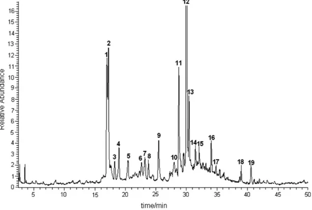
Figure 1 depicts a typical chromatogram obtained from sugarcane juice, identifying the marked peaks. The compounds corresponding to peaks 1 (schaftoside), 2
(isoschaftoside), 5 (vitexin), 9 (4’,5’-dimethyl-luteolin-8-C-glycoside), 11 (tricin-7-O-neohesperoside), and
16 (tricin-7-O-glycoside) were identiied and reported in one of our previous studies of sugarcane juice.11 On the other hand, although the compounds corresponding to peaks 3 (diosmetin-8-C-glycoside), 12 (tricin-7-O -rhamnosylgalacturonide), 15 (tricin-4’-O-(erythro- or
threo-guaiacylglyceryl) ether-7-O-glucopyranoside), and
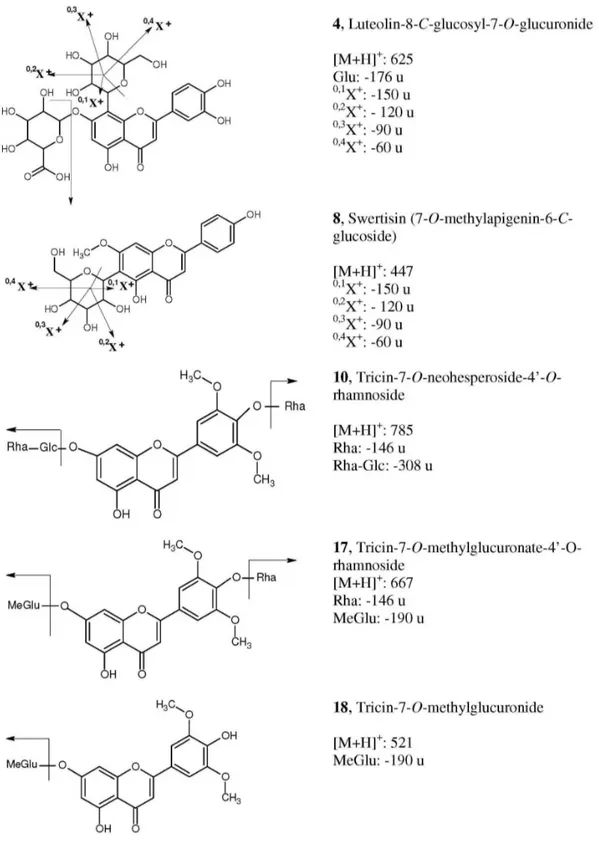
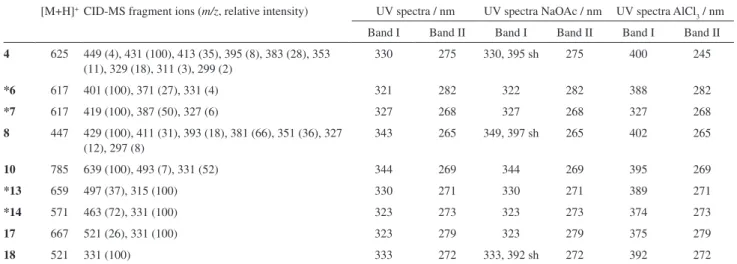
19 (tricin-4’-O-(erythro- or threo-guaiacylglyceryl) ether) were also reported previously, they had been found only in sugarcane leaves or bagasse.12 The structural elucidation of the ive additional minor lavones from sugarcane juice (Figure 2) is discussed below, and the HPLC-APCI-CID-MS and on-line UV data of these compounds are shown in Table 1. Some of the identiications and discussions are also based on the comparison of data herein shown with those obtained in a study using exact mass measurement for unequivocal elucidation of isomeric sugarcane lavones.13
The APCI spectrum of compound 4 presented an [M+H]+ ion at m/z 625 and an abundant fragment at m/z 449 [M+H-176]+. The loss of 176 u is characteristic of a glucuronic acid moiety,14,15 while the ion at m/z 449 suggests a luteolin-C-glucoside derivative. CID-MS/MS of ion m/z 449 generated fragments at m/z 431; 413; 395;
Figure 1. TIC-LC/APCI-MS of sugarcane juice extract. The chromatographic and MS conditions are described in the Experimental section. Peaks identiication: 1 (schaftoside), 2 (isoschaftoside), 3 (diosmetin-8-C-glycoside), 4 (luteolin-8-C-glucosyl-7-O-glucuronide), 5 (vitexin), 8 (swertisin),
9 (4’,5’-dimethylluteolin-8-C-glycoside), 10 (tricin-7-O-neohesperoside-4’-O-rhamnoside), 11 (tricin-7-O-neohesperoside), 12
(tricin-7-O-rhamnosylgalacturonide), 15 (tricin-4’-O-(erythro- or threo-guaiacylglyceryl) ether-7-O-glucopyranoside), 16 (tricin-7-O-glycoside), 17 (tricin-7-O-methylglucuronate-4’-O-rhamnoside), 18 (tricin-7-O-methylglucuronide), 19 (tricin-4’-O-(erythro- or threo-guaiacylglyceryl) ether). Peaks 6, 7, 13 and
Figure 2. Structure of the lavones from sugarcane juice described in this work and fragmentation pathways. Numbering of the compounds as in Figure 1. Fragments nomenclature as in reference 16. Glu = glucuronic acid; Glc = glucose; Rha = rhamnose; MeGlu = methylglucuronide.
383; 353, 329, 311 and 299, which are characteristic of the C-glucoside fragmentation pathway (Figure 2) and are identical to those described for luteolin-8-C-glucoside (orientin).16 The position of the glucuronic acid and glucose moieties was also consistent with the data from the UV
71 nm, indicating a free 5-OH group and the presence of the ortho-dihydroxyl group. Therefore, the glucuronic acid moiety was attributed to the 7- position, also considering that CID-MS/MS data and 0,2X ion16 (Figure 2) pointed to an 8-C-glucoside.
The compound 8 was identiied as being swertisin (7-O-methylapigenin-6-C-glucoside), reported previously in sugarcane molasses and also in other plant sources.4,14 This proposition is conirmed by the [M+H]+ ion at m/z 447 and by the peak at m/z 327, which indicates a loss of 120 u (0,2X ion,16 Figure 2) from the [M+H]+ ion, a characteristic feature of a C-glucoside lavonoid.16 The CID-MS/MS data also indicated a fragmentation pathway of a C-linked glucose moiety (Figure 2; m/z 429, 411, 393, 381, 351, 327 and 297), and analysis of 0,2X ion indicated an 6-C-glucoside.16 The UV analysis with post-column addition of sodium acetate showed the absence of a bathochromic shift of band I, conirming the presence of a blocked 7-OH group and showed also a shoulder in band II indicating a free OH group in position 4’. The UV spectrum obtained with aluminum chloride, while conirming a free 5-OH group, also reveals the absence of
ortho-dihydroxyl groups.
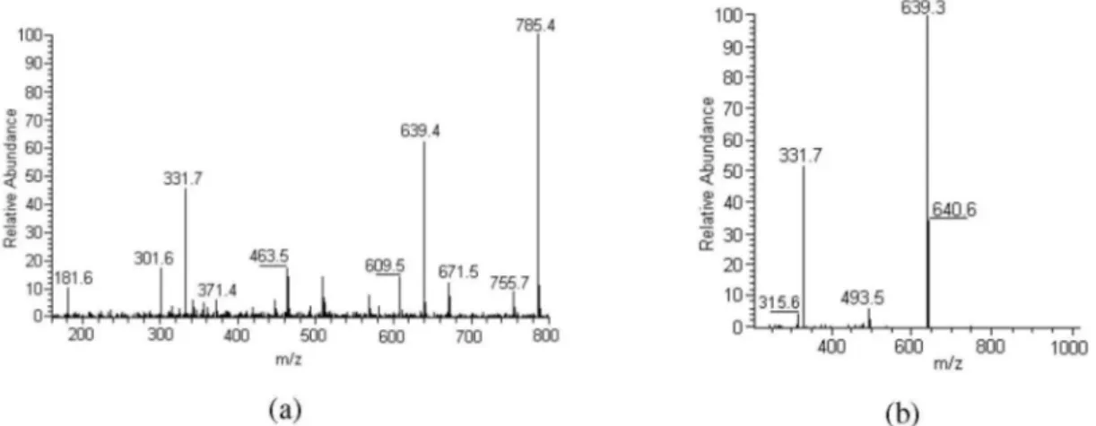
For compound 10, identiied as tricin-7-O -neohes-peroside-4’-O-rhamnoside, the MS and CID-MS/MS analyses showed respectively the [M+H]+ ion at m/z 785 and the fragments at m/z 639, 493 and 331. These fragments have already been described for tricin 7-O-neohesperidoside (compound 11)11 and were also studied by exact mass measurement.13 The fragment at m/z 639 [M+H-146]+ indicated loss of a rhamnose moiety.14,17 The UV data obtained with sodium acetate showed positions 7, 3’ and 4’ blocked, while the 5-OH group was free.
In the case of compound 17, the peak at m/z 667
corresponded to the [M+H]+ ion. The CID-MS/MS fragments at m/z 521 and 331 (the latter indicating the aglycone tricin11,12), showed a loss of 146 and 190 u, respectively. The loss of 146 u is characteristic of a rhamnose moiety,14,17 while the loss of 190 u is reported in the literature as indicative of a methylglucoronic acid group.18,19 The identiication of tricin-7-O-methylglucuronate-4’-O-rhamnoside was also conirmed by the absence of a bathochromic shift in band II with addition of sodium acetate, indicating that position 7 was blocked by the methylglucuronic acid moiety. The absence of a bathochromic shift and shoulder in band I pursuant to the addition of sodium acetate also indicated that positions 3’ and 4’ were blocked.
APCI-MS data of compound 18 showed the [M+H]+ ion located at m/z 521, while the ion at m/z 331 was attributed to the [A+H]+ ion of tricin. In addition, the loss of 190 u was attributed to a methylglucuronic acid moiety (as in
17). The UV analysis with shift reagents indicated the free 4’ position, due to the presence of a shoulder in band I resulting from the addition of sodium acetate, while the absence of a shift in band II indicated a substituted 7-OH group.
The identity of the compounds corresponding to peaks
6, 7, 13 and 14 was not fully elucidated, but some partial propositions about their structure were deduced from their HPLC-UV-MS data (Table 1). All these compounds are glycosilated lavones but the full identiication of the nature and position of the substituent groups was not achieved.
Conclusions
Among the ive compounds fully described here, only swertisin was previously reported in sugarcane molasses.4 Another triglycosilated lavonoid from sugarcane leaves Table 1. On-line HPLC-APCI-CID-MS and UV/DAD data of sugarcane juice lavones described in this work. Numbering according to Figure 1
[M+H]+CID-MS fragment ions (m/z, relative intensity) UV spectra / nm UV spectra NaOAc / nm UV spectra AlCl 3 / nm
Band I Band II Band I Band II Band I Band II
4 625 449 (4), 431 (100), 413 (35), 395 (8), 383 (28), 353 (11), 329 (18), 311 (3), 299 (2)
330 275 330, 395 sh 275 400 245
*6 617 401 (100), 371 (27), 331 (4) 321 282 322 282 388 282
*7 617 419 (100), 387 (50), 327 (6) 327 268 327 268 327 268
8 447 429 (100), 411 (31), 393 (18), 381 (66), 351 (36), 327 (12), 297 (8)
343 265 349, 397 sh 265 402 265
10 785 639 (100), 493 (7), 331 (52) 344 269 344 269 395 269
*13 659 497 (37), 315 (100) 330 271 330 271 389 271
*14 571 463 (72), 331 (100) 323 273 323 273 374 273
17 667 521 (26), 331 (100) 323 279 323 279 375 279
18 521 331 (100) 333 272 333, 392 sh 272 392 272
is described in the literature,4 but this is the irst report of the triglycosilated lavone 10 in sugarcane. Three other compounds were partially identiied as tricin derivatives. Several tricin derivatives have already been described in
Saccharum species.5,11,12,20
The hyphenated HPLC-UV-MS technique proved to be a very important tool for the on-line recording of structural information about minor compounds in a complex matrix such as that of sugarcane juice. Therefore, the on-line structural elucidation of components can offer valuable contributions to the scope of the analytical chemistry (scientiic studies or technological applications such as quality assessment) of extracts of plant foods such as sugarcane and its derivatives.
Acknowledgments
The authors wish to thank the Brazilian institutions FAPESP (fellowship to R.C., 00/11645-2; research grant to J. H. Y., 06/59457-6) and CNPq (fellowship to J. H. Y.) for their inancial support, and the Post-Graduate Dean’s Ofice of the Universidade de São Paulo (travel fellowship to R. C.). Swiss inancial support for this work was provided by the Swiss National Science Foundation and the Fondation Herbette, University of Lausanne.
Supplementary Information
Supplementary data are available free of charge at http://jbcs.sbq.org.br as PDF ile.
References
1. Wolfender, J.-L.; Ndjoko, K.; Hostettmann, K.; J. Chromatogr., A 2003, 1000, 437 and references cited herein.
2. Polasek, J.; Queiroz, E. F.; Hostettmann, K.; Phytochem. Anal.
2007, 18, 13.
3. Fiehn, O.; Plant Mol. Biol. 2002, 48, 155.
4. Smith, P.; Paton, N. H.; Sugar Technol. Rev. 1985, 12, 117. 5. Vila, F. C.; Colombo, R.; de Lira, T. O.; Yariwake, J. H.; J. Braz.
Chem. Soc. 2008, 19, 903 and references cited herein. 6. Ferreira, R. Q.; de Lira, T. O.; Zeraik, M. L.; Yariwake, J. H.;
Avaca, L. A.; J. Agric. Food Chem., submitted.
7. Duarte-Almeida, J. M.; Novoa, A. V.; Linares, A. F.; Lajolo, F. M.; Genovese, M.; Plant Food Hum. Nutr. 2006, 61, 187. 8. Falco, M. C.; Silva-Filho, M. C.; Plant Physiol. Biochem. 2003,
41, 761.
9. de Lira, T. O.; Yariwake, J. H.; Ribeiro, J.; Ferreira, M. M. C., unpublished work.
10. Ducrey, B.; Wolfender, J.-L.; Marston, A.; Hostettmann, K.; Phytochemistry 1995, 38, 129.
11. Colombo, R.; Yariwake, J. H.; Queiroz, E. F.; Ndjoko, K.; Hostettmann, K.; Phytochem. Anal. 2006,17, 337.
12. Colombo, R.; Yariwake, J. H.; Queiroz, E. F.; Ndjoko, K.; Hostettmann, K.; J. Chromatogr., A 2005, 1082, 51.
13. Colombo, R.; Yariwake, J. H.; McCullagh, M.; J. Braz. Chem. Soc.2008, 19, 483.
14. Wolfender, J.-L.; Rodriguez, S.; Hostettmann, K.; J. Chromatogr., A 1998, 794, 299.
15. Schütz, K.; Kammerer, D.; Carle, R.; Schieber, A.; J. Agric. Food Chem. 2004,52, 4090.
16. Waridel, P.; Wolfender, J.-L.; Ndjoko, K.; Hobby, K. R.; Major, H. J.; Hostettmann, K.; J. Chromatogr., A 2001, 926, 29 and references cited herein.
17. de Rijke, E.; Out, P.; Niessen, W. M. A.; Ariese, F.; Gooijer, C.; Brinkman, U. A. T.; J. Chromatogr., A 2006, 1112, 31.
18. Souleles, C.; J. Nat. Prod. 1989, 52, 1311.
19. Huellin, J.; Lin, Y. T.; Huang, Y. J.; Wen, K. C.; Chen, R. M.; Ueng, T. H.; Liao, C. H.; J. Food Drug Anal. 2001, 9, 6. 20. Duarte-Almeida, J. M.; Negri, G.; Salatino, A.; de Carvalho, J.
E.; Lajolo, F. M.; Phytochemistry 2007, 68, 1165.
Received: October 22, 2008 Web Release Date: August 31, 2009
Supplementary Information
*e-mail: [email protected]
On-line Identiication of Minor Flavones from Sugarcane Juice by LC/UV/MS
and Post-Column Derivatization
Renata Colombo,a Janete H. Yariwake,*,a Emerson F. Queiroz,b Karine Ndjokob and Kurt Hostettmannb
aInstituto de Química de São Carlos, Universidade de São Paulo, CP 780, 13560-970 São Carlos-SP, Brazil
bLaboratoire de Pharmacognosie et Phytochimie, École de Pharmacie Genève-Lausanne,
Université de Genève, Quai Ernest Ansermet 30, 1211, Genève 4, Genève, Switzerland
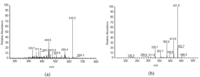
Figure S2. HPLC-APCI-CID-MS spectra of compound 8. (a) CID-MS; (b) CID-MS/MS of ion m/z 447.
Figure S5. HPLC-APCI-CID-MS spectra of compound 18. (a) CID-MS; (b) CID-MS/MS of ion m/z 521.
Figure S4. HPLC-APCI-CID-MS spectra of compound 17. (a) CID-MS; (b) CID-MS/MS of ion m/z 667.