Short Report
Printed in Brazil - ©2017 Sociedade Brasileira de Química0103 - 5053 $6.00+0.00*e-mail: [email protected]
Secondary Metabolites from an Infusion of
Lippia gracilis
Schauer Using the
LC-DAD-SPE/NMR Hyphenation Technique
Valéria R. S. Moraes,*,a,b Sérgio S. Thomasi,b,c Ricardo F. Sprenger,b Vilma M. J. Prado,a
Elizangela M. O. Cruz,d Quezia B. Cass,b Antônio G. Ferreirab and Arie F. Blankd
aDepartamento de Química, Universidade Federal de Sergipe, Av. Marechal Rondon s/n,
49100-000 São Cristovão-SE, Brazil
bDepartamento de Química, Universidade Federal de São Carlos, Rodovia Washington Luiz,
km 235, 13565-905 São Carlos-SP, Brazil
cDepartamento de Química, Universidade Federal de Lavras, Av. Doutor Sylvio Menicucci,
1001, Kennedy 37200-000 Lavras-MG, Brazil
dDepartamento de Agronomia, Universidade Federal de Sergipe, Av. Marechal Rondon s/n,
49100-000 São Cristovão-SE, Brazil
The genus Lippia comprises approximately 200 species of herbs, shrubs and small trees, distributed throughout the South and Central America and tropical Africa. The species Lippia gracilis Schauer is an endemic aromatic plant of the Brazilian Northeast normally found in the states of Bahia, Sergipe and Piauí. The traditional communities of Caatinga, a semi-arid region of the Brazilian Northeast, use its leaves to treat throat and mouth infections, cutaneous diseases, ulcers, vagina disorders, acne, Pityriasis alba, dandruff, burns, wounds, sinusitis, bronchitis, nasal congestion, headache, jaundice and paralysis. Considering that the main investigations into the L. gracilis Schauer species are focused on its volatile constituents, in this report, we describe the isolation, by liquid chromatography with diode array detector-solid phase extraction/nuclear magnetic resonance (LC-DAD-SPE/NMR), of five flavonoids besides free and glycosidically bound carvacrol from an infusion of the leaves of a genotype of this species.
Keywords:Lippia gracilis Schauer, infusion, LC-DAD-SPE/NMR, flavonoids, carvacrol
Introduction
Brazilian plants have a great potential to be explored because, for many of them, there is little or no knowledge of their chemical compositions.1Lippia gracilis Schauer
(Verbenaceae) is a species that is included in this group once, to our knowledge, there is only one report about its non-volatile constituents,2 while most reports are focused
on studies on the composition of essential oils and their biological activities.3-21
The genus Lippia comprises approximately 200 species of herbs, shrubs and small trees, distributed throughout the South and Central America countries and tropical Africa.22
The species L. gracilis Schauer is an endemic aromatic plant to the Brazilian Northeast normally found in the states of Bahia, Sergipe and Piauí. The traditional communities
of Caatinga, a semi-arid region of the Brazilian Northeast, use its leaves to treat throat and mouth infections, cutaneous diseases, ulcers, vagina disorders, acne, Pityriasis alba, dandruff, burns, wounds, sinusitis, bronchitis, nasal congestion, headache, jaundice and paralysis.23-25
Although there is little knowledge about the non-volatile constituents of L. gracilis, there are several phytochemical studies describing the isolation of various compounds from other species of this genus, including iridoid glucosides, phenylethanoid glucosides, phenylpropanoids, triterpenes saponins, flavonoids, naphthoquinones, flavonoids glucosides, lignans, sesquiterpenes and triterpenes.3-24
Previous chemical investigations into the L. gracilis
Schauer species are focused on its volatile constituents where thymol and carvacrol, which showed strong antimicrobial activity against fungi and bacteria, are the main components.12 The chemical composition of the
major components, probably due to genetic conditions depending on the location and conditions under which the plant is grown.
Another study evaluated the chemical composition of essential oils extracted from two different locations of the Caatinga area in the Pernambuco State, Brazil (Buíque and Ouricuri). It was demonstrated that carvacrol and p-cimene are the major constituents of the essential oil from Buíque, while thymol, γ-terpinene and 4-methoxy-acetophenone predominated in the samples from Ouricuri.10 In another
study, the composition of essential oils of L. gracilis from other three states of northeastern Brazil (Ceará, Piauí and Sergipe) was analyzed, and it was demonstrated that thymol and p-cimene predominate in the samples from Ceará15 and
Sergipe,9 while carvacrol and p-cimene predominate in the
sample from Piauí.16
The literature only reports one study on the isolation of fixed constituents from the methanolic extract from the leaves of L. gracilis Schauer, which presented the isolation of the known flavanone: naringenin.2 Two other works only
show the characterization of teas and methanolic extracts from L. gracilis Schauer genotypes cultivated in the rural campus of the Federal University of Sergipe and originated from the states of Sergipe and Bahia (Brazil) by liquid chromatography with diode array detector (LC-DAD) combined with chemometric analysis.22,26
In view of the above, in this study, we report the application of the LC-DAD-SPE/NMR (LC-DAD-solid phase extraction/nuclear magnetic resonance) hyphenation technique to isolate and identify some of the constituents of infusions of the leaves of a genotype of L. gracilis Schauer.
LC-DAD-SPE/NMR is a hyphenated technique that allows the chromatographic peaks to be automatically trapped on SPE cartridges, making multiple trappings possible, which results in a significant increase in the amount of isolated substances, facilitating the NMR analyses.27
This technique is very useful mainly when we are interested in discovering the components present in a mixture, even those in smaller quantities, avoiding the need to isolate them previously by open column chromatography techniques, which would be more time-consuming. Furthermore, a smaller sample handling occurs, avoiding the possibility of generating artifacts by oxidations, solvolysis, and thermal or photochemical degradation.28,29
Experimental
General experimental procedures
The analyses were carried out using an Agilent liquid chromatographic system (1200 series, Agilent
GmbH) equipped with a quaternary pump (G1311A) and a degasser (G1322A) operating at 1.0 mL min-1, a
variable wavelength diode array detector (G1315D), an autosampler (G1329A), and an automatic cartridge exchanger (Bruker Biospin GmbH). The LC system was controlled by the HyStar 2.3 software (Bruker). A Knauer (K120 Knauer Smartline Pump Control 100, Bruker Daltonik GmbH©, V01.11) makeup pump diluted the post column flow with water before the peaks were trapped using a Prospekt 2 SPE unit.
The 1D and 2D NMR spectra of the isolated compounds were acquired at 299 K with the use of a Bruker Avance III instrument (14.1 Tesla, Ultrashield Plus®) fitted with
an automatic sample changer and a triple resonance cryoprobe inverse of 5 mm (1H/13C/15N) with z-field
gradient. Data collection and processing were carried out using the Bruker software Topspin 3.2 version. Each
1H NMR spectrum obtained was recorded using 1D NOESY
(nuclear Overhauser effect spectroscopy) pulse sequence with double presaturation (HDO and deuterated methanol) acquired with 32 scans; acquisition time of 2.7 s; relaxation delay of 2.4 s; 4 dummy scans and 65,536 data points. Spectra were processed without the use of apodization function, TD = 65,536 and using 32,768 data points. COSY (correlation spectroscopy) spectra were acquired with 16 scans; 8 dummy scans; TDF1 = 256 and TDF2 = 4,096. Heteronuclear single quantum correlation (HSQC) spectra were acquired with 32 scans; 16 dummy scans; TDF1 = 256 and TDF2 = 4,096. Heteronuclear multiple bond correlation (HMBC) spectra were acquired with 200 scans; 16 dummy scans; TDF1 = 281, TDF2 = 4,096 and optimized for nJ
C,H = 8 Hz. All the 2D spectra were processed without
the use of apodization function, with number of points in F1 domain of 1,024; number of points in F2 domain of 4,096 and using linear prediction in F1. The spectra were referenced to tetramethylsilane (TMS) at 0.0 ppm and the chemical shifts of all hydrogens and carbons were reported using d values.
The mass spectrometry experiments were carried out using an ESI-ITMS BrukerTM Esquire 6000 equipment and
the data processing was accomplished with the DataAnalysis software. For sample preparation and LC analyses, LC-grade solvents such as acetonitrile (JT Baker, Philipsburg, PA, USA) and methanol (JT Baker, Philipsburg, PA, USA) were used. Deionised water was purified by a Milli-Q® system
Plant material
Leaves of the L. gracilis genotype called 110 (cultivated without irrigation) originated from the state of Sergipe (11°19’21.1’’S; 37°55’14.9’’W) and cultivated at the Experimental Farm of the Rural Campus of the Federal University of Sergipe (UFS) were collected in January 2010. The leaves were identified by PhD Ana Paula Prata, and a voucher specimen was deposited at the Federal University of Sergipe Herbarium, Department of Biology under number 14732. The material was dried in a forced air circulation oven at 40 °C for 5 days.19,26
Sample preparation
The tea was prepared by the infusion method where 200 mL of Milli-Q water at 94 °C (heated in a microwave oven) were added to 2.0 grams of leaves and it was left to rest for 10 minutes. This procedure was performed three times. Then, the tea was filtered through analytical paper, frozen and lyophilized (at the temperature of −54 °C and pressure of 79 µm Hg), providing 527.9 mg of extract.26
LC conditions used in LC-DAD-SPE
For the chromatographic separations, solutions containing 31.9 mg mL-1 of sample were prepared in
methanol and ultrapure water (8:2 v/v) with 4 drops of triflouoroacetic acid (Aldrich); subsequently, it was filtered through a PVDF membrane syringe filter (25 mm, 0.45 µm, Tedia, Brazil) prior to the LC analyses. The chromatographic separations were carried out using an analytical Eurobond Prontosil C18 Luna® column
(125 × 4.0 mm, 5 µm) with 20 µL injection volumes. To isolate compounds 1 and 2, a binary eluent consisting of an aqueous solution of trifluoroacetic acid (TFA, Tedia, Brazil) 0.05% (v/v) (A) and MeOH (B) at 1.0 mL min-1 was used
with a gradient as follow: 5-28% (B) in 14 min, 28-34.5% (B) in 26 min, 34.5-100% (B) in 2 min and, maintained at 100% for 5 min (clean-up) before returning to the initial conditions in 2 min and, then, conditioning for a further 5 min. The elution was monitored based on UV absorption at 206 nm and, for each chromatographic peak, a total of twenty-five trappings were made. To isolate compounds 3
to 7, a binary eluent consisting of an aqueous solution of TFA 0.05% (v/v) (A) and ACN with 0.05% aqueous TFA (B) at 1.0 mL min-1 was used. The gradient employed was:
5-13% (B) in 5 min, 13-28% (B) in 15 min, 28-100% (B) in 15 min, maintained at 100% (B) for 5 min (clean-up). The initial condition was achieved in 1 min and maintained for conditioning for a further 4 min. The elution profile
was monitored based on UV absorption at 200 nm and a total of thirty trappings were carried out. In the end of each chromatographic analysis, the cartridges were dried with nitrogen gas for 30 minutes; the compounds were eluted with methanol-d4 (Deutero, 99.8%) and transferred to NMR
tubes (Bruker, 3 mm o.d.) in order to be analyzed.
Results and Discussion
Considering that one of the main motivations for studying a plant comes from its popular indication for some type of illness, and/or due to any scientifically-proven biological activity, the time and dedication spent isolating the active compounds using open column chromatography may not be worth it, once it commonly results in the isolation of known compounds.30 Thus, for gaining chemical
information with productivity, LC-DAD-SPE/NMR was used for the chemical characterization of the infusion of L. gracilis Schauer genotype 110.
The chromatographic conditions used for the isolation of compounds were based on the chromatographic profile as previously published by our research group,26 with
minor modifications in order to optimize the trapping of the compounds, with high throughput.
To meet this end, the obtained chromatogram profile was divided into two sections; at first, our interest was focused in the two most intense peaks with retention times of 40 and 50 minutes. The substitution of formic acid aqueous solution 0.5% (v/v) as eluent (A) for TFA solution affected the retention times of both peaks and they elluted before 30 min. The gradient profile of the organic modifier MeOH (B) was, thus, adjusted in order to trap orientin (1, tR = 27.0 min,
600 µg),31,32 and isoorientin (2, t
R = 29.0 min, 500 µg)31,32
flavonoids.
For the second section of the original chromatographic profile with compounds that eluted after 50 minutes,26
the exchange of methanol as the modifier for acetonitrile led to higher resolution which improved the trapping process. At the developed chromatographic conditions, it was possible to trap luteolin 4’-O-β-glucopyranoside (3,
tR = 17.5 min, 700 µg),33,34 carvacrol 2-O-β-glucopyranoside
(4, tR = 22.7 min, 500 µg),35 5,7,3’,5’-tetrahydroxy flavanone
(5, tR = 23.2 min, 300 µg),36 naringenin (6, tR = 25.4 min,
500 µg)2 and carvacrol (7, t
R = 29.5 min, 600 µg).31
The SPE trapping method chosen was the UV absorption threshold, which selects the bands based on the intensity of their UV absorptions, this approach minimizes the errors caused by the slight variation in the retention time of the selected bands during the chromatographic runs.
1D NOESY with double saturation for solvent signal suppression) and 2D NMR (COSY, HSQC and HMBC) data as well as by comparing them with data reported in the literature.
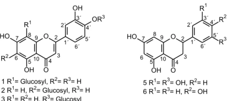
The 1H NMR spectrum of compound 1 showed signals
of aromatic ring B at d 7.56 (s), 7.52 (d, J = 8.1 Hz) and 6.91 (d, J = 8.1 Hz) corresponding to hydrogens on C-2’, 6’ and 5’, respectively. A doublet at d 4.98 (J = 9.8 Hz) and two singlets at d 6.27 and 6.54 were assigned to the anomeric hydrogen (H-1’’) of the glycosyl unit attached to C-8 and to the hydrogens on C-6 and C-3, respectively, confirming the flavone skeleton. The location at C-8 of the sugar moiety was established on the basis of the cross peak from H-1’’ (d 4.98) to C-7 (d 165.9), C-8 (d 106.6) and C-9 (d 159.4) in the HMBC spectrum.
The 1H NMR spectrum of compound 2 was similar
to that of orientin, showing signals for three aromatic hydrogens of the typical ABX system (ring B) at d 7.38 (s), 7.39 (d, J = 8.3 Hz) and 6.91 (d, J = 8.3 Hz) corresponding to hydrogens on C-2’, 6’ and 5’, respectively. A doublet at d 4.90 (J = 9.5 Hz) and two singlets at d 6.51 and 6.56 were assigned to the anomeric hydrogen (H-1’’) of the glycosyl unit attached to C-6 and to the hydrogens on C-8 and C-3, respectively, typical of a flavone skeleton. In this case, the location of the sugar moiety was established at C-6 on the basis of the cross peak from H-1’’ (d 4.90) to C-7 (d 166.6), C-6 (d 110.4) and C-5 (d 163.4) in the HMBC spectrum.
Based on these data and on those obtained by 2D NMR (tables presented in the Supplementary Information section), it was possible to identify the compounds as 3’,4’,5,7-tetrahydroxyflavone-8-glucoside (orientin) and 3’,4’,5,7-tetrahydroxyflavone-6-glucoside (isoorientin).31,32
To further furnish insight in the chemical structures of compounds 1 and 2 they were also submitted to analysis by (−)ESI-ITMS2 (electrospray ionization-ion trap tandem
mass spectrometry). For 1 and 2, the MS2 spectra presented
the product ions m/z 357 [M – H − 90]- and m/z 327
[M – H − 120]-, which are representative of 0,3X and 0,2X
glucose cross-ring cleavage. The differentiation of both is based on the intensity of these ions: for orientin, the base peak is m/z 327, while for isoorientin, it is m/z 357.37
Furthermore, the isoorientin fragmentation spectrum reveals the loss of water (m/z 429 and 411) typical of C-6 and absent from C-8 flavones.38
The 1H NMR spectrum of compound 3 showed a singlet
at d6.61, which presented an HSQC correlation (H−C directly bonded) with a carbon at d 106.4, which is typical of the flavone skeleton. Furthermore, this spectrum displayed signals for three aromatic hydrogens of the typical ABX system(ring B) at d 7.46 (m), 7.47 (d, J = 8.4 Hz) and 7.33 (d, J = 8.4 Hz) corresponding to hydrogens on C-2’, 6’ and
5’, respectively. A meta-coupled AX system for H-6 and H-8 (ring A) was confirmed by the two doublets at d 6.22 (J = 2.1 Hz) and 6.46 (J = 2.1 Hz), respectively. A doublet at d 4.94 (J = 7.5 Hz) was assigned to the anomeric hydrogen (H-1’’) of a glycosyl O-linked to the C-4’ position in the ring B, which was established on the basis of the cross peak from H-1’’ (d 4.94) to C-4’ (d 151.2) in the HMBC spectrum. These results aided by 2D NMR data (see Supplementary Information) allowed the identification of this compound as luteolin-4’-O-β-glucopyranoside.33,34
The 1H NMR spectra of compounds 4 and 7 were very
similar, showing characteristic signals of carvacrol 2-O-β -glucopyranoside and carvacrol, respectively. The presence of an isopropyl group was confirmed by septets at d 2.84 (4: 1H, J = 6.9 Hz) and 2.76 (7: 1H, J = 7.0 Hz) and doublets at d 1.22 (4: 2CH3, J = 6.9 Hz) and 1.19 (7: 2CH3, J = 7.0 Hz)
in each spectrum. In addition, two singlets at d 2.22 (4) and 2.12 (7) corresponding to the methyl group attached to the benzene ring were also observed in each compound. Furthermore, each spectrum showed signals for three aromatic hydrogens of the typical ABX system. For the compound 4, these signals appeared at d 7.01 (d, J = 8.0 Hz, H-6), 7.00 (d, J = 1.8 Hz, H-3) and 6.78 (dd, J = 8.0 and 1.8 Hz, H-5), and for the compound 7 at d 6.93 (dd, J = 7.4 and 0.5 Hz, H-6), 6.59 (dd, J = 7.4 and 1.7 Hz, H-5) and 6.61 (d, J = 1.7 Hz, H-3). A doublet at d 4.86 (J = 7.6 Hz) was assigned to the anomeric hydrogen (H-1’) of a glycosyl
O-linked to the C-2 position in the benzene ring, which was confirmed on the basis of the cross peaks from H-1’ (d 4.86), H-6 (d 7.01) and H-7 (d 2.22) to C-2 (d 158.5) in the HMBC spectrum. On the basis of these analyses and those from the 2D NMR data (see Supplementary Information), the structures of compounds 4 and 7 were confirmed as being carvacrol 2-O-β-glucopyranoside and carvacrol, respectively.31,35
The 1H NMR spectra of compounds 5 and 6 displayed
On the other side, an AA’XX’ system was observed for ring B for compound 6, which was confirmed by the presence of two doublets at d 7.32 (J = 8.7 Hz, H-2’/6’) and 6.82 (J = 8.7 Hz, H-3’/5’). These results combined with 2D NMR data (see Supplementary Information) allowed the identification of compounds 5 and 6 as 5,7,3’,5’-tetrahydroxy flavanoneand naringenin, respectively.2,36
Conclusions
This is the second report about fixed constituents from
L. gracilis Schauer. Herein, we describe the isolation and complete characterization of seven out of ten trapped compounds by LC-DAD-SPE/NMR from the infusion of the leaves of genotype 110 of this species: orientin 1, isoorientin 2, luteolin 4’-O-β-glucopyranoside luteolin 3, carvacrol 2-O-β-glucopyranoside 4, 5,7,3’,5’-tetrahydroxy flavanone 5, naringenin 6, and carvacrol 7.
Furthermore, to our knowledge, the compounds 1 to
5 have never been reported for this genus. In this way, the use of the LC-DAD-SPE/NMR approach allowed greater knowledge about the chemical composition of the compounds present in the infusions of L. gracilis with only 45 hours of chromatographic run time.
Supplementary Information
The tables containing the NMR data, 1H NMR and UV
spectra (acquired using DAD) for each compound isolated and identified are available free of charge at http://jbcs.sbq. org.br as PDF file.
Acknowledgments
The authors thank the Brazilian funding agencies FAPITEC/SE, FAPESP (2011/11021-3 and 201301710-1), CNPq and CAPES for their financial support and scholarships, as well as INCT-Biorational Control of Plague Insects and Northeast Biotechnology Network (RENORBIO).
References
1. Cecílio, A. B.; Faria, D. B.; Oliveira, P. C.; Caldas, S.; Oliveira, D. A.; Sobral, M. E. G.; Duarte, M. G. R.; Moreira, C. P. S.; Silva, C. G.; Almeida, V. L.; J. Ethnopharmacol. 2012, 141, 975.
2. Guimarães, A. G.; Gomes, S. V. F.; Moraes, V. R. S.; Nogueira, P. C. L.; Ferreira, A. G.; Blank, A. F.; Santos, A. D. C.; Viana, M. D.; Silva, G. H.; Quintans, L. J.; J. Nat. Med. 2012, 66, 428. 3. Pascual, M. E.; Slowing, K.; Carretero, E.; Mata, D. S.; Villar,
A.; J. Ethnopharmacol. 2001,76, 201.
4. Bitu, V.; Botelho, M. A.; Costa, J. G. M.; Rodrigues, F. F. G.; Veras, H. N. H.; Martins, K. T.; Lyra, A.; Coluchi, G. G.; Ruela, R. S.; Queiroz, D. B.; Siqueira, J. S.; Quintans, L. J.; Rev. Bras. Farmacogn. 2012,22, 69.
5. Guilhon, C. C.; Raymundo, L. J. R. P.; Alviano, D. S.; Blank, A. F.; Arrigoni-Blank, M. F.; Matheus, M. E.; Cavalcanti, S. C. H.; Alviano, C. S.; Fernandes, P. D.; J. Ethnopharmacol. 2011,
135, 406.
6. Cavalcanti, S. C. H.; Niculau, E. S.; Blank, A. F.; Câmara, C. A. G.; Araújo, I. N.; Alves, P. B.; Bioresour. Technol. 2010,101, 829.
7. Neto, R. M.; Matos, F. J. A.; Andrade, V. S.; Melo, M. C. N.; Carvalho, C. B. M.; Guimarães, S. B.; Pessoa, O. D. L.; Silva, S. L.; Silva, S. F. R.; Vasconcelos, P. R. L.; Rev. Bras. Farmacogn.
2010,20, 261.
8. Mendes, S. S.; Bomfim, R. R.; Jesus, H. C. R.; Alves, P. B.; Blank, A. F.; Estevam, C. S.; Antoniolli, A. R.; Thomazzi, S. M.; J. Ethnopharmacol. 2010,129, 391.
9. Teles, T. V.; Bonfim, R. R.; Alves, P. B.; Blank, A. F.; Jesus, H. C. R.; Quintans-Jr, L. J.; Serafini, M. R.; Bonjardim, L. R.; Araujo, A. A. S.; Afr. J. Biotechnol. 2010,9, 8800.
10. Neves, I. A.; Oliveira, J. C. S.; Camara, C. A. G.; Schwartz, M. O. E.; J. Essent. Oil Res. 2008,20, 157.
11. Lima, M. C. L.; Lemos, T. L. G.; Pessoa, O. D. L.; Santiago, G. M. P.; Matos, F. J. A.; Arriaga, A. M. C.; Oliveira, J. P. P.; Sant’ana, A. E. G.; Chem. Nat. Comp. 2008,44, 254. 12. Albuquerque, C. C.; Camara, T. R.; Mariano, R. L. R.;
Willadino, L.; Marcelino Júnior, C.; Ulisses, C.; Braz. Arch. Biol. Technol. 2006,49, 527.
13. Santiago, G. M. P.; Lemos, T. L. G.; Pessoa, O. D. L.; Arriaga, A. M. C.; Matos, F. J. A.; Lima, M. A. S.; Santos, H. C.; Lima, M. C. L.; Barbosa, F. G.; Luciano, J. H. S.; Silveira, E. R.; Menezes, G. H. A.; Nat. Prod. Commun. 2006,1, 573. 14. Pessoa, O. D. L.; Carvalho, C. B. M.; Silvestre, J. O. V. I.;
Lima, M. C. L.; Neto, R. M.; Matos, F. J. A.; Lemos, T. L. G.;
Fitoterapia2005,76, 712.
15. Lemos, T. L. G.; Monte, F. J. Q.; Matos, F. J. A.; Alencar, F. W.; Craveiro, A. A.; Barbosa, R. C. S. B.; Lima, E. D.; Fitoterapia
1992,63, 266.
16. Matos, F. J. A.; Plantas da Medicina Popular do Nordeste - Propriedades Atribuídas e Confirmadas, 1ª ed.; Ed. UFC: Fortaleza, Brasil, 1999.
17. Melo, J. O.; Fachin, A.; Rizo, W. F.; Jesus, H. C. R.; Arrigoni-Blank, M. F.; Alves, P. B.; Marins, M. A.; França, S. C.; Arrigoni-Blank, A. F.; Genet. Mol. Res. 2014,13, 2691.
18. Cruz, E. M. O.; Costa-Junior, L. M.; Pinto, J. A. O.; Santos, D. A.; Araujo, S. A.; Arrigoni-Blank, M. F.; Bacci, L.; Alves, P. B.; Cavalcanti, S. C. H.; Blank, A. F.; Vet. Parasitol. 2013,
195, 198.
19. Cruz, E. M. O.; Pinto, J. A. O.; Fontes, S. S.; Arrigoni-Blank, M. F.; Bacci, L.; Jesus, H. C. R.; Santos, D. A.; Alves, P. B.; Blank, A. F.; Sci. World J. 2014,2014, 1.
20. Melo, J. O.; Bitencourt, T. A.; Fachin, A. L.; Cruz, E. M. O.; Jesus, H. C. R.; Alves, P. B.; Arrigoni-Blank, M. F.; Franca, S. C.; Beleboni, R. O.; Fernandes, R. P. M.; Blank, A. F.; Scher, R.; Acta Tropica 2013,128, 110.
21. Carvalho, R. R. C.; Laranjeira, D.; Carvalho, F. J. L. S.; Souza, P. E.; Blank, A. F.; Alves, P. B.; Jesus, H. C. R.; Warwick, D. R. N.; Quim. Nova2013,36, 241.
22. Gomes, S. V. F.; Santos, A. D. C.; Moraes, V. R. S.; Martins, L. R. R.; Viana, M. D.; Blank, A. F.; Pereira-Filho, E. R.; Cass, Q. B.; Nogueira, P. C. L.; Alves, P. B.; Chromatographia2010,
72, 275.
23. Lorenzi, H.; Matos, F. J. A.; Plantas Medicinais no Brasil: Nativas e Exóticas, 1a ed.; Plantarum: São Paulo, Brazil, 2002.
24. Gomes, S. V. F.; Nogueira, P. C. L.; Moraes, V. R. S.; Ecletica Quim. 2011,36, 64.
25. Albuquerque, U. P.; Medeiros, P. M.; Almeida, A. L. S.; Monteiro, J. M.; Lins, N. E. M. F.; Melo, J. G.; Santos, J. P.; J. Ethnopharmacol. 2007,114, 325.
26. Prado, V. M. J.; Moraes, V. R. S.; Nogueira, P. C. L.; Cruz, E. M. O.; Blank, A. F.; Pereira-Filho, E. R.; Martins, L. R. R.;
Quim. Nova. 2012,35, 1814.
27. Lambert, M.; Stærk, D.; Hansen, S. H.; Sairafianpour, M.; Jaroszewski, J. W.; J. Nat. Prod. 2005, 68, 1500.
28. Clarkson, C.; Stærk, D.; Hansen, S. H.; Jaroszewski, J. W.;
Anal. Chem. 2005, 77, 3547.
29. Xu, Y.-J.; Capistrano, I. R.; Dhooghe, L.; Foubert, K.; Lemière, F.; Maregesi, S.; Baldé, A.; Apers, S.; Pieters, L.; Planta Med.
2011, 77, 1139.
30. Jaroszewski, J. W.; Planta Med. 2005, 71, 691.
31. Wen, P.; Han, H.; Wang, R.; Wang, N.; Yao, X.; Asian J. Tradit. Med. 2007,2, 149.
32. Burns, D. C.; Ellis, D. A.; March, R. E.; Magn. Res. Chem.
2007,45, 835.
33. Krenn, L.; Miron, A.; Pemp, E.; Petr, U.; Kopp, B.; J. Biosci.
2003,58C, 6.
34. Patora, J.; Klimek, B.; Acta Pol. Pharm. 2002,59, 139. 35. Kamel, M. S.; Assaf, M. H.; Hasanean, H. A.; Ohtani, K.; Kasai,
R.; Yamasaki, K.; Phytochemistry 2001,58, 1149.
36. Gohari, A. R.; Ostad, S. N.; Moradi-Afrapoli, F.; Malmir, M.; Tavajohi, S.; Akbari, H.; Saeidnia, S.; Sci. World J. 2012, 2012, 1.
37. Ferreres, F.; Silva, B. M.; Andrade, P. B.; Seabra, R. M.; Ferreira, M. A.; Phytochem. Anal.2003,14, 352.
38. Waridel, P.; Wolfender, J. L.; Ndjoko, K.; Hobby, K. R.; Major, H. J.; Hostettmann, K.; J. Chromatogr. A 2001,926, 29.