http://dx.doi.org/10.1590/s2175-97902017000216098
A
r
*Correspondence: C. K. C. Malheiros. Faculdade de Farmácia. Universidade Federal do Pampa. BR 472, Km 592, Caixa Postal 118, 97508-000 - Uruguaiana - RS, Brazil. E-mail: camila.kcm@gmail.com
Preliminary
in vitro
assessment of the potential toxicity and
antioxidant activity of
Ceiba speciosa
(A. St.-Hill) Ravenna
(Paineira)
Camila Krüger Cardoso Malheiros
1,*, Joyce Sayonara Barbosa Silva
1, Tânia Cristiane Hofmann
1,
Thiane Martins Messina
1, Vanusa Manfredini
1, Jacqueline da Costa Escobar Piccoli
1, Débora
Faoro
2, Luís Flávio Souza Oliveira
1, Michel Mansur Machado
1, Fabiane Moreira Farias
11Faculty of Pharmacy, Federal University of Pampa, Uruguaiana, RS, Brazil, 2 Federal University of Santa Maria, Cachoeira do Sul, RS, Brazil
The bark tea of Ceiba speciosa, a tropical tree of the Malvaceae family, is used in the Northwestern Region of Rio Grande do Sul state, Brazil, to reduce blood cholesterol levels. However, there are no scientiic data on the eicacy and safety of this plant. The aim of the present study was to evaluate the
in vitro antioxidant and toxic potential of bark extracts of C. speciosa. We performed a preliminary phytochemical analysis by high-performance liquid chromatography-diode array detection (HPLC-DAD) and evaluated the oxidative damage to proteins and lipids, the radical scavenging efect, and genotoxicity of the lyophilized aqueous extract (LAECs) and the precipitate obtained from the raw ethanol extract (Cs1). The phytochemical proile demonstrated the presence of phenolic and flavonoid compounds. The LAECs and Cs1 prevented damage to lipids and proteins at concentrations of 50 and 10 µg/mL. They also showed a scavenging efect on 2,2-diphenyl-1-pricril-hydrazyl (DPPH) radicals in a concentration-dependent manner. Furthermore, no genotoxic efect was observed at concentrations of 10, 5 and 2 µg/mL in the Comet assay. The present study is the irst evaluation regarding the characterization of C. speciosa and its safety, and the results demonstrate its antioxidant potential and suggest that its therapeutic use may be relatively safe.
Uniterms:Ceiba speciose/phytochemistry.Ceiba speciose/antioxidant activity. Phenolic content. Comet assay. DPPH scavenging capacity. Natural medicine. Safety.
INTRODUCTION
Given the importance of medicinal plants as sources of new drugs, ethnopharmacology is an important tool for the discovery of species with potential biological activity (Carlini et al., 2006), even in the absence of scientific
information on the safety and eicacy of these species.
This branch of science studies traditional knowledge and use of plants as alternatives to essential resources for survival, such as food and medicine (Balick, Cox, 1996). Studies of medicinal plants are also of fundamental importance since many of them have a strictly local use and may offer an alternative treatment for various
diseases. The popularization of medicinal plants has made them easier to acquire; however, there remains a lack of adequate information about the properties of the plants used and their possible interactions with prescribed
drugs, as well as problems in botanical identiication, and
Policy on Medicinal Plants and Herbal Medicines (Brasil, 2007), aimed at the implementation of the therapeutic
use of medicinal plants in the Brazilian Uniied Health
System (SUS). The main aims of this policy were to encourage research focused on ensuring the proper and safe use of these species. In addition, the National List of Medicinal Plants of Interest to Brazilian Public Health System (RENISUS), containing 71 species for therapeutic purposes (Brasil, 2009), was created in order to guide studies and research that support the development of a list of medicinal plants and herbal medicines to be made available for use by the general population.
One of the most signiicant contributors to toxicity
and the development of various diseases are free radicals; however, they are fundamental to biochemical processes and represent an essential part of aerobic life and metabolism. Under normal conditions, reactive oxygen species (ROS) are an essential part of such processes, and the most common ROS include the superoxide anion (O2−•), hydrogen peroxide (H2O2), peroxyl radicals (ROO•), and reactive hydroxyl radical (HO•) (Benhammou, Bekkara, Panovska, 2009; Gaur et al., 2009). The imbalance between free radical production and endogenous detoxification systems (predominantly non-enzymatic and enzymatic antioxidants) can lead to oxidative stress. Damage occurs mainly through the interaction of these radical species and macromolecules such as lipids, proteins, and deoxyribonucleic acid (DNA) (Benhammou, Bekkara, Panovska, 2009). The interaction of free radicals with DNA can lead to mutations and may therefore promote carcinogenesis, and accentuate the processes involved in the development of atherosclerosis, arthritis, and
ischemia. ROS detoxiication systems include enzymatic
and non-enzymatic antioxidants. Enzymatic systems include superoxide dismutase, catalase, glutathione
peroxidase, and other enzymes that act speciically against
ROS (El-Shenawy, Mohammadden, Al-Fahmie, 2012). Non-enzymatic systems, either endogenous in origin or obtained through the diet, include glutathione, vitamin C, indoles, catechols and polyphenols, bioflavonoids, vitamin E, and carotenoids (Kharrazi et al., 2008). The demand for antioxidants means that plant materials are a
signiicant resource in the ight against diseases associated
with oxidative stress.
Despite their popular medicinal use for many different purposes, especially in South America, there have beenfew pharmacological studies of species of the genus Ceiba, which is not listed by RENISUS (Said, Nahla, Ehsan, 2013). Popularly known as silk tree, kapok and “barriguda”, trees of this genus belong to the family Malvaceae and are mainly found in rainforest areas.
Ceiba speciosa (A. St. Hill) is a tropical tree species with a wide geographical distribution that is usually found in the mesophytic semideciduous forests of Paraguay, Argentina and Brazil. In Brazil, they occur abundantly in the states of Rio de Janeiro and Minas Gerais (Beleski-Carneiro, Suqui, Reicher, 2002). In the state of Rio Grande do Sul, this plant is found mainly in the forest region of Alto Uruguai. This species is also a popular ornamental tree in parks and gardens because of its undemanding soil requirements, achieving satisfactory growth even in dry and sandy soils with poor chemical fertility (Veloso et al.,
1997; Cappelatti, Schmitt, 2009). The scientiic literature
describes various popular medicinal uses of several species of the genus Ceiba (Cartaxo, Souza, Albuquerque, 2010; Scarpa, 2004; Ladeji, Omekarah, Solomon, 2003). There are reports of the popular use of this plant in the northwest region of the state of Rio Grande do Sul for the reduction of serum cholesterol, triglyceride and glucose levels. The present study aimed to evaluate the in vitro antioxidant potential and toxicity of C. speciosa, contributing to our knowledge about this poorly studied plant; and to increase ethnopharmacological knowledge by collecting some phytochemical, pharmacological, and toxicological data about the species.
MATERIAL AND METHODS
Chemicals
All chemicals used were of analytical grade. High-performance liquid chromatography (HPLC) solvents were purchased from Merck (Darmstadt, Germany). All other reagents were obtained from Sigma Chemical Co. (St. Louis, MO, USA).
Plant material
The plant material was collected in May 2012 in the municipality of Santo Antônio das Missões, Rio Grande
do Sul, Brazil. The species was identiied and a voucher
specimen (RSPF 12367) was deposited in the Herbarium of the University of Passo Fundo, Passo Fundo, Rio Grande do Sul, Brazil.
Preparation of aqueous extract
The extract was iltered and the iltrate was lyophilized
resulting in the lyophilized aqueous extract of C. speciosa (LAECs).
Preparation of ethanolic extract
The raw ethanolic extract of C. speciosa was obtained by maceration until exhaustion at room temperature after which the solvent was removed under reduced pressure at 45 ºC using a rotary evaporator. During this process, a large amount of brown precipitate formed (Cs1). Little material remained in the ethanol extract after Cs1 separation, and it was thus discarded. Like the aqueous extract, Cs1 was rich in phenolic compounds and was therefore used for experiments. Further investigations on the complete ethanolic extract are being carried out.
Phytochemical analysis
Determination of total polyphenolic content
Measurement of total polyphenols was performed in accordance with the method established by Chandra
and Mejia (2004) using a inal concentration of 0.150 mg/
mL. The standard curve was prepared using gallic acid. All analyses were performed in triplicate.
Determination of total flavonoid content
The total flavonoid content of LAECs and Cs1 was
determined in accordance with Zhishen, Mengcheng and Jianming (1999), with a starting concentration of 1 mg/ mL. The standard curve was prepared using quercetin, and all analyses were performed in triplicate.
Analysis by high-performance liquid chromatography with diode array detection (HPLC / DAD)
Lyophilized aqueous extract of C. speciosa (LAECs) and Cs1 were submitted to preliminary chromatographic
analysis to determine their chromatographic proiles and
the spectra of their main compounds in the ultraviolet
region with emphasis on the quantiication of flavonoids
and phenolic compounds (quercetin, rutin, caffeic acid, gallic acid, chlorogenic acid, rosmarinic acid and kaempferol). High performance liquid chromatography (HPLC) of the samples was performed with an HPLC system (Shimadzu, Kyoto, Japan) consisting of a prominence auto sampler (SIL-20A), equipped with Shimadzu LC-20 AT reciprocating pumps connected to a DGU 20A5 degasser with a CBM 20A integrator, SPD-M20A UV-VIS-DAD (diode-detector), and Software LC solution 1.22 SP1, according to the method described by Boligon et al. (2009) with minor modiications. The
analyses were carried out with a C18 analytical column (5 µm; 4.6 x 250 mm) and a gradient combining solvent A
(1% formic acid in water) and B (acetonitrile) with a flow
rate of 0.7 ml/min (Table I). The UV absorption spectra of standards as well as the samples were recorded in the range of 230-400 nm. Table II shows the detection wavelengths of phenolic compounds analyzed.
In vitro toxicological and antioxidant evaluation
Leukocyte and plasma samples
The experimental protocols used for the cytotoxicity and genotoxicity analyses, including venous blood collection, were approved by the Research Ethics Committee of the Federal University of Pampa (Universidade Federal do Pampa [UNIPAMPA]), under registration number 27045614.0.0000.5323.
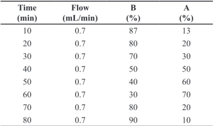
The venous blood was collected by venipuncture after 12 hours’ overnight fasting using top purple Vacutainer® (BD diagnostics, Plymouth, UK) tubes with heparin from a single healthy non-medication-using donor aged over 18 years old. The blood sample was centrifuged at 3000 rpm for 10 minutes and the white TABLE I - Gradient elution used for the evaluation of lyophilized aqueous extract (LAECs) and precipitate obtained from the raw ethanol extract (Cs1). Solvent A (1% formic acid in water). Solvent B (acetonitrile)
Time (min)
Flow (mL/min)
B (%)
A (%)
10 0.7 87 13
20 0.7 80 20
30 0.7 70 30
40 0.7 50 50
50 0.7 40 60
60 0.7 30 70
70 0.7 80 20
80 0.7 90 10
TABLE II - Wavelengths used for the detection of phenolic compounds by High-Performance Liquid Chromatography with Diode Array Detection (HPLC/DAD)
Phenolic Compound Wavelengths
(nm)
Gallic Acid 254
Cafeic, Ellagic, and Chlorogenic Acids 325
pellet (leukocytes) was transferred to another test tube
with phosphate-bufered saline (PBS; pH 7.4). The number
of leukocytes was standardized (8×10³ cells/mL) for measurements of cell viability and oxidative damage of DNA (Pereira et al., 2015).
Cell viability test
Cell viability in human leukocytes was assessed by the loss of membrane integrity using the Trypan Blue method (Burow et al., 1998) to determine the concentrations of LAECs and Cs1 to be used in in vitro tests. Stock solutions of LAECs and Cs1 (2 µg/mL) were prepared in PBS. From these, aliquots were transferred to Eppendorf tubes and diluted in a suspension of leukocytes to achieve concentrations of 1000, 750, 500, 250, 100,
50, 10, 5 and 2 µg/mL. Phosphate-bufered saline with
H2O2 was used as the positive control. The negative control consisted of a PBS and leukocyte suspension. The concentrations chosen for this study showed cell viability above 85%. The analysis was performed in triplicate.
Evaluation of oxidative damage and antioxidant potential
Radical-scavenging capacity – DPPH assay
Evaluation of in vitro antioxidant capacity was performed by measuring the abilities of LAECs and Cs1 to quench the 2,2-diphenyl-1-pricril-hydrazyl (DPPH) radical, according to the method of Sharma and Bhat (2009). The analysis was performed in triplicate.
Lipid peroxidation test
Assessment of lipid damage was performed by quantification of thiobarbituric acid reactive species (TBARS) (Ohkawa, Ohishi, Yagi, 1979). Initially, LAECs and Cs1 solutions were prepared at the previously chosen concentrations. Two hundred microliters of plasma were mixed with 100 µL H2O2 100 µmol/L (1:10 dilution) and 200 µL LAECs or Cs1 solutions. For the positive control 200 µL H2O2 was used in place of the LAECs and Cs1 solutions. In the negative control, they were replaced by saline solution. The analysis was performed in triplicate.
Determination of protein carbonyl groups
Determination of protein carbonyl groups was performed according to the method of Morabito et al. (2004). Initially, LAECs and Cs1 solutions were prepared at the previously chosen concentrations. To perform the test, 200 µL plasma was added to an Eppendorf tube and mixed with 100 µL H2O2 solution 100 µmol/L (1:10 dilution), and 200 µL LAECs or Cs1 solutions. As a
positive control, 200 µL H2O2 was used in place of the LAECs and Cs1 solutions. In the negative control, they were replaced by 200 µL PBS. The analysis was performed in triplicate.
Evaluation of genotoxicity
Comet assay
The Comet assay was used to measure the single and double strand breaks in DNA (Singh et al., 1995). An Eppendorf tube containing 200 µL extract and 200 µL leukocyte suspension was prepared. All steps after blood collection were performed in the dark or under dimmed red light to prevent additional DNA damage.
The Comet assay was performed in triplicate, analyzing 100 nuclei per sample of each concentration s t u d i e d . T h e s e 1 0 0 c e l l s w e r e t h e n e x a m i n e d microscopically at 40x magnification and classified according to tail damage: 0 being no damage to 4 being maximum damage. The sum of these values was used to ascertain a damage index (0–400) for each treatment. The analysis was performed in triplicate.
Statistical analysis
Data were expressed as mean ± standard deviation. Concentrations of flavonoid and phenolic compounds obtained by HPLC were analyzed using one-way analysis of variance (ANOVA) followed by the Tukey test. Other results were analyzed using one-way ANOVA followed by the Bonferroni test. Results were considered
statistically signiicant when p<0.05. Statistical analysis
was performed using the software GraphPadPrism 5.0.
RESULTS
Phytochemical analysis
Total flavonoid and total polyphenolic contents
Total flavonoid contents, expressed as microgram equivalents of quercetin per gram of extract sample, were 240 µg and 229 µg of quercetin/g for LAECs and Cs1, respectively. Total polyphenolic contents, expressed as microgram equivalents of gallic acid per gram of extract sample, were 425 µg and 470 µg of gallic acid/g for LAECs and Cs1, respectively.
HPLC / DAD
in Table III. The retention times (tRs) of identified compounds were: gallic acid (tR=9.93 min; peak 1), chlorogenic acid (tR = 19.36 min; peak 2), caffeic acid (tR=24.57 min; peak 3), ellagic acid (tR=30.19 min; peak 4), rutin (tR=37.41 min; peak 5), quercetin (tR=46.23 min; peak 6), and kaempferol (tR=54.13 min; peak 6) (Figure 1). Table IV shows the amounts of these compounds at each concentration used in in vitro tests. LAECs contained higher concentrations of these compounds except for quercetin.
In vitro toxicological and antioxidant evaluation
Cell viability test
The aim of cell viability testing (Figure 2) was to choose concentrations for the in vitro studies.
Concentrations that maintained the cell viability above 85% were 50 µg/mL (92%), 10 µg/mL (98%), 5 µg/mL (99%), and 2 µg/mL (100%) for LAECs and 50 µg/mL (87%), 10 µg/mL (96%), 5 µg/mL (98%), and 2 µg/mL (99%) for Cs1.
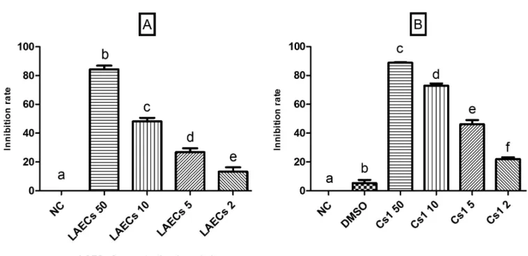
Radical-scavenging capacity – DPPH assay
LAECs and Cs1 demonstrated signiicant radical scavenging activity in the DPPH assay (Figure 3). They exhibited a scavenging effect of 85.13% and 88.95% respectively at a concentration of 50 µg/mL. The inhibition rate was concentration-dependent, being 49.12%, 27.52%, and 13.32% for LAECs and 73.12%, 45.35%, and 21.36% for Cs1, both at concentrations of 10, 5 and 2 µg/mL, respectively. Dimethyl sulfoxide (DMSO) was only used to help dissolve Cs1, and it was included in the analysis
TABLE III - Composition of lyophilized aqueous extract of C. speciosa (LAECs) and raw ethanol extract (Cs1)
Compounds LAECs Cs1 LOD LOQ
mg/g % mg/g % µg/mL µg/mL
Gallic Acid 8.67 ± 0.03 a 0.86 9.37 ± 0.01 a 0.93 0.015 0.049
Chlorogenic Acid 43.19 ± 0.01 b 4.31 16.43 ± 0.02 b 1.54 0.009 0.029
Cafeic Acid 41.70 ± 0.02 b 4.17 30.28 ± 0.02 c 3.02 0.024 0.078
Ellagic Acid 6.53 ± 0.02 c 0.65 3.56 ± 0.03 d 0.35 0.013 0.042
Rutin 19.82 ± 0.01 d 1.98 3.91 ± 0.01 d 0.39 0.027 0.090
Quercetin 22.39 ± 0.03 e 2.23 41.80 ± 0.01 e 4.18 0.019 0.063
Kaempferol 65.41 ± 0.02 f 6.54 46.32 ± 0.03 f 4.63 0.026 0.085
Results are expressed as mean ± standard deviation of three determinations. Means followed by diferent letters difer by Tukey test (p <0.05). LOD - Limit of Detection; LOQ - Limit of Quantiication.
FIGURE 1 - Representative high performance liquid chromatography proiles of lyophilized aqueous extract of C. speciosa (LAECs)
(A) and raw ethanol extract (Cs1) (B). UV detection at 325 nm. Gallic acid (1), chlorogenic acid (2), cafeic acid (3), ellagic acid
and igure to demonstrate that it did not have a statistically signiicant efect, and therefore no efect on the results.
Lipid peroxidation test
LAECs and Cs1 had an almost constant efect on
lipid peroxidation at all concentrations tested, with similar
efects to those of the negative control (Figure 4).
Determination of protein carbonyl groups
At concentrations of 50 and 10 µg/mL, LAECs demonstrated a carbonylation index statistically similar to that of the negative control (Figure 5); however, LAECs at 5 and 2 µg/mL and Cs1 at all concentrations showed results statistically similar to that of the positive control.
Evaluation of genotoxicity
Comet assay
Lyophilized aqueous extract of C. speciosa at concentrations of 10, 5, and 2 µg/mL and Cs1 at concentrations of 5 and 2 µg/mL produced statistically similar results to the negative control, showing no
signiicant DNA damage; however, LAECs at 50 µg/mL and Cs1 at 50 and 10 µg/mL presented a score of 1 for DNA damage (Figure 6).
DISCUSSION
Studies of the toxicity and the protective efects of
species used in traditional medicine are essential. In this TABLE IV - Composition of lyophilized aqueous extract of C. speciosa (LAECs) and raw ethanol extract (Cs1), at the concentrations used for the in vitro tests
Compound LAECs Cs1
µg/50 µg µg/10 µg µg/5 µg µg/2 µg µg/50 µg µg/10 µg µg/5 µg µg/2 µg
Gallic Acid 0.43 0.09 0.043 0.02 0.47 0.09 0.05 0.02
Chlorogenic Acid 2.16 0.43 0.21 0.09 0.82 0.16 0.08 0.03
Cafeic Acid 2.08 0.42 0.21 0.08 1.51 0.30 0.15 0.06
Ellagic Acid 0.33 0.06 0.03 0.01 0.18 0.03 0.02 0.01
Rutin 0.99 0.20 0.09 0.04 0.19 0.04 0.02 0.01
Quercetin 1.12 0.22 0.11 0.04 2.09 0.42 0.20 0.08
Kaempferol 3.27 0.65 0.33 0.13 2.32 0.46 0.23 0.09
FIGURE 2 - Efects of lyophilized aqueous extract (LAECs) (A) and raw ethanol extract (Cs1) (B) on cell viability of human
leukocytes. 0 µg/mL – Negative control (phosphate bufered saline). Data are expressed as mean ± SD. Columns with diferent
sense, this work reports the phenolic composition of bark extracts of C. speciosa and their in vitro toxicity and antioxidant activity. The phytochemical analysis of Cs1
and LAECs showed a high concentration of polyphenolic
compounds, comprising mainly flavonoids and phenolic
acids. These compounds, which most commonly occur in FIGURE 3 - 2,2-diphenyl-1-pricril-hydrazyl (DPPH) radical scavenging activity. (A) lyophilized aqueous extract (LAECs) at 50, 10, 5 and 2 µg/mL; (B) raw ethanol extract (Cs1) at 50, 10, 5 and 2 µg/mL; NC – Negative Control (distilled water); Data are expressed as mean ± SD. Columns with diferent letters are signiicantly diferent (p<0.05).
FIGURE 4 - Efects of lyophilized aqueous extract (LAECs) and raw ethanol extract (Cs1) on lipid peroxidation test induced by
hydrogen peroxide (H2O2 100 µmol/L) in plasma. NC – Negative Control (phosphate bufered saline); PC – Positive Control; (H2O2 100 µmol/L); (A) LAECs (50, 10, 5 and 2 µg/mL); (B) Cs1 (50, 10, 5 and 2 µg/mL). Data are expressed as mean ± SD. Columns
plants, represent an important class of phytochemical as they play a protective role against pathogens and predators. In humans they are known to have diverse physiological
roles since they have antifungal, anti-inflammatory and
antioxidant properties (Ayala-Zavala et al., 2012). Our results are consistent with those reported by Krishnaveni FIGURE 5 - Efects of lyophilized aqueous extract (LAECs) and raw ethanol extract (Cs1) on protein carbonylation induced by
hydrogen peroxide (H2O2 100 μmol/L) in plasma. NC – Negative Control (phosphate bufered saline); PC – Positive Control; (H2O2 100 µmol/L1); (A) LAECs (50, 10, 5 and 2 µg/mL); (B) Cs1 (50, 10, 5 and 2 µg/mL). Data are expressed as mean ± SD. Columns
with diferent letters are signiicantly diferent (p<0.05).
FIGURE 6 - Efects of lyophilized aqueous extract (LAECs) and raw ethanol extract (Cs1) in Comet assay of human leukocytes.
NC – Negative Control (phosphate bufered saline, PBS); PC – Positive Control; (H2O2 100 µmol/L); (A) LAECs (50, 10, 5 and
et al. (2013), who found a high concentration of flavonoids (7.7 mg quercetin/g of extract) and phenolic compounds (3.3 mg gallic acid/g of extract) in C. speciosa leaves. LAECs showed higher concentrations of phenolic compounds than Cs1, with the exception of quercetin.
In the present study, a cell viability assay was performed to determine the concentrations to be used in the analysis (Koko et al., 2008). The cell viability remained above 85% at relatively high concentrations, indicating that LAECs and Cs1 were non-toxic to human leukocytes. Antioxidant tests were performed using a DPPH assay; this is a widely accepted method for evaluation of in vitro antioxidant activity of extracts and is based on the capacity of antioxidant compounds present in the sample to capture DPPH radicals (Benhammou, Bekkara, Panovska, 2009; Ayala-Zavala et al., 2012; Mohsin, Mahadevan, Kurup, 2014; Wan-Ibrahim, Sidik, Kuppusamya, 2010). The results showed that LAECs and Cs1 inhibited ≥ 50% of radicals at concentrations of 10 µg/mL and 50 µg/ mL, showing that both samples appear to protect the studied biological matrix, since the concentrations tested were able to neutralize the radical DPPH. In a study by Ayala-Zavala et al. (2012), the authors examined several parameters to assess the antioxidant activity of Phellinus gilvus, Phellinus rimosus, and Phellinus badius,and found a correlation between the total polyphenolic content and DPPH radical scavenging activity; therefore, the inhibitory potential found in this work indicates an elevated concentration of these phytochemicals and their high antioxidant power.
Following determination of the antioxidant potential of C. speciosa, the in vitro efects of diferent concentrations of LAECs and Cs1 on some important parameters of oxidative stress were evaluated. An imbalance between production and inactivation of ROS can lead to oxidative stress and damage to various biological macromolecules, and proteins exposed to ROS can suffer oxidative modifications that can lead
to changes in their functions, and, consequently, afect
cellular metabolism (Gupta, Ballal, 2015). In this context, measurement of protein carbonyl groups in biological samples is a reliable parameter with which to assess ROS-mediated protein oxidation (Levine et al., 1990). In this study, LAECs at concentrations of 10 and 50 µg/ mL showed a protein carbonyl content similar to the negative control, demonstrating no evidence of oxidative damage, and suggesting that these samples prevented H2O2-induced protein carbonylation. However, the results for LAECs at concentrations of 2 and 5 µg/mL were similar to those observed for the positive control, suggesting the occurrence of oxidative damage. At all
concentrations tested, Cs1 demonstrated a carbonyl group content similar to the positive control, which indicates the
occurrence of protein damage. These results may reflect
the high concentrations of quercetin in Cs1 because,
despite the beneicial actions of flavonoids, studies have
demonstrated that quercetin has an in vitro pro-oxidant action (Choi, Chee, Lee, 2003; Heim, Tagliaferro, Bobilya, 2002; Yang et al., 2012). Studies have reported that, in general, phenolic compounds exhibit pro-oxidant activity in vitro but not in vivo, and the conditions for this arise mainly in the presence of transition metals and acidic pH (Eghbaliferiz, Iranshahi, 2016). This suggests that further studies are necessary to determine whether these factors are present in our samples. In other species, ROS can target lipids and initiate the lipid peroxidation process, which may lead to molecular cell damage (Gupta & Ballal, 2015). In the present study, LAECs and Cs1
showed no diferences compared to the negative control
at all concentrations tested in the H2O2-induced lipid peroxidation assay, which indicates the absence of lipid oxidative damage and hence the potential antioxidant action of C. speciosa, consistent with the results of the DPPH test. These results are an important indication of the safety of this species, because the occurrence of lipid peroxidation is directly related to various disease processes such as carcinogenesis and atherosclerosis.
Besides protein and lipids, DNA is also a signiicant
target for oxidative damage mediated by ROS. Samples were subjected to the Comet assay, which is considered a sensitive method for the detection of single and double DNA strand breaks (Collins et al., 2008). LAECs at concentrations of 2, 5 and 10 µg/mL, and Cs1 at
concentrations of 2 and 5 µg/mL, showed no signiicant
DNA damage. Both samples at higher concentrations (50 µg/mL of LAECs and 10 and 50 µg/mL of Cs1) showed low levels of DNA damage (score 1). These results indicate that further genotoxic studies are required in order to deepen knowledge about the safety of C. speciosa.
CONCLUSION
This study revealed that C. speciosa extracts have promising antioxidant potential, which may be related to their high polyphenolic content. Phenolic compounds are the constituents responsible for the antioxidant potential of natural products (Krishnaveni et al., 2013; Loganayaki, Siddhuraju, Manian, 2013) from species of the genus Passiflora and Citrus, both known for being rich in
flavonoids and with well-established antioxidant activity.
extracts showed either no damage or at the most a low degree of DNA damage (grade 1) in the Comet assay and they also prevented lipid peroxidation at all concentrations tested. Aqueous extract (LAECs), which is used in folk medicine, showed no damage to proteins at concentrations of 10 and 50 µg/mL.
The use of plants is widely accepted by the public, and this fact is closely related to the popular belief that
natural products are free of adverse efects. However, the
popularization of the use of medicinal plants, their ease of access, and lack of professional guidance on their correct
use, represent a health risk (Al-Arii, 2013; Jeong et al., 2012). This study describes a preliminary evaluation of the biological activity and safety of C. speciosa and shows that this species exhibits in vitro antioxidant potential and low toxicity, which may be related at least in part to its high concentration of polyphenols. The results provide an important foundation for knowledge about this species, which is popularly used for the reduction of serum cholesterol, triglyceride and glucose levels. Further phytochemical analyses are necessary to determine the chemical compounds responsible for the antioxidant activity ofC. speciosa and to correlate them with other
possible pharmacological or even toxicological efects of
this very promising plant.
ACKNOWLEDGEMENTS
This work was supported by Conselho Nacional
de Desenvolvimento Cientíico e Tecnológico (CNPq)
and Fundação de Amparo à Pesquisa do Rio Grande do Sul (FAPERGS). The authors thank the researchers of
Laboratório de Biotecnologia da Reprodução (Biotech –
UNIPAMPA) for their collaboration in the experiments.
REFERENCES
AL-ARIFI, M.N. Availability and needs of herbal medicinal information resources at community pharmacy, Riyadh region, Saudi Arabia. Saudi Pharm J. v.21, n.4, p.351-360, 2013.
AYALA-ZAVALA, J.F.; SILVA-ESPINOZA, B.A.; CRUZ-VALENZUELA, M.R.; VILLEGAS-OCHOA, M.A.; E S Q U E D A , M . ; G O N Z Á L E Z - A G U I L A R , G . A . ; CALDERÓN-LÓPEZ, Y. Antioxidant and antifungal potential of methanol extracts of Phellinus spp. from Sonora, Mexico. Rev. Iberoam. Micol., v.29, n.3, p.132-138, 2012.
BALICK, M.J.; COX, P.A. Plants, people and culture: the science of ethnobotany. New York: HPHLP, 1996. 228p.
BELESKI-CARNEIRO, E.; SUQUI, J.; REICHER, F. Structural and biological features of a hydrogel from seed coats of
Chorisia speciosa. Phytochemistry., v.61, n.2, p.157-163, 2002.
BENHAMMOU, N.; BEKKARA, F.A.; PANOVSKA, T.K. Antioxidant activity of methanolic extracts and some bioactive compounds of Atriplex halimus. Comptes Rendus Chimie, v.12, n.12, p.1259-1266, 2009.
BOLIGON, A.A.; FELTRIN, A.C.; MACHADO, M.M.; JANONIK, V.; ATHAYDE, M.L. HPLC analysis and phytoconstituents isolated from ethyl acetate fraction of
Scutia buxifolia Reiss. leaves. Lat. Am. J. Pharm., v.28, n.1, p.121-124, 2009.
BRASIL. Ministério da Saúde. Política nacional de plantas
medicinais e itoterápicos. Brasília: Ministério da Saúde,
2007. 60p. Disponível em: <http://bvsms.saude.gov.br/bvs/
publicacoes/politica_nacional_itoterapicos.pdf>. Access:
19 Set. 2016.
BRASIL. Ministério da Saúde. RENISUS - Relação Nacional de Plantas Medicinais de Interesse ao SUS. Brasília: Secretaria de Ciência, Tecnologia e Insumos Estratégicos, 2009. 3p.
Disponível em: <http://bvsms.saude.gov.br/bvs/sus/pdf/
marco/ms_relacao_plantas_medicinais_sus_0603.pdf>. Access: 19 Set. 2016.
BUROW, M.E.; WELDON, C.B.; TANG, Y.; NAVAR, G.L.; KRAJEWSKI, S.; REED, J.C.; HAMMOND, T.G.; CLEJAN, S.; BACKMAN, B.S. Differences in
susceptibility to tumor necrosis factor α-induced apoptosis
among MCF-7 breast cancer cell variants. Cancer Res., v.58, n.21, p.4940-4946, 1998.
CALIXTO, J.B. Twenty-ive years of research on medicinal plants
in Latin America: a personal review. J. Ethnopharmacol., v.100, n.1-2, p.131-134, 2005.
CAPPELATTI, L.; SCHMITT, L.J. Caracterização da flora
arbórea de um fragmento urbano de floresta estacional
CARLINI, E.A.; RODRIGUES, E.; MENDES, F.R.; TABACH, R.; GIANFRATTI, B. Treatment of drug dependence with Brazilian herbal medicines. Rev. Bras. Farmacogn., v.16, p.690-695, 2006.
CARTAXO, S.L.; SOUZA, M.M.A.; ALBUQUERQUE, U.P. Medicinal plants with bioprospecting potential used in semi-arid northeastern Brazil. J. Ethnopharmacol., v.131, n.2, p.326-342, 2010.
CHANDRA, S.; MEJIA, E.G. Polyphenolic compounds, antioxidant capacity, and quinone reductase activity of an aqueous extract of Ardisia compressa in comparison to mate (Ilex paraguariensis) and green (Camellia sinensis) teas. J. Agr. Food Chem., v.52, n.11, p.3583-3589, 2004.
CHOI, E.J.; CHEE, K.M.; LEE, B.H. Anti- and prooxidant
efects of chronic quercetin administration in rats. Eur. J. Pharmacol., v.482, n.1-3, p.281-285, 2003.
COLLINS, A.R.; OSCOZ, A.A.; BRUNBORG, G.; GAIVÃO, I.; GIOVANNELLI, L.; KRUSZEWSKI, M.; SMITH, C.C.; STETINA, R. The comet assay: topical issues. Mutagenesis, v.23, n.3, p.143-151, 2008.
EGHBALIFERIZ, S.; IRANSHAHI, M. Prooxidant activity
of polyphenols, flavonoids, anthocyanins and carotenoids:
Updated review of mechanisms and catalyzing metals: prooxidant activity of polyphenols and carotenoids.
Phytother Res., v.30, n.9, p.1379-91, 2016.
EL-SHENAWY, N.S.; MOHAMMADDEN, A.; AL-FAHMIE, Z.H. Using the enzymatic and non-enzymatic antioxidant defense system of the land snail Eobania vermiculata as biomarkers of terrestrial heavy metal pollution. Ecotox. Environ. Safe., v.84, p.347-354, 2012.
GAUR, K.; KORI, M.L.; TYAGI, L.K.; NEMA, R.K.; SHARMA, C.S.; TRIPATHI, P. In-vitro antioxidant activity of leaves of Ipomoea istulosa Linn. Acad. J. Plant. Sci., v.2, n.2, p.60-64, 2009.
GUPTA, A.; BALLAL, A. Unraveling the mechanism responsible for the contrasting tolerance of Synechocystis
and Synechococcus to Cr(VI): Enzymatic and non-enzymatic antioxidants. Aquat. Toxicol., v.164, p.118-125, 2015.
HEIM, K.E.; TAGLIAFERRO, A.R.; BOBILYA, D.J. Flavonoid antioxidants: chemistry, metabolism and structure-activity relationships. J. Nutr. Biochem., v.13, n.10, p.572-584, 2002.
JEONG, T.Y.; PARK, B.K.; CHO, J.H.; KIM, Y.I.; AHN, Y.C.; SON, C.G. A prospective study on the safety of herbal medicines, used alone or with conventional medicines. J. Ethnopharmacol., v.143, n.3, p.884-888, 2012.
K H A R R A Z I , H . ; VA I S I - R AY G A N I , A . ; R A H I M I , Z.; TAVILANI, H.; AMINIAN, M.; POURMOTABBED, T. Association between enzymatic and non-enzymatic antioxidant defense mechanism with apolipoprotein E genotypes in Alzheimer disease. Clin. Biochem., v.41, n.12, p.932-936, 2008.
KOKO, W.S.; MESAIK, M.A.; YOUSAF, S.; GALAL, M.; CHOUDHARY, M.I. In vitro immunomodulating properties of selected Sudanese medicinal plants. J. Ethnopharmacol., v.118, n.1, p.26-34, 2008.
KRISHNAVENI, M.; AMSAVALLI, L.; CHANDRASEKAR, R.; MADHAIYAN, P.; DURAIRAJ, S. Antioxidant activity of plants at Govt. College of Engineering Campus, Salem, Tamil nadu, India. Int. J. Pharm. Sci. Rev. Res., v.21, n.1, p.160-163, 2013.
LADEJI, O.; OMEKARAH, I.; SOLOMON, M. Hypoglycemic properties of aqueous bark extract of Ceiba pentandra in streptozotocin-induced diabetic rats. J. Ethnopharmacol., v.84, n.2-3, p.139-142, 2003.
LEVINE, R.L.; AMSAVALLI, L.; CHANDRASEKAR, R.; MADHAIYAN, P.; DURAIRAJ, S. Damage to proteins and lipids tissues under oxidative stress. Method Enzymol., v.186, p.464-478, 1990.
LOGANAYAKI, N.; SIDDHURAJU, P.; MANIAN, S. Antioxidant activity and free radical scavenging capacity of phenolic extracts from Helicteres isora L. and Ceiba pentandra L. J. Food Sci. Tech., v.50, n.4, p.687-695, 2013.
MASHWANI, Z.U.; KHAN, M.A.; ULLAH, Z.; CHAUDHARY, H.J. An ethno botanical perspective of traditional medicinal plants from the Khattak tribe of Chonthra Karak, Pakistan.
MEDEIROS, P.M.; LADIO, A.H.; ALBUQUERQUE, U.P. Sampling problems in Brazilian research: a critical evaluation of studies on medicinal plants. Rev. Bras. Farmacogn., v.24, n.2, p.103-109, 2015.
MOHSIN, S.; MAHADEVAN, R.; KURUP, G.M.
Free-radical-scavenging activity and antioxidant efect of ascophyllan
from marine brown algae Padina tetrastromatica. Biom. Prev. Nutr., v.4, n.1, p.75-79, 2014.
MORABITO, F.; CRISTIANI, M.; SAIJA, A.; STELITANO, C.; CALLEA, V.; TOMAINO, A.; MINCIULLO, P.L.; GANGEMI, S. Lipid peroxidation and protein oxidation in patients affected by Hodgkin’s Lymphoma. Mediat.
Inlamm., v.13, n.5/6, p.381-383, 2004.
OHKAWA, H.; OHISHI, H.; YAGI, K. Assay for lipid peroxide in animal tissues by thiobarbituric acid reaction. Ann. Clin. Biochem., v.95, n.2, p.351-358, 1979.
P E R E I R A , L . P. ; S I LVA , F. E . B . ; F L O R E S , E . M . M . ; SCHREKKER, H.S.; MACHADO, M.M.; OLIVEIRA, L.F.S. In vitro ZnCl2 cytotoxicity and genotoxicity in
human leukocytes: Zero-order kinetic cellular zinc influx. Acta Scient. Health Sci., v.37, n.1, p.63-68, 2015.
SAID, W.M.; NAHLA, O.M.; EHSAN, N.S.K. Comparative study of three species of Malvatheca (Bombacoideae) and Malvoideae (Malvaceae sensu lato) using morphological, anatomical and RAPD-PCR analyses. Adv. Environ. Biol., v.7, n.2, p.415-426, 2013.
SCARPA, G.F. Medicinal plants used by the Criollos of Northwestern Argentine Chaco. J. Ethnopharmacol., v.91, n.1, p.115-135, 2004.
SHARMA, O.P.; BHAT, T.K. DPPH antioxidant assay revisited.
Food Chem., v.113, n.4, p.1202-1205, 2009.
SINGH, N.; MCCOY, M.; TICE, R.; SCHNEIDER, E. A simple
technique for quantiication of low levels of DNA damage in
individual cells. Exp. Cell Res., v.175, n.1, p.184-191, 1995.
VELOSO, D.P.; PAULA, V.F.; BARBOSA, L.C.A.; DEMUNER, A.J. A química da família Bombacaceae. Quím Nova., v.20, n.6, p.627-630, 1997.
WAN-IBRAHIM, W.I.; SIDIK, K.; KUPPUSAMYA, U.R. A high antioxidant level in edible plants is associated with genotoxic properties. Food Chem., v.122, n.4, p.1139-1144, 2010.
YANG, B.; CHEN, F.; HUA, Y.; HUANG, S.S.; LIN, S.; WEN, L.; JIANG, Y. Prooxidant activities of quercetin, p-courmaric acid and their derivatives analysed by quantitative structure-activity relationship. Food Chem., v.131, p.508-512, 2012.
ZHISHEN, J.; MENGCHENG, T.; JIANMING, W. The
determination of flavonoid contents in mulberry and their
scavenging efects on superoxide radicals. Food Chem.,
v.64, n.4, p.555-559, 1999.