Heart Rate and its Variability Assessed by Spectral Analysis in Elderly
Subjects with Orthostatic Hypotension: A Case-Control Study
Rose Mary Ferreira Lisboa da Silva, Carlos Eduardo de Souza Miranda, Maira Tonidandel Barbosa, Maria
Aparecida Camargos Bicalho
Faculdade de Medicina da Universidade Federal de Minas Gerais, Belo Horizonte, MG - Brazil
Mailing Address: Rose Mary Ferreira Lisboa da Silva • Avenida Prof. Alfredo Balena, 190, sala 246, Centro, Belo Horizonte, MG - Brazil
E-mail: [email protected], [email protected]
Manuscript received May 02, 2017, revised manuscript October 12, 2017, accepted November 09, 2017
DOI: 10.5935/abc.20180043
Abstract
Background: The prevalence of orthostatic hypotension (OH) increases with age and is associated with changes in autonomic regulation of blood pressure (BP) and heart rate (HR).
Objective: to assess HR and HR variability (HRV) in elderly subjects with OH and determine OH predictors.
Methods: a total of 105 patients aged ≥ 60 years, 39 with OH (case group) and 66 without OH (control group) (age-matched)
were studied. Patients underwent clinical assessment, electrocardiogram, biochemistry tests and Holter monitoring for spectral analysis of HRV (Fourier transform) in the supine and orthostatism positions to identify low frequency (LF) and high frequency (HF) components, as well as the LF/HF ratio.
Results: median age was 73.0 years, 64 patients were women. In all participants, there was a reduction in HF (133.0 versus 76.0 ms2, p = 0.001) and increase in LF/HF (1.6vs 2.1; p < 0.001) and no change in LF (233.0 versus 218.0 ms2,
p = 0.080). Between-group comparisons revealed significant differences in the median values of HR in the supine position (62.0 vs. 69.0 bpm, p = 0.001) and LF in the supine position (157.0 in case group vs. 275.0 ms2 in the control group,
p = 0.014). Spearman’s correlation coefficient of 0.27 was found between the groups. Multivariate analysis revealed that HR in the supine position was an independent variable for OH (p = 0.001- 95%CI = -0.022 and -0.006). Using the operating characteristic curve, the best cutoff point was 61 bpm, with a sensitivity of 77.3% and specificity of 51.3%, positive predictive value of 61.3%, and negative predictive value 69.3%. Odds ratio was 3.23 for OH in patients with a HR lower than 61 bpm.
Conclusions: lower LF and HR in the supine position were found in patients with OH, regardless of age and gender. The independent predictor for OH was HR in the supine position, with an odds ratio of 3.23 for values lower than 61 bpm. (Arq Bras Cardiol. 2018; 110(4):303-311)
Keywords: Heart Rate; Hypotension, Orthostatic; Accidental Falls; Syncope; Aged; Dizziness.
Introduction
Orthostatic hypotension (OH), also known as postural hypotension is defined as a sustained fall in blood pressure (of at least 20 mm Hg in systolic pressure and/or at least 10 mm Hg in diastolic pressure) occurring within
3 minutes of standing.1,2 OH has been associated with
falls, presyncope, syncope, functional impairment in the elderly, cardiovascular events, and increased mortality.3-5
Its prevalence varies from 6 to 35%,4 and can achieve 41%
in individuals aged 80 years or older.6
With aging, there are changes in autonomic regulation of heart rate (HR) and blood pressure. Middle-aged women
have a more dominant parasympathetic whereas men have a more sympathetic regulation of heart rate.7 In addition,
increased levels of norepinephrine and reduced sensitivity of beta-adrenergic receptors are found in elderly subjects. There is a decrease in vasomotor response mediated by alpha receptors, with decline in venous capacitance response of the lower limbs and in baroreflex response, which is also due to artery stiffness.8,9 This altered regulation lead to autonomic
dysfunction and may cause OH. Autonomic nervous system, involved in the physiopathology of OH, may be examined by measurements of the heart rate variability (HRV) by Holter monitoring,10 a non-invasive, low-cost method.
Studies on the autonomic nervous system, including those on baroreflex sensitivity and head-up tilt test have been performed in hypertensive and normotensive elderly patients who were compared with young subjects. These studies included up to 80 elderly subjects, 64 with hypertension.11-13
In the largest study, involving 362 volunteers, there were 38 men and 51 women aged between 57 and 88 years,13 but
Methods
This was an observational, prospective, cross-sectional study. Our sample was composed of 105 outpatients aged 60 years or older, included during the period from February 2013 to August 2014. Patients with dementia, autonomic dysfunction-related neurologic diseases, persistent or permanent atrial fibrillation, pacemakers, institutionalized patients, and those using antiarrhythmic agents (Class I, III or IV agents according to Vaughan Williams classification) or digoxin were excluded.
The study was approved by the local Research Ethics Committee and all participants signed the informed consent form.
For sample estimation, a one-tailed test was used, with significance level at 5%, power of 90%, two controls per case, frequency of OH of 30%. Two groups, age matched, were studied – a case group (n = 39) with OH, and a control group (n = 66) without OH.
Participants underwent clinical assessment, clinical pathology tests, 12-lead electrocardiography, measurements of blood pressure (BP) at supine position at the 5th minute of rest and at
the 3rd minute of orthostatism or before, in case they had OH
symptoms according to well-established conditions,2 monitored
by the Holter system. Measurements were performed at a temperature-controlled room, in the afternoon, at least two hours after lunch to exclude the possibility of post-prandial hypotension. Holter monitoring was performed using a three-channel digital recorder (Cardioflash) (modified V1 and V5 and DIII) version 1.0, at supine and orthostatic positions for 15 and 10 minutes, respectively, for analysis of HRV in the frequency domain by the fast Fourier transform method. Measurements of high-frequency (HF) and low-frequency (LF) components that indicate parasympathetic and sympathetic activities, respectively, as well as the LF/HF ratio10 were calculated.
This analysis was performed after manual edition of recordings to remove artifacts and correct arrhythmias. Measures were obtained during 5 minutes at 10th minute of supine position
and 5th minute of orthostatic position. Results of the spectral
analysis were expressed in ms2.
The Framingham14 and the PROCAM15 risk scores were also
calculated using clinical and laboratory data, which included plasma levels of cholesterol and its fractions, triglycerides, and fasting glucose levels.
Statistical analysis
For data analysis, we used the International Business Machines (IBM) Statistical Package for Social Sciences (SPSS) Statistics 19. Results were expressed as numbers and proportions for categorical variables and as central tendency (mean and median) and dispersion measures for continuous variables. Associations between categorical variables were assessed by the chi-square test or the Fisher’s exact test, as appropriate. Data normality was not tested. The Mann-Whitney test was used for comparisons between continuous variables, and correlations between categorical variables were assessed by the Spearman's rank correlation test. The Wilcoxon test was used to compare the two periods HRV components in the spectral analysis (supine and orthostatic positions). Stepwise multivariate analysis was performed to evaluate predicting values of OH,
considering the variables with a p ≤ 0.10 in the univariate
analysis. Receiver operating characteristic curve was analyzed for the stable variable postural response. The level of significance was set at 5%.
Results
General characteristics of the population
Mean and median age were 71.9 and 73.0 years, respectively; 64 (61%) were women. Clinical variables of the study population are described in Table 1.
With respect to cardiovascular risk factors, systemic arterial hypertension (SAH) and dyslipidemia were the most frequent, found in 80 (76.2%) and 42 (40%) patients, respectively. Diabetes was found in 17.1% of patients.
Thiazide diuretics were the most used antihypertensive drugs; 42 patients (40%) used them isolated or in combination with other antihypertensive agents. Following thiazide diuretics, angiotensin II receptor blockers (29.8%), angiotensin-converting enzyme (ACE inhibitor) (28.6%) and beta-blockers (27.6%) were the most common, with similar frequencies of use. Also, 14.3% of patients were using calcium antagonists (amlodipine or nifedipine).
Symptoms characterized by previous history of dizziness, falls, and presyncope and/or syncope were reported by 64 patients (61%).
Impaired conduction in the left bundle branch was detected at electrocardiography in 9.5% of patients, with mean PR and QT intervals of 166.9 ms (120-280) and 403.0 ms (320-520), respectively.
Comparison between case and control groups
No difference was found in age (mean of 73.5 ± 8.0 years; median of 74.0 in the case group and 71.0 ± 6.8 years and 72.0 years in the control group, p = 0.119), but a significant difference in sex was observed between the groups (56.4% of men in the case group and 27.8% of men in the control group, p = 0.005). No correlation was found between these two variables (Spearman’s coefficient correlation of 0.274).
Results of other comparisons between the two groups are described in Table 2. No patient had dizziness, presyncope or syncope in orthostatism when BP was measured. No difference considered abnormal in BP between the upper limbs was detected in seated position.
Significant differences were found in the frequency of previous symptoms (dizziness, prepsyncope and syncope) – 77% in the case group versus 51.5% in the control group (p < 0.001). However, no difference between patients with and without previous symptoms were found in age
(mean or median) – 71.4 ± 7.4 years; 72.0 years versus
72.7 ± 7.8 years, 74.0 years; respectively (p = 0.38) – nor in BP measured in the supine position.
Table 1 – Anthropometric and clinical variables of studied patients
Variables Median Interquartile range Q1 - Q3 Minimum value Maximum value
Age (years) 73.0 65.5 – 77.0 60.0 91.0
Weight (kg) 62.0 56.0 – 72.0 44.0 102.0
Height (m) 1.58 1.51 – 1.62 1.41 1.80
BMI (kg/m2) 25.7 22.5 – 29.7 17.8 40.9
WC (cm) 87.3 80.3 – 96.0 68 116
HR supine position (bpm) 68.0 60.0 – 76.0 38.0 105.0
HR orthostatic (bpm) 72.0 64.0 – 80.0 44.0 109.0
SAP supine position (mmHg) 140.0 127.0 – 152.0 92.0 196.0
DAP supine position (mmHg) 80.0 75.0 – 87.0 60.0 104.0
SAP orthostatic position (mmHg) 130.0 120.0 – 142.0 60.0 220.0
DAP orthostatic position (mmHg) 80.0 70.0 – 90.0 30.0 100.0
SAP seated position (mmHg) 135.0 120.0 – 150.0 100.0 194.0
DAP seated position (mmHg) 80.0 70.0 – 90.0 60.0 106.0
BMI: body mass index; WC: waist circumference; HR: heart rate; bpm: beats per minute; SAP: systolic arterial pressure; DAP: diastolic arterial pressure; mmHg: millimeter of mercury; Q1: 25th percentile; Q3: 75th percentile
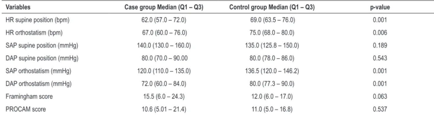
Table 2 – Between-group comparison of heart rate, blood pressure and cardiovascular risk scores
Variables Case group Median (Q1 – Q3) Control group Median (Q1 – Q3) p-value
HR supine position (bpm) 62.0 (57.0 – 72.0) 69.0 (63.5 – 76.0) 0.001
HR orthostatism (bpm) 67.0 (60.0 – 76.0) 75.0 (68.0 – 80.0) 0.006
SAP supine position (mmHg) 140.0 (130.0 – 160.0) 135.0 (125.8 – 150.0) 0.189
DAP supine position (mmHg) 80.0 (70.0 – 90.00 80.0 (78.0 – 86.0) 0.543
SAP orthostatism (mmHg) 120.0 (110.0 – 135.0) 136.5 (120.0 – 146.2) 0.001
DAP orthostatism (mmHg) 72.0 (60.0 – 84.0) 80.0 (77.3 – 90.0) 0.001
Framingham score 15.5 (6.0 – 24.3) 12.0 (6.0 – 17.0) 0.063
PROCAM score 10.6 (5.01 – 21.4) 11.0 (5.0 – 16.8) 0.537
SD: standard deviation; HR: heart rate; bpm: beats per minute; SAP: systolic arterial pressure; DAP: diastolic arterial pressure; mmHg: millimeter of mercury. Mann‑Whitney test; Q1: 25th percentile; Q3: 75th percentile
in the frequency of diabetes (7 patients in the case group and 11 in the control group; p = 0.86) or coronary arterial disease (5% in the case group and 9% in the control group) between the groups. All patients were stable, without chest pain.
Regarding the main groups of antihypertensives, higher percentage of users of ACE inhibitors was observed in the case group (41.0%) than in the control group (21.2%) (p = 0.030). No difference was found in other antihypertensive agents.
Heart rate variability
Medians and interquartile ranges of HRV components in supine position were – LF 233.0 ms2 (130.5 – 422.5),
HF 133.0 ms2 (62.0 – 347.5), LF/HF 1.6 (0.8 – 3.0) – and
in orthostatic position were – LF 218.0 ms2 (110.5 – 359.7),
HF 76.0 ms2 (32.0 – 227.0) and LF/HF 2.1 (1.1 e 4.8).
Comparisons of HRV components between supine and
orthostatic positions performed by the Wilcoxon test showed no difference in LF (p = 0.080), but significant differences in HF (p = 0.01) and LF/HF (p < 0.001).
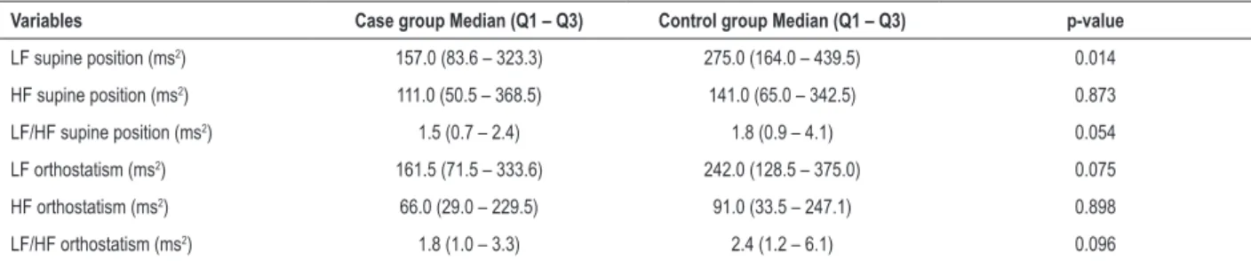
When HRV absolute values were compared between case and control groups by the Mann-Whitney test, significant difference was found in LF in supine position (Table 3). No difference between the groups was found in other components. Due to HRV data interval, a logarithmic transformation of HRV components was performed, and the same p-values were maintained. For analysis of HRV with change of position, median differences in LF component were compared between case and control groups (i.e. between the supine and the orthostatic position, median -0.27 ms2) by the
Mann Whitney test (p = 0.43). Median differences of HF and
LF/HF components were 33.0 ms2 and 0.53, respectively,
Figures 1 and 2 depict the analysis of HRV components of a patient with OH in the supine and orthostatic positions, respectively. Figures 3 and 4 show the analysis of HRV components of a patient without OH in the supine and orthostatic positions, respectively.
Stepwise multivariate analysis
Variables with a p ≤ 0.10 in the univariate analysis –
sex, use of ACE inhibitors, presence of previous symptoms, HR, LF and LF/HF in supine and orthostatic positions, and Framingham score – were considered for the multivariate analysis. The independent variable with statistical significance was HR in the supine position, with p = 0.001, 95% confidence interval of -0.0022 – -0.006.
Analysis of the receiver operating characteristic curve Using the receiver operating characteristic curve for the stable variable postural response without OH, and considering the variable HR in the supine position, an area under the
curve of 0.70 was obtained (Figure 5), with a p = 0.001 (95% confidence interval of 0.595-0.796). The best cutoff point was 61 bpm, with a sensitivity of 77.3% and specificity of 51.3%. Positive predictive value was 61.3%, and negative predictive value 69.3%. Odds ratio was 3.23 for OH in patients with a HR lower than 61 bpm.
Discussion
The main finding of this study was that, in contrast to HRV components, HR in the supine position was an independent predictor for the occurrence of OH in the study population. Median HR in the supine position was significantly lower in the case group than in the control group in the same position. Although this variable was a predictor of OH, with an odds ratio of 3.23 for patients with HR < 61 bpm, it was not considered a good diagnostic test, as confirmed by the analysis of the receiver operating characteristic curve.
Aging is one of the main predicting factors for OH, which may be explained by changes in the autonomic regulation
Table 3 – Comparison of heart rate spectral analysis between case and control groups
Variables Case group Median (Q1 – Q3) Control group Median (Q1 – Q3) p-value
LF supine position (ms2) 157.0 (83.6 – 323.3) 275.0 (164.0 – 439.5) 0.014
HF supine position (ms2) 111.0 (50.5 – 368.5) 141.0 (65.0 – 342.5) 0.873
LF/HF supine position (ms2) 1.5 (0.7 – 2.4) 1.8 (0.9 – 4.1) 0.054
LF orthostatism (ms2) 161.5 (71.5 – 333.6) 242.0 (128.5 – 375.0) 0.075
HF orthostatism (ms2) 66.0 (29.0 – 229.5) 91.0 (33.5 – 247.1) 0.898
LF/HF orthostatism (ms2) 1.8 (1.0 – 3.3) 2.4 (1.2 – 6.1) 0.096
SD: standard deviation; LH: low frequency; HF: high frequency; LH/HF: low frequency/high frequency ratio; ms: milliseconds. Mann‑Whitney test; Q1: 25th percentile;
Q3: 75th percentile
Figure 1 – Spectral analysis of a male patient (67 years of age) with orthostatic hypotension in supine position. RR: number of QRS in sinus rhythm; VLF: very low
frequency; LF: low frequency; HF: high frequency; HFnu: HF normalized unit.
18000
13500
4500 9000
0
0.0 0.1 0.2 0.3 0.4 0.5
Frequency (c/b)
Duration: 5 min Total RR: 188
corrected: 0 RR average: 1135 ms
53 bpm Total potential: 1579 VLF Power: 906
(0.00–0.04)Hz LF Power: 434
(0.04–0.15)Hz LFnu: 64.6 HF Power: 237
Figure 3 – Spectral analysis of a female patient (62 years of age) without orthostatic hypotension in supine position. RR: number of QRS in sinus rhythm; VLF: very low frequency; LF: low frequency; HF: high frequency; HFnu: HF normalized unit.
10000
7500
2500 5000
0
0.0 0.1 0.2 0.3 0.4 0.5
Frequency (c/b)
Duration: 5 min Total RR: 366
corrected: 0 RR average: 820 ms
73 bpm Total potential: 484 VLF Power: 205
(0.00–0.04)Hz LF Power: 233
(0.04–0.15)Hz LFnu: 83.5 HF Power: 35
(0.15–0.40)Hz HFnu: 12.4 LF/HF: 6.75
Figure 2 – Spectral analysis of a male patient (67 years of age) with orthostatic hypotension (same of Figure 1) in orthostatic position, showing a decrease in low
frequency (LF) component and in the LF/high frequency (HF) ratio, in relation to supine position. RR: number of QRS in sinus rhythm; VLF: very low frequency; HFnu: HF normalized unit.
26 K
19 K
13 K
6 K
0
0.0 0.1 0.2 0.3 0.4 0.5
Frequency (c/b)
Duration: 5 min Total RR: 280
corrected: 0 RR average: 1074 ms
56 bpm Total potential: 2424 VLF Power: 1828
(0.00–0.04)Hz LF Power: 342
(0.04–0.15)Hz LFnu: 57.5 HF Power: 244
(0.15–0.40)Hz HFnu: 41.0 LF/HF: 1.40
of BP and HR. Increases in norepinephrine plasma levels, decreased sensitivity of beta-adrenergic receptors, decreased vasomotor response mediated by alpha receptors, and reduced baroreflex response and parasympathetic tone contribute to the occurrence of OH in the elderly.8,9 Therefore, while
approximately 5% of middle-aged adults have OH,16 the
prevalence increases to nearly 41% in those aged 80 years or older.6 In the present study, patients with and without
OH were age-matched and all of them were younger than 60 years, and hence results were not affected by age.
Previous studies have shown different results in the prevalence of OH and its association with gender, according to age and study setting. In a study conducted in the 90’s decade, Rutan et al.17 evaluated a population of 5,201 elderly subjects (≥65 years), with an OH prevalence of 18.2% and
no difference between the sexes. In patients hospitalized for OH, the prevalence was higher in men (55.3% in the age range of 65 – 74 years), although 54% of patients aged 75 years or older were women, with a total of 15,858 admissions for OH in a year.18 In our population, there was
a predominance of men in the group of patients with OH; however, Spearman’s correlation coefficient was 0.27, i.e., of small magnitude, which suggest that there was no statistical difference in sex between the groups. Also, sex was not an OH predictor in multivariate analysis.
Studies have demonstrated the importance of rises in HR in the orthostatic position, its association with increased sympathetic activity and higher orthostatic tolerance.19 Nevertheless, the role
Figure 5 – Receiver operating characteristic curve of heart rate in the supine position (blue line), considering the stable variable postural response without orthostatic hypotension.
1.0
1.0 0.8
0.8 0.6
0.6 0.4
0.4 0.2
0.0
0.0 0.2
Sensitivity
1 – Specificity
Figure 4 – Spectral analysis of a female patient (62 years of age) without orthostatic hypotension (the same of Figure 3) in orthostatic position, showing an increase
in low frequency (LF) component and in the LF/high frequency (HF) ratio, in relation to supine position. VLF: very low frequency; RR: number of QRS in sinus rhythm; VLF: very low frequency; HFnu: HF normalized unit.
26 K
19 K
13 K
6 K
0
0,0 0,1 0,2 0,3 0,4 0,5
Frequency (c/b)
Duration: 5 min Total RR: 410
corrected: 0 RR average: 732 ms
82 bpm Total potential: 1201 VLF Power: 628
(0,00–0,04)Hz LF Power: 548
(0,04–0,15)Hz LFnu: 95,8 HF Power: 17
(0,15–0,40)Hz HFnu: 3,0 LF/HF: 32,16
dysfunction20 may be associated with reduced HR in supine
position in elderly patients, regardless of OH. In the study population, when this variable was analyzed, median HR in supine position in the case group was significantly lower than in control group. In addition to the changes already described, another factor that may be associated with differences between the groups would be the use of beta-blockers. These medications have a negative chronotropic effect on HR.21 However, in the present
study, no difference was found in the use of most of these drugs between the groups. Significant difference was found in the use of ACE inhibitors, which decrease vascular resistance but have no substantial effect on HR, despite restoration of parasympathetic tone with the use of the drug in hypertensive patients.22
In light of this, analysis of HRV was important for the study of HR profile in elderly subjects with OH in supine position. With change of position, as expected,10 there was a decrease
in HF and increase in LF/HF in all patients, without an increase in the LF component. There is a decline in baroreflex and HRV with age in both sexes, resulting in autonomic dysfunction.6,8,13
A U-curve has been used to describe the progressive decrease of HRV with aging, which reaches its nadir in the sixth or seventh decade of life, followed by progressive increase, which is determinant for longevity from those decades on.23-26
infer from their results that autonomic system and baroreflex
dysfunction are associated with OH. Harrington et al.11
evaluated baroreflex sensitivity by digital plethysmography in the elderly – 75 normotensive and 64 hypertensive patients, without medications. The authors reported reduced baroreflex,
with impairment of HF component. Kawaguchi et al.12
shown a decrease in the LF/HF ratio and decreased cerebral perfusion measured by infrared spectroscopy in a group of 80 elderly subjects after passive standing. A more recent study on hypertensive patients,27 18 with OH and 64 without OH
(mean age of 74.2 years), demonstrated that, despite lower systolic volume in those with OH, no significant differences in change in the LF/HF ratio after orthostatism were found, which is in agreement with our results. Barantke et al.13
demonstrated a decline in all components of HRV with age, and an association between LF and baroreflex sensitivity in orthostatic position. This evidence that LF reflects baroreflex function rather than sympathetic innervation measured by 6-[18F] fluorodopamine has also been demonstrated by other authors.28,29 Consequently, our findings suggest the hypothesis
that the lack of increase in HR and LF with orthostatism may be related to baroreflex dysfuntion,27,30 which predisposes to OH.
Blood pressure may also influence the prevalence of OH. Studies on hypertensive elderly patients demonstrated higher prevalence of OH in those with higher BP levels. Gangavati et al.31 followed 722 elderly patients and found a
prevalence of OH of 19% in participants with uncontrolled
hypertension (BP ≥ 140/90 mmHg) and of 5% in those
with controlled hypertension (BP < 140/90 mmHg). Mean age was 78 years in both groups. Valbusa et al.32 reported
similar findings but with different OH prevalence – the authors evaluated 994 patients with mean age of 88 years; the prevalence of OH was 13% in hypertensive patients with
BP ≤ 140/90 mmHg and 23% in those with BP > 140 mmHg.
In the present study, no difference between the groups was found in baseline BP in the supine position or in the prevalence of hypertension.
Regarding the association of OH with the use of medications, in a study with 189 patients aged 75 or older with OH, the prevalence of OH was of 35%, 58%, 60% and 65% in those patients using none, one, two, three or more medications, respectively. Although the study included medications other than anti-hypertensive agents, hydrochlorothiazide was associated with higher prevalence of OH (65%).33 Analysis of a cohort of 3,775 women aged
between 60 and 80 years demonstrated that the use of three or more anti-hypertensive agents had a 2.2 greater chance of developing OH in comparison with patients taking no medications.34 In the present study, although the use of ACE
inhibitors was significantly higher in patients with OH, this drug was not a predictor of this condition, which may be explained by its role on autonomic modulation.22
Diabetes mellitus may also result in autonomic dysfunction.4
In our study, its prevalence was 17.1% in the population, with no difference between the groups and, thereby, had no influence on the results.
As previously reported, clinical manifestations of OH that may lead to falls, fractures, presyncope and syncope cause functional impairment in the elderly, which is known as frailty
syndrome.3-5 In the current study, previous symptoms including
dizziness, presyncope and syncope were more frequent in patients in the case group and, according to the literature, these symptoms may be associated with frailty syndrome and lower BP values after orthostatism.4,35
Data in the literature on the association between frailty and risk for cardiovascular disease are scarce. A study on 1,622 elderly men aged between 71 and 92 years showed an association between frailty and increased risk factors, including waist circumference, lipid profile and SAH, despite similar prevalence of these factors between frail and non-frail elderly persons. Cardiovascular risk scores were not calculated, but this association was independent of established cardiovascular disease.36 In the present study,
patients were assessed for cardiovascular risk using the
Framingham14 and PROCAM scores,15 with no difference
between the groups. It is worth mentioning that 75 years is the age limit for the use of these scores.
Limitations
The main limitations of this study were the number of patients and the fact that they were assessed only once, which made the evaluation of reproducibility of results impossible. The use of digital plethysmography for measurement of BP levels in orthostatic position would enable the early detection of OH. Besides, we did not evaluate the very low frequency (VLF) component of HRV, associated with renin-angiotensin-aldosterone system, thermoregulation and peripheral vasomotor tone.
Conclusions
In the study population, lower LF and HR in the supine position was found in patients with OH, regardless of gender, BP in supine position and use of beta-blockers. HR in the supine position was an independent predictor for OH with an odds ratio of 3.23 for values lower than 61 bpm.
Author contributions
Conception and design of the research and Statistical analysis: Silva RMFL; Acquisition of data and Analysis and interpretation of the data: Silva RMFL, Miranda CES, Barbosa MT, Bicalho MAC; Writing of the manuscript: Silva RMFL, Miranda CES; Critical revision of the manuscript for intellectual content: Silva RMFL, Miranda CES, Barbosa MT.
Potential Conflict of Interest
No potential conflict of interest relevant to this article was reported.
Sources of Funding
There were no external funding sources for this study.
Study Association
Ethics approval and consent to participate
This study was approved by the Ethics Committee of the Federal University of Minas Gerais under the protocol
number 01933812.0.0000.5149. All the procedures in this study were in accordance with the 1975 Helsinki Declaration, updated in 2013. Informed consent was obtained from all participants included in the study.
1. Moya A, Sutton R, Ammirati F, Blanc JJ, Brignole M, Dahm JB, et al; Task Force for the Diagnosis and Management of Syncope; European Society of Cardiology (ESC); European Heart Rhythm Association (EHRA); Heart Failure Association (HFA); Heart Rhythm Society (HRS). Guidelines for diagnosis and management of syncope (version 2009). Eur Heart J. 2009;30(21):2631-71. doi: 10.1093/eurheartj/ehp298.
2. Freeman R, Wieling W, Axelrod FB, Benditt E, Benarroch E, Biaggioni I, et al. Consensus statement on the definition of orthostatic hypotension, neurally mediated syncope and the postural tachycardia syndrome. Clin Auton Res. 2011;21(2):69-72. doi: 10.1007/s10286-011-0119-5.
3. Ricci F, Fedorowski A, Radico F, Romanello M, Tatasciore A, Di Nicola M, et al. Cardiovascular morbidity and mortality related to orthostatic hypotension: a meta-analysis of prospective observational studies. Eur Heart J. 2015;36(25):1609-17. doi: 10.1093/eurheartj/ehv093.
4. Ricci F, De Caterina R, Fedorowski A. Orthostatic hypotension: epidemiology, prognosis, and treatment. J Am Coll Cardiol. 2015;66(7):848-60. doi: 10.1016/j.jacc.2015.06.1084.
5. Finucane C, Kenny RA. Falls risk, orthostatic hypotension, and optimum blood pressure management: is it all in our heads? Am J Hypertens. 2017;30(2):115-7. doi: 10.1093/ajh/hpw129.
6. Finucane C, O’Connell MD, Fan CW, Savva GM, Soraghan CJ, Nolan H, et al. Age-related normative changes in phasic orthostatic blood pressure in a large population study: findings from The Irish Longitudinal Study on Ageing (TILDA). Circulation. 2014;130(20):1780-9. doi: 10.1161/ CIRCULATIONAHA.114.009831.
7. Kuo TB, Lin T, Yang CC, Li CL, Chen CF, Chou P. Effect of aging on gender differences in neural control of heart rate. Am J Physiol. 1999;277(6 Pt 2):H2233-9. PMID: 10600841.
8. Monahan KD. Effect of aging on baroreflex function in humans. Am J Physiol Regul Integr Comp Physiol. 2007;293(1):R3-R12. doi: 10.1152/ ajpregu.00031.2007.
9. Mattace-Raso FU, van den Meiracker AH, Bos WJ, van der Cammen TJ, Westerhof BE, Elias-Smale S, et al. Arterial stiffness, cardiovagal baroreflex sensitivity and postural blood pressure changes in older adults: the Rotterdam Study. J Hypertens. 2007; 25(7):1421-6. doi: 10.1097/ HJH.0b013e32811d6a07.
10. Task force of the European Society of Cardiology and the North American Society of Pacing and Electrophysiology. Heart rate variability: standards of measurement, physiological interpretation and clinical use. Circulation. 1996;93(5):1043-65. doi: https://doi.org/10.1161/01. CIR.93.5.1043.
11. Harrington F, Murray A, Ford GA. Relationship of baroreflex sensitivity and blood pressure in an older population. J Hypertens. 2000;18(11):1629-33. PMID: 11081776.
12. Kawaguchi T, Uyama O, Konishi M, Nishiyama T, Iida T. Orthostatic hypotension in elderly persons during passive standing: a comparison with young persons. J Gerontol A Biol Sci Med Sci. 2001;56(5):M273-80. PMID: 11320106.
13. Barantke M, Krauss T, Ortak J, Lieb W, Reppel M, Burdorf C, et al. Effects of gender and aging on differential autonomic responses to orthostatic maneuvers. J Cardiovasc Electrophysiol. 2008;19(12):1296-303. doi: 10.1111/j.1540-8167.2008.01257.x.
14. Wilson PW, D`Agostino RB, Levy D, Belanger AM, Silbershatz H, Kannel WB. Prediction of coronary heart disease using risk factor categories. Circulation. 1998;97(18):1837-47. doi: https://doi.org/10.1161/01.CIR.97.18.1837.
15. Assmann G, Cullen P, Schulte H. Simple scoring scheme for calculating the risk of acute coronary events based on the 10-year follow-up of the Prospective Cardiovascular Munster (PROCAM) study. Circulation. 2002;105(3):310-5. doi: https://doi.org/10.1161/hc0302.102575. Erratum in: Circulation. 2002;105(7):900.
16. Rose KM, Eigenbrodt ML, Biga RL, Couper DJ, Light KC, Sharrett AR, et al. Orthostatic hypotension predicts mortality in middle-aged adults: the Atherosclerosis Risk In Communities (ARIC) Study. Circulation. 2006;114(7):630-6. doi: 10.1161/CIRCULATIONAHA.105.598722.
17. Rutan GH, Hermanson B, Bild DE, Kittner SJ, LaBaw F, Tell GS. Orthostatic hypotension in older adults. The Cardiovascular Health Study. CHS Collaborative Research Group. Hypertension. 1992;19(6 Pt 1):508-19. doi: https://doi.org/10.1161/01.HYP.19.6.508.
18. Shibao C, Grijalva CG, Raj SR, Biaggioni I, Griffin MR. Orthostatic hypotension-related hospitalizations in the United States. Am J Med. 2007;120(11):975-80. doi: 10.1016/j.amjmed.2007.05.009.
19. Convertino VA. Neurohumoral mechanisms associated with orthostasis: reaffirmation of the significant contribution of the heart rate response. Front Physiol. 2014 Jun 30;5:236. doi: 10.3389/fphys.2014.00236.
20. Jensen PN, Gronroos NN, Chen LY, Folsom AR, deFilippi C, Heckbert SR, et al. Incidence of and risk factors for sick sinus syndrome in the general population. J Am Coll Cardiol. 2014;64(6):531-8. doi: 10.1016/j.jacc.2014.03.056.
21. Ram CV. Beta-blockers in hypertension. Am J Cardiol. 2010;106(12):1819-25. doi: 10.1016/j.amjcard.2010.08.023.
22. Menezes Ada S Jr, Moreira HG, Daher MT. Analysis of heart rate variability in hypertensive patients before and after treatment with angiotensin II-converting enzyme inhibitors. Arq Bras Cardiol. 2004;83(2):169-72; 165-8. doi: http://dx.doi.org/10.1590/S0066-782X2004001400008.
23. Piccirillo G, Bucca C, Bauco C, Cinti AM, Michele D, Fimognari FL, et al. Power spectral analysis of heart rate in subjects over a hundred years old. Int J Cardiol. 1998;63(1):53-61. doi: https://doi.org/10.1016/S0167-5273(97)00282-9.
24. Zulfiqar U, Jurivich DA, Gao W, Singer DH. Relation of high heart rate variability to healthy longevity. Am J Cardiol. 2010;105(8):1181-5. doi: 10.1016/j.amjcard.2009.12.022. Erratum in: Am J Cardiol. 2010;106(1):142.
25. Nicolini P, Ciulla MM, De Asmundis C, Magrini F, Brugada P. The prognostic value of heart rate variability in the elderly, changing the perspective: from sympathovagal balance to chaos theory. Pacing Clin Electrophysiol. 2012;35(5):622-38. doi: 10.1111/j.1540-8159.2012.03335.x.
26. Almeida-Santos MA, Barreto-Filho JA, Oliveira JL, Reis FP, da Cunha Oliveira CC, Sousa AC. Aging, heart rate variability and patterns of autonomic regulation of the heart. Arch Gerontol Geriatr. 2016 Mar-Apr;63:1-8. doi: 10.1016/j.archger.2015.11.011.
27. Toba A, Ishikawa J, Harada K. Orthostatic hypotension and association of arterial compliance in elderly patients with hypertension: a pilot study. Blood Press Monit. 2017;22(5):274-8. doi: 10.1097/MBP.0000000000000274.
28. Moak JP, Goldstein DS, Eldadah BA, Saleem A, Holmes C, Pechnik S, et al. Supine low-frequency power of heart rate variability reflects baroreflex function, not cardiac sympathetic innervation. Heart Rhythm. 2007;4(12):1523-9. doi: 10.1016/j.hrthm.2007.07.019.
This is an open-access article distributed under the terms of the Creative Commons Attribution License 29. Rahman F, Pechnik S, Gross D, Sewell L, Goldstein DS. Low frequency power
of heart rate variability reflects baroreflex function, not cardiac sympathetic innervations. Clin Auton Res. 2011;21(3):133-41. doi: 10.1007/s10286-010-0098-y.
30. Joseph A, Wanono R, Flamant M, Vidal-Petiot E. Orthostatic hypotension: a review. Nephrol Ther. 2017 Apr;13 Suppl 1:S55-S67. doi: 10.1016/j. nephro.2017.01.003.
31. Gangavati A, Hajjar I, Quach L, Jones RN, Kiely DK, Gagnon P, et al. Hypertension, orthostatic hypotension, and the risk of falls in a community-dwelling elderly population: the maintenance of balance, independent living, intellect, and zest in the elderly of Boston Study. J Am Geriatr Soc. 2011;59(3):383-9. doi: 10.1111/j.1532-5415.2011.03317.x. Erratum in: J Am Geriatr Soc. 2011;59(5):960.
32. Valbusa F, Labat C, Salvi P, Vivian ME, Hanon O, Benetos A; PARTAGE investigators. Orthostatic hypotension in very old individuals living in nursing homes: the PARTAGE study. J Hypertens. 2012;30(1):53-60. doi: 10.1097/ HJH.0b013e32834d3d73.
33. Poon O, Braun U. High prevalence of orthostatic hypotension and its correlation with potentially causative medications among elderly veterans. J Clin Pharm Ther. 2005;30(2):173-8. doi: 10.1111/j.1365-2710.2005.00629.x.
34. Kamaruzzaman S, Watt H, Carson C, Ebrahim S. The association between orthostatic hypotension and medication use in the British Women’s Heart and Health Study. Age Ageing . 2010;39(1):51-6. doi: 10.1093/ageing/afp192.
35. O’Connell MD, Savva GM, Fan CW, Kenny RA. Orthostatic hypotension, orthostatic intolerance and frailty: the Irish Longitudinal Study on Aging-TILDA. Arch Gerontol Geriatr. 2015;60(3):507-13. doi: 10.1016/j. archger.2015.01.008.