Original article (short paper)
Relationship between resting heart rate and anthropometric,
metabolic and hemodynamic parameters in the elderly
aged 80 years and over
Fabrício E. Rossi
Universidade Estadual Paulista “Júlio de Mesquita Filho”, Rio Claro, SP, Brasil
Ana Laura Ricci-Vitor
Universidade Federal de São Paulo, São Paulo, SP, Brasil
Igor C. Gomes
Universidade de São Paulo, São Paulo, SP, Brasil.
Vanessa R. Santos
Universidade Estadual Paulista “Júlio de Mesquita Filho”, Rio Claro, SP, Brasil
João Paulo J. Sabino Luiz Guilherme S. Branco
Universidade de São Paulo, Ribeirão Preto, SP, Brazil
Diego G. D. Christofaro Luiz Carlos M. Vanderlei
Ismael F. Freitas Junior
Universidade Estadual Paulista “Júlio de Mesquita Filho”, Presidente Prudente, SP, Brasil.
Abstract––This study examined the relationship between resting heart rate (RHRr) and anthropometric, metabolic and hemodynamic parameters in subjects aged 80 years and over. One hundred thirteen individuals were divided into two
groups (RHR:<66 beats/min and ≥66 beats/min). Anthropometric parameters (weight, height, body mass index and
waist circumference (WC) were measured. Hemodynamic parameters (systolic (SBP) and diastolic (DBP) pressure) were measured and pulse pressures (PP) were obtained. Metabolic parameters were fasting blood glucose, triglycerides
and total cholesterol. In elderly aged 80 and over, RHR inluenced the changes observed in DBP, PP and triglycerides. Additionally, subjects with RHR≥66 beats/min had higher DBP, glucose, total cholesterol and lower PP as compared
with elderly with RHR<66 beats/min. Men demonstrated greater weight, height, and WC than women while women had higher percentage of body fat, trunk fat, and higher total cholesterol. Thus, subjects with 80 years old and over who
present RHR≥66 have higher DBP and lower PP and heart rate variability compared with the elderly with RHR<66.
Keywords: heart rate, glucose, arterial pressure.
Introduction
The life expectancy of the population, especially the elderly, has increased. It is estimated that by 2050, one in five in-dividuals will be elderly (Hashmi et al., 2014). Associated with aging, an increase in the prevalence of cardiovascular and cerebrovascular diseases is also found that is consid-ered the leading cause of death in this population (Santana et al., 2013).
The identification of risk factors is of great importance regarding predictors of mortality (Benetos, Rudnichi,
Thomas, Safar, & Guize, 1999), as well as enabling the implementation of intervention strategies for the preven-tion of cardiovascular disease (Girotto, Andrade, Cabrera, & Ridão, 2009), and its complications (Nogueira, Ribeiro, & Cruz, 2009).
factor (Palatini, 2009). Studies with subjects of different age groups found a positive relationship between RHR and total cholesterol, triglycerides, dyslipidemia (Park et al., 2010), arterial stiffness (Benetos et al.,1999) and disorders of the carbohydrate metabolism (Kowalski et al., 2012), as well as an increase in cardiovascular diseases (Cooney, Vartiainen, Laatikainen, Juolevi, Dudina, & Graham, 2010). However, in the elderly aged 80 years and over, it is not yet clear whether the relationship between RHR and anthropometric, metabolic and hemodynamic parameters is similar to that in individuals of other age groups.
Thus, we hypothesized that elderly subjects aged 80 years and over who presented lower RHR values would have bet-ter health components (hemodynamic, anthropometric and metabolic) compared to the elderly with higher RHRs. Thus, the objective of this study was to compare the hemodynamic, anthropometric and metabolic parameters with the RHR in elderly subjects aged 80 years and over.
Method
Participants
This was a transversal, randomized study, which was conducted from October 2009 to May 2010 in the city of Presidente Prudente, São Paulo, Brazil. The name, address and telephone number of all subjects aged 80 and over were obtained from the Public Health System registers in Presidente Prudente-SP, Brazil. In order to ensure that all individuals who lived in the urban area presented the same chance of being part of the study, a simple randomized draw was conducted and all contacts were made by phone. The following exclusion criteria were adopted: 1) Bedridden elderly; 2) Living in rural areas; 3) Using pace-makers; 4) Not performing the pre-established evaluations. Thus, the final sample selected for analysis consisted of 113 subjects, 39 men and 74 women.
All study participants were informed about the study procedures and only those who signed the informed consent were included in the sample. All protocols were reviewed and approved by the Ethics Committee of the São Paulo State University (Protocol no. 26/2009).
Procedures
Hemodynamic parameters
Resting heart rate
The RHR assessments were performed individually in a room with an ambient temperature of between 21 and 23°C and relative humidity of between 40 and 60% (Palatini et al., 2006). The volunteers were instructed not to consume alcohol and/or stimulant drinks such as coffee and tea for a period of 12 hours prior to the assessment, and any par-ticipants who used medications, were requested to suspend
them for a period of 12 hours prior the evaluation (Rossi, Ricci-Vitor, Sabino, Vanderlei & Freitas, 2014).
On the day of data collection, the volunteers were in-structed to remain in silence, awake, resting, spontaneously breathing whilst sitting comfortably for five minutes. After the explanation of the procedures for data collection, an elastic strap was positioned around the chest, at the height of the xiphoid process, and a heart rate receiver, Polar S810i (Polar Electro, S810i model, Finland), was placed on the pulse. For the data recording, two samples of 30 seconds duration were taken with a three-minute interval between them and the average of the two recordings was used for analysis. Data were recorded in beats per minute (beats/min).
For classiication of individuals with higher or lower
RHR, after verifying that the data did not have normal distri-bution using the Kolmogorov-Smirnov test, and taking into account the lack of cut-off points for this variable, the median values of the total sample were used. Thus, individuals who
presented RHR ≥ 66 beats/min were classiied with higher
RHR and those who presented RHR < 66 beats/min were
classiied with lower RHR.
Arterial pressure
Prior to the measurement of blood pressure (BP), the
participants remained seated at rest for ive minutes, follow -ing the recommendations of the American Heart Association (Pickering et al., 2005). To measure the BP, an automatic blood pressure monitor (Omron Healthcare brand, Inc., Intellisense, Model HEM 742 INT, Bannockburn, Illinois, USA), previ-ously validated for use in adults (Coleman, Freeman, Steel, & Shennan, 2005) was used. Two measurements were made with a two-minute interval between them. Thus, the means of systolic blood pressure (SBP) and diastolic blood pressure (DBP) were obtained. Mean blood pressure (MBP) was calculated using the following formula: MBP = DBP + 1/3 (SBP - DBP). In addition, pulse pressure (PP) was obtained from the difference between the SBP and DBP (PP = SBP - DBP).
Heart rate variability
Heart rate variability (HRV) was analyzed in both time and frequency domains. In the time domain HRV was
quantiied by the average and the standard deviation of the
differences between consecutive heart beats. For analysis in the frequency domain, the time series of the pulse intervals were interpolated at 10 Hz and divided into segments of 512 continuous beats, overlapping by 50%. Each segment was subjected to a Hanning window type and analysis was performed by the Fourier rapid transformer (FFT). The
oscillatory components found were quantiied into bands of
(Kochiadakis, Kanoupakis, Rombola, A. T., Igoumenidis, N. E., Chlouverakis, G. I., & Vardas, 1998).
Anthropometric parameters
Anthropometry and Body Composition
Height was measured using a ixed stadiometer (Sanny,
Brazil), with an accuracy of 0.1 cm and a maximum length of 2.2 m. The evaluated subjects were barefoot, wearing light clothing and standing at the base of the stadiometer, position-ing themselves with their backs to the machine, touchposition-ing their shoulder blades, buttocks and heels to the equipment´s verti-cal support. The head was positioned in the Frankfurt plane,
eyes ixed towards the horizon, allowing the movable part of
the stadiometer could be positioned correctly. The body mass measurement was performed using a digital scale (Filizola, Brazil), with an accuracy of 0.1 kg and a maximum capacity of 180 kg. Participants were barefoot and wearing light clothing so as not to interfere with the measurement. At the time of the measurement, the volunteers were positioned standing immobile without support on the scale platform, facing the researcher (Freitas Jr, 2009).
Waist circumference (WC) was measured around the small-est circumference between the upper edge of the iliac crsmall-est and the last rib, using a metal tape (Sanny, Brazil) with a 1 millimeter
precision. The cut-offs used were ≥ 88 cm for women and ≥ 102
cm for men (Lean, Han, & Morrison, 1995).
For the analysis of body composition and body fat distri-bution the Dual-Energy X-ray Absorptiometry (DXA) scanner version 4.7 (General Electric Healthcare, Lunar DPX-NT; England, UK) technique was used. The examination lasted for approximately 15 minutes. The participants were positioned on the unit in a supine position and remained motionless throughout the examination. This method estimates body composition by dividing the body into three anatomic components: fat free mass, fat mass and bone mineral content. Thus the percentage of body fat (% BF) and percentage trunk fat (% TF) were estimated.
The classiication of overweight was according WHO (2004)
(BMI= >24.9) and waist circumference (102 cm to men and 88 cm for women) (Lean, Han, Morrison, 1995). The
meta-bolic syndrome classiication was according the International
Diabetes Federation (IDF): 1- concentration of triglycerides
≥150 mg/dl; 2- blood pressure systolic ≥130 mmHg or diastolic blood pressure ≥85 mmHg; 3- fasting plasma glucose concen
-tration ≥100 mg/dl.
Metabolic Parameters
Blood collection was after 12 hrs of overnight fasting. Blood samples were collected in vacuum tubes without anticoagulant separating gel; after collection, the blood was centrifuged for 10 minutes at 3,000 rpm to separate the serum from the other blood components, and the serum was used for analysis.
For determination of glucose (GLU), triglycerides (TG) and, total cholesterol (TC) an enzymatic colorimetric kit was used and processed in an Autohumalyzer A5 (Human, & Jones, 2004).
Statistical Analysis
For the statistical treatment, data distribution was checked using the Kolmogorov-Smirnov test and, based on the data set, nonparametric statistics were performed and the data were de-scribed as median and interquartile range, according to sex. The Mann-Whitney test was used for comparison of hemodynamic and metabolic variables according to RHR and sex. The Spearman rank correlation (rho) was used to analyze the relationship be-tween RHR and the independent variables (SBP, DBP, PP, TG, TC and GLU) and linear regression was used with adjust for age, sex, and % BF. Statistical analysis was performed using the SPSS statistical package version 17.0 (SPSS, Inc. Chicago, IL, USA)
software and the level of statistical signiicance was set at 5%.
Results
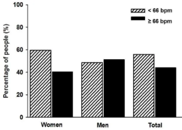
We assessed 113 individuals over 80 years old. The total prevalence of overweight was (n=69, 61.1%), central obesity (n= 49, 40.2%), hypertension (n= 99, 87.6%), hyperglycaemia (n= 29, 25.7%), hyper triglycerides (n= 34, 30.1%). Additionally, Figure 1 shows the prevalence of RHR in the group of elderly patients, according to gender. It can be observed that 40.5% of the women had RHR values equal to or greater than 66 beats/ min, while in the men this percentage was 51.3%, equivalent to 44.2% of the total sample (p=0.882).
Figure 1. Prevalence of resting heart rate according to gender in elderly aged 80 years and over.
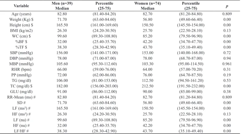
Table 1. Comparison of variables in relation to anthropometric, metabolic, hemodynamic and heart rate variability according to gender in the elderly aged 80 years old and over.
Variable Men (n=39) Median
Percentile (25-75)
Women (n=74) Median
Percentile
(25-75) p
Age (years) 82.80 (81.40-84.20) 82.70 (81.20-84.80) 0.809
Weight (Kg) $ 71.70 (63.60-84.60) 56.80 (49.60-66.40) 0.00
Height (cm) $ 165.50 (161.00-169.60) 150.50 (145.50-154.00) 0.00
BMI (kg/m2) 26.30 (24.20-30.50) 25.70 (22.50-28.10) 0.13
WC (cm) $ 99.60 (89.30-108.80) 85.20 (79.50-96.90) 0.00
%BF $ 32.00 (25.40-35.70) 42.20 (34.70-47.70) 0.00
%TF $ 38.30 (28.30-42.90) 43.70 (35.10-49.40) 0.00
SBP (mmHg) 156.00 (141.00-171.00) 153.00 (140.00-168.00) 0.72
DBP (mmHg) 78.00 (71.00-87.00) 78.00 (68.70-87.00) 0.94
MBP (mmHg) 105.60 (95.30-112.60) 103.30 (95.00-114.50) 0.961
RHR (bpm) 66.00 (59.00-76.00) 64.00 (57.00-70.20) 0.31
PP (mmHg) 72.00 (62.00-86.00) 76.00 (64.70-87.50) 0.19
TG (mg/dl) 106.00 (81.00-153.00) 112.50 (94.50-161.20) 0.53
TC (mg/dl) $ 182.00 (156.00-203.00) 212.50 (191.50-232.00) 0.00
GLU (mg/dl) 91.00 (86.00-112.00) 90.00 (85.00-99.00) 0.38
RR-Mean (ms) # 82.80 (81.40-84.20) 82.70 (81.20-84.80) 0.809
SD # 71.70 (63.60-84.60) 56.80 (49.60-66.40) 0.00
LF (ms²) # 165.50 (161.00-169.60) 150.50 (145.50-154.00) 0.00
HF (ms²) # 26.30 (24.20-30.50) 25.70 (22.50-28.10) 0.13
LF (nu) # 99.60 (89.30-108.80) 85.20 (79.50-96.90) 0.00
HF (nu) # 32.00 (25.40-35.70) 42.20 (34.70-47.70) 0.00
LF/HF # 38.30 (28.30-42.90) 43.70 (35.10-49.40) 0.00
Legend: $= statistical differences between genders. BMI = Body mass index; WC = Waist circumference; %BF = Body fat percentage; %TF = Percentage of trunk fat; SBP = Systolic blood pressure; DBP = Diastolic blood pressure; MBP = Mean blood pressure; RHR = Resting heart rate; PP = pulse pressure; TG = Triglycerides; TC = total cholesterol; GLU = Fasting glucose; RR = interval between consecutive heart beats; SD = standard deviation of RR; HF = high frequency spectral component; LF= low frequency spectral component; nu = normalized units; # = 65 subjects were analyzed.
Table 2 presents the comparisons of hemodynamic and metabolic variables according to RHR. It can be observed that elderly patients with an RHR < 66 beats/min had lower DBP and GLU and higher PP compared with elderly patients with an
RHR ≥ 66 beats/min. Also, according to the HRV, the individuals
with RHR < 66 had higher RRmean and SD, HFms², HFnu, and lower LFnu and LF/HF relation.
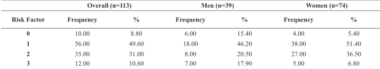
Table 3 shows a lower prevalence of metabolic syndrome in all individuals of the sample and according to gender.
Table 4 shows the differences in GLU, TG, SBP, DBP, WC
and BMI in the comparison between all the variable quantiied
in this study according to the presence of metabolic syndrome and no-metabolic syndrome eldw erly aged 80 years and over
with higher values in the irst group.
When the relationship between variables was verified, Spearman’s correlation showed positive relationship be-tween RHR and DBP (r= 0.27; p= 0.004), and negative correlation between RHR with PP (r= -0.23; p=0.013).
Table 5 shows the values of the linear regression between the hemodynamic (SBP, DBP, and PP) and met-abolic (TG, TC and GLU) variables and RHR. Of note, regardless of age, sex, and %BF, RHR influenced the changes observed in DBP, PP and TG; however, in relation to SBP, TC and GLU the changes were not statistically significant.
Discussion
The results of this study suggest that increased levels of RHR are associated with unfavorable hemodynamic and metabolic parameters in elderly subjects aged 80 years and over.
In terms of the hemodynamic and metabolic parameters,
there was no statistically signiicant difference between gen -ders, except for TC which showed the highest concentrations in females. Additionally, we obtained a lower value of PP in the group with an RHR less than 66 beats/min. In general, lower HR and lower PP are associated with better prognosis (Perret-Guillaume et al., 2009; Protogerou, et al., 2007); however, in this study, higher HR was associated with lower PP which was reasonable since its calculation is based on the difference be-tween SBP and DBP. Additionally, PP is an important indicator of arterial stiffness, and the behavior of blood circulation, which is a strong predictor of cardiovascular morbidity and mortality (Protogerou, et al., 2007).
Table 2. Comparison of variables between higher and lower resting heart rate, anthropometric, metabolic, hemodynamic and heart rate variability according to gender in the elderly aged 80 years and over.
Variable
Overall RHR < 66 beats.min-1
N=63
Overall RHR > 66 beats.min-1
N=50
RHR < 66 beats. min-1 Men (n=19)
RHR > 66 beats. min-1 Men (n=20)
RHR < 66 beats. min-1 Women
(n=44)
RHR > 66 beats. min-1 Women
(n=30)
Difference Between
Overall P
Weight (Kg) 63.67 (13.62) 63.89 (15.08) 70.00 (23.50) 71.85 (19.90) 59.30 (17.40) 55.30 (17.00) 0.93 BMI (kg/m2) 26.31 (4.33) 25.78 (3.96) 26.10 (8.10) 26.76 (6.10) 26.16 (4.80) 25.12 (6.90) 0.50 WC (cm) 91.18 (12.49) 91.37 (13.31) 98.50 (20.50) 100.25 (17.80) 85.50 (16.60) 84.80 (20.90) 0.94 %BF 38.31 (9.64) 35.04 (9.61) 34.20 (6.00) 28.70 (11.10) 42.80 (10.80) 39.80 (16.80) 0.08 %TF 40.89 (10.57) 37.44 (10.52) 40.80 (8.70) 34.60 (14.00) 44.30 (10.70) 41.30 (18.90) 0.09 SBP (mmHg) 158.38 (27.49) 151.78 (19.82) 161.00 (45.00) 151.50 (25.00) 155.00 (31.10) 151.00 (32.00) 0.14 DBP (mmHg) 75.87 (12.54) 80.94 (11.52) 74.00 (18.00) 83.00 (14.00)* 75.00 (20.00) 79.00 (19.00)* 0.03 MBP (mmHg) 103.37 (15.61) 104.55 (12.58) 101.33 (26.30) 106.17 (15.00) 102.00 (19.30) 102.67 (19.70) 0.66 PP (mmHg) 82.51 (22.52) 70.84 (16.60) 75.00 (37.00) 69.00 (27.00)* 80.00 (27.00) 74.00 (24.00)* 0.00 TG (mg/dl) 123.13 (60.20) 147.980 (70.55) 106.00 (50.00) 124.50 (102.00) 104.00 (58.00) 137.00 (111.00) 0.05 TC (mg/dl) 196.71 (37.70) 206.46 (43.61) 189.00 (64.00) 180.50 (50) 203.00 (36.00) 216.00 (48.00) 0.21 GLU (mg/dl) 94.48 (17.96) 104.42 (33.88) 88.00 (13.00) 96.50 (40) 90.00 (13.00) 90.00 (15.00) 0.05 RR-Mean (ms) # 997.00 (216.00) 823.50 (185.00) 1096.00 (232.00) 823.50 (130.00)* 993.00 (173.00) 824.00 (227.00)* <0.00
SD (ms) # 18.00 (9.00) 14.00 (9.00) 22.00 (22.00) 17.00 (9.00) 17.00 (9.00) 12.50 (10.00)* 0.03 LF (ms²) # 34.83 (76.35) 33.94 (41.86) 83.58 (85.98) 33.94 (22.60) 34.78 (62.50) 31.66 (57.40) 0.28 HF (ms²) # 58.98 (135.84) 39.09 (52.46) 78.63 (182.20) 48.93 (132.20) 52.53 (115.20) 27.99 (35.50)* 0.01 LF (nu) # 34.00 (20.00) 46.50 (26.00) 35.00 (29.00) 42.50 (36.00) 33.00 (20.00) 49.00 (19.50)* 0.03 HF (nu) # 66.00 (20.00) 53.50 (26.00) 65.00 (29.00) 57.50 (36.00) 67.00 (20.00) 51.00 (19.50) * 0.03 LF/HF # 0.54 (0.61) 0.97 (1.21) 0.56 (1.15) 0.76 (1.81) 0.53 (0.62) 1.05 (1.00) * 0.04
* = statistically signiicantly differences within gender; IQR = Interquartile range; BMI = Body mass index; WC = Waist circumference; %BF = Body fat percentage; %TF = Percentage of trunk fat; SBP = Systolic blood pressure; DBP = Diastolic blood pressure; MBP = Mean blood pressure; PP = pulse pressure; TG = Triglycerides; TC = total cholesterol; GLU = Fasting glucose; RR = interval between consecutive heart beats; SD = standard deviation of RR; HF = high frequency spectral component; LF= low frequency spectral component; nu = normalized units; # = n of 65 subjects were analyzed.
Table 3. Prevalence of Metabolic Syndrome in elderly aged 80 years and over.
Overall (n=113) Men (n=39) Women (n=74)
Risk Factor Frequency % Frequency % Frequency %
0 10.00 8.80 6.00 15.40 4.00 5.40
1 56.00 49.60 18.00 46.20 38.00 51.40
2 35.00 31.00 8.00 20.50 27.00 36.50
3 12.00 10.60 7.00 17.90 5.00 6.80
with both high blood pressure and high RHR demonstrated a greater probability of death from cardiovascular diseases, in-cluding coronary artery disease, when compared with people with high blood pressure and normal or decreased RHR.
The increased blood pressure and RHR can be attributed to functional changes, such as, autonomic imbalance, resulting from an increase in the sympathetic nervous system and a reduc-tion in the parasympathetic nervous system (Jose, & Collison, 1970). Previous studies have shown that patients with autonomic imbalance may have a higher probability of death (Olshansky, Sabbah, Hauptman, & Colucci, 2008; Zucker, Patel, & Schultz, 2012). An increase in RHR results in an autonomic imbalance, which could provide increased myocardial oxygen consumption,
thereby reducing the reserves of high-energy substrates, which hinders subendocardial perfusion and may result in a lowering of the atrial threshold and hence an increase in the incidence of ventricular arrhythmias (Zucker, Wang, Brandle, Schultz, & Patel, 1995). Thus, these responses may be involved in the increased mortality of those with sympathetic hyperactivity and tachycardia. In this study, an increase in DBP and RHR
were found. Thus, our indings suggest a possible functional
changes since we found and increase in sympathetic activity and autonomic imbalance.
Table 5. Linear regression between the resting heart rate, systolic and diastolic blood pressure, pulse pressure and blood glucose after adjusting for age, gender and body fat percentage in the elderly aged 80 years old and over.
RHR-adjusted for age RHR-adjusted for age and gender RHR-adjusted for age. gender and %BF
β p CI-95% β p CI-95% β p CI-95%
SBP (mmHg) -0.03 0.50 (-0.12; 0.06) -0.02 0.57 (-0.12; 0.06) -0.02 0.68 (-0.11; 0.07)
DBP (mmHg) 0.27 0.00 (0.09; 0.44) 0.27 0.00 (0.09; 0.44) 0.26 0.00 (0.09; 0.43)
PP (mmHg) -0.13 0.01 (-0.24; -0.03) -0.13 0.02 (-0.23; -0.02) -0.12 0.03 (-0.22; -0.01)
TG (mg/dl) 0.03 0.08 (-0.00; 0.06) 0.03 0.07 (-0.00; 0.06) 0.04 0.03 (0.00; 0.07)
TC (mg/dl) 0.04 0.29 (-0.03; 0.08) 0.04 0.16 (-0.02; 0.10) 0.05 0.10 (-0.01; 0.10)
GLU (mg/dl) 0.04 0.38 (-0.05; 0.12) 0.03 0.42 (-0.05; 0.12) 0.04 0.33 (-0.04; 0.12) RHR = resting heart hate; SBP = Systolic blood pressure; DBP = Diastolic blood pressure; PP = pulse pressure; TG = Triglycerides; TC = total cholesterol; BF = percentage of body fat.
Table 4.Comparison between metabolic syndrome and no-metabolic syndrome elderly aged 80 years old and over.
Variable
No-MS Median (IQR)
N= 99
MS Median (IQR)
N=12
p
Weight (Kg) 61.10 (18.80) 74.75 (21.20) 0.00
BMI (kg/m2) 25.77 (5.50) 30.30 (5.40) <0.00
WC (cm) 89.00 (17.40) 105.80 (14.20) <0.00
%BF 36.20 (13.00) 39.90 (20.30) 0.44
%TF 41.00 (12.70) 45.20 (15.60) 0.10
SBP (mmHg) 152.00 (31.00) 165.00 (26.00) 0.03
DBP (mmHg) 76.00 (20.00) 86.00 (13.00) 0.01
PP (mmHg) 74.00 (23.00) 74.50 (25.00) 0.49
TG (mg/dl) 103.00 (64.00) 212.00 (68.00) <0.00
TC (mg/dl) 201.00 (47.00) 214.00 (63.00) 0.17
GLU (mg/dl) 89.00 (12.00) 129.50 (51.00) <0.00
HR (bmp) 63.00 (14.00) 70.50 (18.00) 0.07
RR-Mean (ms) # 920.00 (221.00) 913.00 (353.00) 0.40
SD # 17.00 (9.00) 19.50 (27.00) 0.31
LF (ms²) # 33.74 (59.36) 66.14 (54.79) 0.36
HF (ms²) # 45.62 (57.23) 43.59 (112.52) 0.44
LF (nu) # 41.00 (21.00) 57.00 (27.00) 0.83
HF (nu) # 59.00 (21.00) 43.00 (27.00) 0.83
LF/HF # 0.77 (0.72) 1.59 (3.28) 0.89
MS = metabolic syndrome; IQR = Interquartile range; BMI = Body mass index; WC = Waist circumference; %BF = Body fat percentage; %TF = Percentage of trunk fat; SBP = Systolic blood pressure; DBP = Diastolic blood pressure; MBP = Mean blood pressure; PP = pulse pressure; TG = Triglycerides; TC = total cholesterol; GLU = Fasting glucose; RR = interval between consecutive heart beats; SD = standard deviation of RR; HF = high frequency spectral component; LF= low frequency spectral component; nu = normalized units; # = n of 65 subjects were analyzed.
response at rest for both groups. Similarly, no differences were observed when the groups with and without metabolic syndrome were evaluated. However, when the groups were compared between the differences in the RHR values, it can be observed higher parasympathetic modulation by the increase in the HF values in ms² and nu, better balance in the relation LF/HF and low value to the index that represent the predominance of sympa-thetic branch - LF (nu) (Tarvainen, Niskanen, Lipponen, Ranta-Aho, & Karjalainen, 2009; Vanderlei, Pastre, Hoshi, Carvalho,
& Godoy, 2009). These indings reinforce the RHR results.
Regarding the metabolic parameters, the present study showed that elderly subjects with higher RHR values have
altered GLU and a trend in the TG compared to those with lower RHR, the changes in TG being independent of age, sex and %BF
as shown in Table 3. However, after adjustment, the inluence on
the GLU ceased to exist. Regarding the values of TG between the two groups, these values are within normal limit. Kowalski et al. (2012) in a recent study conducted with subjects with a
average age of 57.4 years found that an RHR of ≥ 72.5 beats/
min is an independent risk factor for disorders in the absorption of carbohydrates. These same authors also added that subjects with carbohydrate absorption disorders demonstrated an RHR
hemodynamic and metabolic parameters in elderly subjects aged 80 years and over. Thus, we suggest for future studies investigate lower values of RHR as independent risk factors.
In relation to the anthropometric parameters, the women had higher % BF and TF, but a smaller WC compared with the men; however, no statistical difference regarding RHR. It is known that weight gain, BF, particularly in the trunk region (Donato, Fuchs, Oppermann, Bastos, & Spritzer, 2006), and a sedentary lifestyle are conditions that impair the quality of life during the aging process (Ford, Li, Zhao, Pearson, Tsai, & Churilla, 2010) since they are associated with the development of morbidities such as diabetes mellitus, hypertension, dyslipidemia and met-abolic syndrome (Gruen, Hao, Piston, & Hasty, 2007). So, the
indings of this study suggest that women could present higher
impairment arising from the aging process.
Despite the importance of the indings presented here,
some limitations need to be mentioned such as the character-istics of the individuals assessed regarding the risk factors and cardiovascular diseases and medication not being evaluated. However, they were described according with risk factors for the development of cardiovascular diseases and the volunteers were requested to suspend medications for a period of 12 hours prior to the autonomic evaluation. This is important because Menown et al. (2013) indicated that RHR is associated with an increment in the risk for all-cause morbidity and mortality, including cardiovascular mortality, in populations with or without risk factors or cardiovascular disease. And in another diseases, that have decrease of heart rate variability as with age, this measurement is independent of the use of some medications (Bédard, Marquis, Poirier, & Provencher, 2010). A strong aspect of the study which stands out is the randomization of the sampling process, which ensured that each elderly subject contained the same chance of being drawn. The sample was composed only of elderly people considered long-lived (> 80 years), which is another highlight of the study, since there are few studies involving RHR and anthropometric, metabolic and hemodynamic parameters in this population. Finally, it is sug-gested that longitudinal studies should be performed to verify
the inluence of different forms of intervention on RHR and
the hemodynamic, metabolic and anthropometric parameters in this population.
Conclusion
A positive relationship between RHR and some metabolic and hemodynamic parameters and elderly aged 80 years and over was found. Subjects with 80 years old and over who
pres-ent RHR ≥ 66 have higher DBP and lower PP and heart rate
variability compared with the elderly with RHR < 66.
References
Bédard, M.E., Marquis, K., Poirier, P., & Provencher, S. (2010). Reduced heart rate variability in patients with chronic obstructive
pulmonary disease independent of anticholinergic or β-agonist
medications. COPD: Journal of Chronic Obstructive Pulmonary Disease, 7, 391-397. doi: 10.3109/15412555.2010.528083. Benetos, A., Rudnichi, A., Thomas, F., Safar, M., & Guize, L. (1999).
Inluence of heart rate on mortality in a French population: role
of age, gender, and blood pressure. Hypertension, 33, 44-52. doi: 10.1161/01.HYP.33.1.44.
Coleman, A., Freeman, P., Steel, S., & Shennan, A. (2005). Validation of the Omron MX3 Plus oscillometric blood pressure monitoring device according to the European Society of Hypertension inter-national protocol. Blood Pressure Monitoring, 10, 165-168. doi: 00126097-200506000-00009.
Cooney, M. T., Vartiainen, E., Laatikainen, T., Juolevi, A., Dudina, A., & Graham, I.M. (2010). Elevated resting heart rate is an inde-pendent risk factor for cardiovascular disease in healthy men and women. American Heart Journal, 159, 612-619. doi: 10.1016/j. ahj.2009.12.029.
Donato, G. B., Fuchs, S. C., Oppermann, K., Bastos, C., & Spritzer, P. M. (2006). Association between menopause status and cen-tral adiposity measured at different cutoffs of waist circum-ference and waist-to-hip ratio. Menopause, 13, 280-285. doi: 00042192-200613020-00018.
Freitas, I.F, Jr. (2009). Medidas: estatura, peso, comprimento dos segmentos. Padronização de técnicas antropométricas. Presidente Prudente (SP): Cultura Acadêmica, 23-5.
Freitas, I.F, Jr., Monteiro, P.A., Silveira, L.S., Cayres, S.U., Antunes, B.M., Bastos, K.N., ... & Fernandes, R.A. (2012). Resting heart rate as a predictor of metabolic dysfunctions in obese children and ado-lescents. BMC pediatrics, 12(1), 5. doi: 10.1186/1471-2431-12-5. Ford, E.S., Li, C., Zhao, G., Pearson, W.S., Tsai, J., & Churilla, J.R.
(2010). Sedentary behavior, physical activity, and concentrations of insulin among US adults. Metabolism, 59, 1268-1275. doi: 10.1016/j.metabol.2009.11.020.
Gillman, M.W., Kannel, W.B., Belanger, A., & D’Agostino, R.B.
(1993). Inluence of heart rate on mortality among persons with
hypertension: the Framingham Study. American Heart Journal, 125, 1148-1154.
Girotto, E., Andrade, S.M., Cabrera. M.A.S., & Ridão, E.G. (2009). Prevalência de fatores de risco para doenças cardiovasculares em hipertensos cadastrados em unidade de saúde da família. Acta Scientiarum Health Sciences, 31, 77-82. doi: 10.4025/actasci-healthsci.v31i1.4492.
Gruen, M.L., Hao, M., Piston, D.W., & Hasty, A.H. (2007). Leptin requires canonical migratory signaling pathways for induction of monocyte and macrophage chemotaxis. American Journal Physiology- Cell Physiology, 293, 1481-1488. doi: 10.1152/ ajpcell.00062.2007
Hashmi, A., Ibrahim-Zada, I., Rhee, P., Aziz, H., Fain, M.J., Friese, R.S., & Joseph, B. (2014). Predictors of mortality in geriatric trauma patients: a systematic review and meta-analysis. Journal of Trauma and Acute Care Surgery, 76(3), 894-901. doi: 10.1097/ TA.0b013e3182ab0763.
Human, R.P., & Jones, G.A. (2004). Evaluation of swab transport sys-tems against a published standard. Journal of Clinical Pathology, 57, 762-773. doi:10.1136/jcp.2004.016725.
Kochiadakis, G.E., Kanoupakis, E.M., Rombola, A.T., Igoumenidis, N.E., Chlouverakis, G.I., & Vardas, P.E. (1998). Reproducibility of tilt table testing in patients with vasovagal syncope and its relation to variations in autonomic nervous system activity. Pacing and clinical electrophysiology,21(5), 1069-1076.
Kowalski, J., Brylik, A., Irzmański, R., Pawlicki, L., Ciećwierz, J.,
Jarzabek, K., & Barylski, M. (2012). (Resting heart rate in subjects with carbohydrate disorders). Polski merkuriusz lekarski: organ Polskiego Towarzystwa Lekarskiego, 32(188), 93-97.
Lauria, A.A., Santos, T.M., Amorim, P.R.S., Marques, F.A.D., Lima, J.R.P. Prediction of 8 baseline heart rate of individuals with differ-ent levels of habitual physical activity. 9 Rev Bras Med Esporte. 2013;19(1).
Lean, M.E., Han, T.S., & Morrison, C.E. (1995). Waist circumference as a measure for indicating need for weight management. British Medical Journal, 311, 158-161. doi: http://dx.doi.org/10.1136/ bmj.311.6998.158
Menown, I., Davies, S., Gupta, S., Kalra, P.R., Lang, C.C., Morley, C., & Padmanabhan, S. (2013). Resting heart rate and outcomes in patients with cardiovascular disease: where do we current-ly stand? Cardiovascular therapeutics, 31(4), 215-223. doi: 10.1111/j.1755-5922.2012.00321.x.
Nogueira, M.C., Ribeiro, L.C., & Cruz, O.G. (2009). Social inequalities in premature cardiovascular mortality in a medium-size Brazilian city. Cadernos de Saúde Pública, 25, 2321-2332.
Olshansky, B., Sabbah, H.N., Hauptman, P.J., & Colucci, W.S. (2008). Parasympathetic nervous system and heart failure: pathophysiology and potential implications for therapy. Circulation, 118, 863-871. doi: 10.1161/CIRCULATIONAHA.107.760405
Palatini, P., Benetos, A., Grassi, G., Julius, S., Kjeldsen, S.E., Mancia, G., ... & Zanchetti, A. (2006). European Society of Hypertension
Identiication and management of the hypertensive patient with el -evated heart rate: statement of a European Society of Hypertension Consensus Meeting. Journal Hypertension, 24(4), 603-610. doi: 00004872-200604000-00001.
Palatini, P. (2009). Elevated Heart Rate: A “New” Cardiovascular Risk Factor? Progress in Cardiovascular Diseases, 52, 1-5. doi:10.1016/j.pcad.2009.06.001
Park, B-J., Lee, H.R., Shim, J.Y., Lee, J.H., Jung, D.H., & Lee, Y.J. (2010). Association between resting heart rate and arterial stiffness in Korean adults. Archives of Cardiovascular Disease, 103, 246-252. doi: 10.1016/j.acvd.2010.03.004
Pickering, T.G., Hall, J.E., Appel, L.J., Falkner, B.E., Graves, J., Hill, M.N., ... & Roccella, E.J. (2005). Subcommittee of Professional and Public Education of the American Heart Association Council on High Blood Pressure Research. Recommendations for blood pressure measurement in humans and experimental animals: Part 1: blood pressure measurement in humans: a statement for professionals from the Subcommittee of Professional and Public Education of the American Heart Association Council on High Blood Pressure Research. Hypertension, 45(1), 142-161. Perret-Guillaume C1, Joly L, Benetos A. Heart rate as a risk factor
for cardiovascular disease. American Heart Association Council on High Blood Pressure Research. Prog Cardiovasc Dis. 2009 Jul-Aug;52(1):6-10. doi: 10.1016/j.pcad.2009.05.003.
Protogerou, A.D., Papaioannou, T.G., Blacher, J., Papamichael, C.M., Lekakis, J.P., & Safar, M.E. (2007). Central blood pressures: do
we need them in the management of cardiovascular disease? Is it a feasible therapeutic target? Journal Hypertension, 25, 265-272. doi: 00004872-200702000-00002.
Recommendations for blood pressure measurement in humans and ex-perimental animals: Part 1: blood pressure measurement in humans: a statement for professionals from the Subcommittee of Professional and Public Education of the American Heart Association Council on High Blood Pressure Research. Hypertension, 45(1), 142-161. Rossi, F.E., Ricci-Vitor, A. ., Sabino, J. ., Vanderlei, L., & Freitas, I.
F., Jr. (2014). Autonomic modulation and its relation with body composition in swimmers. The Journal of Strength & Conditioning Research, 28(7), 2047-2053. doi: 10.1519/JSC.0000000000000344. Santana, J.S., Franco Filho, J.C.S., Neto, S., Melo, E.V.D., Santana,
N.O.D., Barreto, A.T.F., ... & Oliveira, J.L.M. (2013). Prognostic value of chronotropic incompetence in elderly patients undergoing exercise echocardiography. Arquivos brasileiros de cardiologia, 100(5), 429-436.
Vanderlei, L.C.M., Pastre, C.M., Hoshi, R.A., Carvalho, T.D.D., & Godoy, M.F.D. (2009). Noções básicas de variabilidade da frequên-cia cardíaca e sua aplicabilidade clínica. Rev Bras Cir Cardiovasc, 24(2), 205-17.
Tarvainen, M.P., Niskanen, J.P., Lipponen, J.A., Ranta-Aho, P.O., & Karjalainen, P.A. (2009). Kubios HRV—a software for advanced heart rate variability analysis. In 4th European Conference of the International Federation for Medical and Biological Engineering (pp. 1022-1025). Springer Berlin Heidelberg.
Zucker, I.H., Wang, W., Brandle, M., Schultz, H.D., & Patel, K.P. (1995). Neural regulation of sympathetic nerve activity in heart failure. Progress in Cardiovascular Diseases, 37, 397-414. doi: 10.5935/abc.20130091.
Zucker, I.H., Patel, K.P., & Schultz, H.D. (2012). Neurohumoral stimu-lation. Heart Fail Clinical, 8, 87-99. doi: 10.1016/j.hfc.2011.08.007. WHO. World Health Organization Consequences of overweight and obesity on the health of adults and children. In: WHO. Obesity Preventing and Controlling Global Epidemic. 1. Ed. Sao Paulo: ROCA, 2004.
Author’s note
Fabrício E. Rossi and Vanessa R. dos Santos are afiliated with the
Institute of Bioscience, São Paulo State University, Rio Claro, São Paulo, Brazil.
Ana Laura Ricci-Vitor is afiliated with the Department of Physio -therapy, University Federal of São Paulo, São Paulo, SP, Brazil.
Igor C. Gomes is afiliated with the Departament of Epidemiology,
University of São Paulo, São Paulo, SP, Brazil.
João Paulo J. Sabino and Luiz Guilherme S. Branco are afiliated with
the Department of Physiology, Dental School of Ribeirão Preto, Uni-versity of São Paulo, Ribeirão Preto, Brazil.
Luiz Carlos M. Vanderlei is afiliated with the Department of Physiother -apy, São Paulo State University, Presidente Prudente, São Paulo, Brazil.
Diego G. D. Christofaro and Ismael F. Freitas Junior are afiliated
Corresponding author
Fabrício Eduardo Rossi
Department of Physical Education, University, Estadual Paulista, Presidente Prudente, São Paulo, Brazil
Rua Roberto Simonsen, nº 305 – Centro Educacional, Presidente Prudente, CEP 19060-900, São Paulo, Brasil.
Email: [email protected]
Manuscript received on June 16, 2015 Manuscript accepted on December 12, 2015