The microbiome and inborn errors of metabolism: Why we should look
carefully at their interplay?
Karina Colonetti
1,2, Luiz Fernando Roesch
3and Ida Vanessa Doederlein Schwartz
1,2,4 1Programa de Pós-Graduação em Genética e Biologia Molecular, Universidade Federal do Rio Grande do
Sul, Porto Alegre, RS, Brazil.
2
Laboratory of Basic Research and Advanced Investigations in Neurosciences (BRAIN), Hospital de Clínicas
de Porto Alegre, Porto Alegre, RS, Brazil.
3
Interdisciplinary Research Center on Biotechnology-CIP-Biotec, Universidade Federal do Pampa, Bagé,
RS, Brazil.
4
Medical Genetics Service, Hospital de Clínicas de Porto Alegre, Porto Alegre, RS, Brazil.
Abstract
Research into the influence of the microbiome on the human body has been shedding new light on diseases long known to be multifactorial, such as obesity, mood disorders, autism, and inflammatory bowel disease. Although in-born errors of metabolism (IEMs) are monogenic diseases, genotype alone is not enough to explain the wide phenotypic variability observed in patients with these conditions. Genetics and diet exert a strong influence on the microbiome, and diet is used (alone or as an adjuvant) in the treatment of many IEMs. This review will describe how the effects of the microbiome on the host can interfere with IEM phenotypes through interactions with organs such as the liver and brain, two of the structures most commonly affected by IEMs. The relationships between treatment strat-egies for some IEMs and the microbiome will also be addressed. Studies on the microbiome and its influence in indi-viduals with IEMs are still incipient, but are of the utmost importance to elucidating the phenotypic variety observed in these conditions.
Keywords:Inborn errors of metabolism, microbiome, microbiota, diet, treatment.
Received: July 28, 2017; Accepted: January 19, 2018.
Introduction
The human body host a large amount of non-human genetic material, the microbiome, defined as the set of mi-croorganisms, their genes, and the surrounding environ-mental conditions (Marchesi and Ravel, 2015). The human gut microbiome is believed to play an important role in the development of basic physiological systems, such as the di-gestive, immune, and nervous systems, and constitutes a virtual metabolic organ of unquestionable importance (Lopez-Legarreaet al., 2014; Suezet al., 2014; Maukonen and Saarela, 2015). The gastrointestinal (GI) tract is a meta-bolically rich environment that harbors approximately three-quarters of the body’s immune cells, contains vagal afferent endings which respond to immune cells and im-mune and bacterial products (cytokines, proteases, 5-Hydroxytryptimaine and CRH for corticotropin-releasing hotmone, CRH, histamine), and has receptors for com-pounds produced by neuroendocrine cells (Omran and
Aziz, 2014). Diet and genes related to the immune system and metabolism are among the key factors with potential to alter the bacterial community present in the gut. Thus, the associations of diet, metabolism, the central nervous sys-tem, and the immune system with the development and composition of the gut microbiome has become the object of intense interest among the scientific community (Mayer et al., 2014).
Inborn errors of metabolism (IEM) are rare mono-genic genetic diseases characterized by absent or deficient activity of a given enzyme and which can sometimes be managed with dietary strategies. The phenotypic heteroge-neity found in IEMs is manifested mainly by the age at on-set of symptoms, presence (or absence) of neurological compromise, and response to the treatment. In untreated phenylketonuria (PKU) and in propionic and methyl-malonic acidemia patients, for instance, the neurological and behavior impairment are highly variable. The develop-ment of liver disease is common to several IEMs, such as tyrosinemia type 1 and urea cycle disorders. Also, the re-sponse to the treatment is not the same among patients with the same genotype.
Genetics and Molecular Biology, 41, 3, 515-532 (2018)
Copyright © 2018, Sociedade Brasileira de Genética. Printed in Brazil DOI: http://dx.doi.org/10.1590/1678-4685-GMB-2017-0235
Send correspondence to Karina Colonetti. Universidade Federal do Rio Grande do Sul, Rua Ramiro Barcelos, 2350, 90035-903 Porto Alegre, RS, Brazil. E-mail: [email protected].
Convergent efforts of professionals in different fields have enabled the discovery of new mechanisms and pro-cesses whereby the microbiome can exert local and sys-temic effects. In this non-systematic review of the literature, we will focus on how the gut microbiome could influence the context of treatable IEMs.
The human gut microbiome
Among the various microbial habitats found in the human body, the GI tract harbors the vast majority of mi-crobial cells (Senderet al., 2016). The composition of the microbiota varies along the GI tract, both quantitatively and qualitatively, depending on the environmental conditions (pH, oxygen, etc.) (Donaldsonet al., 2016). In the small bowel (particularly the duodenum), the composition is sim-ilar to that of the stomach, while the large bowel (especially the colon) contains the majority of the gut’s microbial pop-ulation, as it is the site of fermentation, due to the availabil-ity of nutrients obtained from digestion (Madigan and Martinko, 2006).
Prior to the development of next-generation sequenc-ing (NGS) techniques, the gene profile of these microor-ganisms had never been determined accurately (Grenham et al., 2011). The ability to obtain a large number of gene sequences in a short period of time and at relatively low cost led to the acquisition of an immense volume of data to which biological significance could then be ascribed (Cho and Blaser, 2012). Advances in these techniques, coupled with the development of bioinformatics tools, have allowed analysis of the gut microbiome to an extent that would have been impossible with bacterial cultures alone (Hiergeistet al., 2015). Furthermore, the use of NGS and bioinformatics techniques, with the aid of databases and computational and statistical algorithms, has allowed complex studies for the detection, quantification, and functional analysis of the human microbiome and its physiological associations, thus expanding knowledge of microbial ecology beyond simple pathogenvs.host relationships.
Initiatives such as the Human Microbiome Project, created in the United States in 2008, have sought to charac-terize the microbial communities of various sites in the hu-man body, with a focus on analyzing the role of these microorganisms in sickness and in health (Human Micro-biome Project Consortium, 2012). In Europe, a similar ef-fort known as MetaHIT, which took place from 2008 to 2012, sought to study the association of the gut microbiome with several states of health and illness, prioritizing obesity and inflammatory bowel disease (Metagenomics of the Hu-man Intestinal Tract, MetaHIT.
The results of the aforementioned initiatives have led to a new appreciation for the human microbiome from taxo-nomic and functional points of view. The microbiota is both functionally relevant and uniquely personal, differing even between monozygotic twins, what suggests that childhood exposure to different environmental factors is a
determi-nant of development of the adult microbiota (Turnbaugh and Gordon, 2009). Despite great interpersonal variation in the microbiota, the metabolic roles of its microorganisms are highly conserved: enriching the biosynthesis of co-factors and vitamins, in addition to a key role in central car-bohydrate metabolism, aromatic amino acids (AA), and ATP synthesis in the lower GI tract (Segataet al., 2011; Human Microbiome Project Consortium, 2012). This has given rise to the notion of a “functional core” of microor-ganisms rather than a core set of microbial taxa, as the same essential roles can be played by different taxa (Lloyd-Price et al., 2016).
The gut microbiota is influenced by the environment and affected by diet, medications, age, geographic factors, surgical interventions, and host genetics, particularly genes related to the immune system and metabolism (Yatsunenko et al., 2012; Dabrowska and Witkiewicz, 2016; Goodrichet al., 2016). The gut microbiome suffers drastic changes dur-ing the first three years of life (Yatsunenkoet al., 2012). After that, diet is one of the main factors that shape the gut microbiota (De Filippoet al., 2010; Davidet al., 2014), and the microbiome continues to evolve all lifelong (Ottmanet al., 2012; Odamakiet al., 2016). Once diet is strongly cor-related with cultural habits and is affected by geographic factors, such as availability of nutrients and source of car-bohydrates, fibers and fat, one can also consider that culture affects the patterns found in the microbiome (Yatsunenko et al., 2012). To study the microbiome is also to study ecol-ogy. From an ecological point of view, maintaining suffi-cient bacterial diversity and richness is important for gut microbiota functional redundancy, adaptability and to pro-vide a certain tolerance against environmental challenges, resilience (Gillet al., 2006). Western diets, rich in calories and refined sugar, are associated with lower richness in mi-crobial communities at individual level (alpha diversity) and higher variation among individuals (beta diversity) when compared with diets high in fiber and relatively low in calories (Martínezet al., 2015). Individuals who con-sume a Western type diet with high-energy and high-fat in-take present changes in metabolic and immune biomarkers, such as a higher body mass index and higher levels of in-flammatory markers than those who follow a high-fiber, low-calorie diet (Caniet al., 2009). Taken together, these facts have led to associations between microbial richness and health. Once microbial richness is strongly associated with diet patterns (De Filippoet al., 2010; Cotillardet al., 2013; Sonnenburg and Sonnenburg 2014), both the compo-sition and energy content of one’s diet are important modu-lators of the microbiota (Oriach et al., 2016). Diet is a crucial driver of the composition of the microbial commu-nity from childhood to old age (Kashtanovaet al., 2016) and has the potential to alter the bacterial metabolite pro-file, thus influencing the host’s metabolism both directly and indirectly.
The major bacterial metabolites known to influence the host include short-chain fatty acids (SCFAs) and vita-mins. SCFAs are organic monocarboxylic acids with six or fewer carbon atoms, generated by anaerobic fermentation of indigestible dietary fibers (such as cellulose, xylans, and inulin) in the gut. The main SCFAs produced as a result of these fermentation processes are butyrate, acetate, and pro-pionate. SCFAs are absorbed by the host and are important energy sources, corresponding to 10% of the energy source in a Western diet. Portal and hepatic veins contain large amounts of SCFAs (Cummingset al., 1987). SCFAs also stimulate growth of bacteria in the genera Lactobacillus
and Bifidobacterium, these playing a key role in colon
physiology and metabolism (Royet al., 2006) and influenc-ing the immune and inflammatory responses (Maslowski and Mackay, 2011; Tremaroli and Bäckhed, 2012; Lopez-Legarreaet al., 2014).In vitro, SCFAs increase the produc-tion of anti-inflammatory cytokines, such as IL-10, and de-crease production of proinflammatory cytokines, such as IL-1b, IL-6, and TNF-a(Vinoloet al., 2011). Production of SCFAs also promotes transcription of thePTH1gene, which encodes tryptophan hydroxylase, the rate-limiting enzyme of serotonin synthesis in the gut (Reigstadet al., 2015). SCFAs are also generally involved in G-protein sig-naling, modulation of cell sigsig-naling, cell–cell interactions, gene expression, immune function, and neurotransmitter synthesis and release (Nakaoet al., 1998; Le Poulet al., 2003; Nguyenet al., 2007; Hanet al., 2014; Nankovaet al., 2014). Several physiological effects, including regulation of energy homeostasis, obesity, immune system functions, cancer, and cerebral function, as well as histone deace-tylase (HDAC) inhibition, have been associated with butyr-ate (Koh et al., 2016). Specific host transporters and receptors are available for butyrate, and it is also used by colon cells as a source of energy through beta-oxidation (Stillinget al., 2016). Furthermore, acetate and propionate can be used by the liver for lipogenesis and gluconeo-genesis, respectively (Janssen and Kersten, 2015). The po-tential for modulation of host metabolism and genetics by the gut microbiota suggests that the role of this factor war-rants closer attention. This is especially true in IEMs in which metabolic pathways are originally altered, as the microbiome may act to reinforce metabolic pathways that are advantageous or disadvantageous to the host, with a di-rect impact on phenotype.
The evidence for a role of the composition of the hu-man gut microbiota and its metabolites in health and illness becomes increasingly stronger (Sharonet al., 2014; Cole-man and Nunes, 2016; Rooks and Garrett, 2016). Changes in the GI tract microbiota induce metabolic changes with systemic effects (Tremaroli and Bäckhed, 2012; Ochoa-Repáraz and Kasper, 2014; Sharonet al., 2014), and current research seeks to characterize microbiota–host interactions to elucidate the depth and breadth of this influence.
Some conditions, such as liver and bowel diseases
and Clostridium difficile infection, are already being
treated with microbiota-modifying therapies. These in-clude probiotics, prebiotics, antibiotics, and fecal trans-plant (Sheth et al., 2016; Young, 2017). Probiotics are living microorganisms that, when administered at an appro-priate concentration, can confer health benefits to the host, while prebiotics are indigestible components of foods that benefit the host by promoting growth or activity of a spe-cific bacterial species or community in the colon. Fecal transplant is the administration of fecal matter from a healthy donor to a diseased individual, with the objective of restoring the typical microbial community of the healthy gut. These strategies can be used jointly or in isolation to re-store the balance of the intestinal microbial community in the event of dysbiosis, which is any change to the composi-tion of resident commensal communities relative to the community found in healthy individuals.
Inborn Errors of Metabolism (IEM)
IEMs are individually rare diseases, but as a group they are fairly common. Currently, more than 600 known human diseases are classified as IEMs (Alfadhel et al., 2016). Classically, IEMs are defined as a set of monogenic (single-gene) diseases that cause protein dysfunction, with partial or total loss of enzyme activity; however, IEMs can be pleiotropic, and may involve virtually any organ or sys-tem. Clinical onset may occur from even before birth up to adulthood (Sharer, 2011), and environmental triggers may be crucial determinants of individual phenotype (Lanpher et al., 2006). In an individual IEM, one primary metabolite flux is affected. In complex disease, however, a whole net-work of metabolite fluxes might be subtly altered to con-tribute to the overall phenotype. This concept of metabolic flux is essential in the translation of genetic and environ-mental factors into the phenotype or threshold for disease (Lanpheret al., 2006). Even a single metabolite defect can affect several secondary metabolic pathways, with a greater or lesser degree of environmental influence, to contribute to each patient’s specific phenotype.
The treatment and management of IEMs are always individualized, based on each patient’s diagnosis and phe-notype, and there is broad heterogeneity even within each category (Argmannet al., 2016). Despite this heterogeneity in management approaches, the specific treatment usually falls into one of three classes: (I) enzyme replacement ther-apy, to replenish the deficient enzyme; (II) substrate reduc-tion therapy; or (III) dietary treatment, although organ transplantation is also used in some cases (Ezgu, 2016). Additional non-specific treatment may be necessary, de-pending on the presence of comorbidities, such as neuro-psychiatric disorders in PKU patients (Bilderet al., 2017), or renal and neurologic impairment in patients with tyro-sinemia type I (Santraet al., 2008; Chinskyet al., 2017). Given the importance of diet to the microbiome, we will
primarily address dietary therapy in this review, with a sec-ondary focus on the importance of the microbiome in allo-geneic hematopoietic stem-cell transplantation (HSCT).
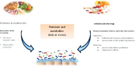
Dietary treatment for IEMs may be employed as monotherapy or adjuvant therapy. Its purpose is to elimi-nate or reduce whichever toxic compound that accumulates in the body (Schwartzet al., 2008). However, this form of therapy has several limitations, including overload and/or deficiency of certain food groups and nutrients (Crenn and Maillot, 2007; Boyeret al., 2015). Theoretically, diets re-stricted or excessively rich in certain nutrients may prompt a state of intestinal dysbiosis with systemic effects, leading to malnutrition, obesity (Henao-Mejiaet al., 2012), type 1 (Wenet al., 2008) or type 2 diabetes (Larsenet al., 2010), inflammatory bowel disease (Ashtonet al., 2017; Geirnaert et al., 2017) and liver disease (Lee and Sokol, 2015), as well as a variety of disorders featuring an inflammatory component, symptoms of autism spectrum disorders (De Angeliset al., 2015), and even cancer (Jacquelineet al., 2017; Xu and Jiang, 2017). Studies seeking to identify the effects of dietary treatment and nutrient supplementation on the microbiome of patients with IEMs are still scarce. A summary of this research will be presented below and in Table 1.
Organ transplantation (mainly liver transplantation and HSCT) is also a treatment option for several IEMs (Sirrset al., 2013; Boelens et al., 2014). Within this con-text, the microbiome was recently noted as a key factor in graft-vs.-host disease (GVHD). Acute GVHD is character-ized by rupture of the intestinal barrier, caused by the con-ditioning regimen administered before HSCT and by leakage of microbe-associated molecular patterns (MAMPs, also known as pathogen-associated molecular patterns or PAMPs), particularly lipopolysaccharide (LPS). The proinflammatory response mounted against these mol-ecules leads to systemic inflammation. Antibiotic treatment in the perioperative period of allogeneic HSCT has been as-sociated with a higher likelihood of GVHD and lower odds of survival, which suggests a potentially pathogenic role of antibiotics through depletion of gut microbiome diversity. The finding that fecal transplant successfully treats GVHD by reconstituting the microbiota has reinforced this theory (Balmeret al., 2014; Melis et al., 2014; Kakihanaet al., 2016; Rashidiet al., 2017; Routyet al., 2017; Spindelboeck et al., 2017). Efforts to characterize the influence of the microbiome in complications resulting from organ trans-plantation are paving the way for new avenues of treatment. Administration ofLactobacillus, for instance, appears to be a promising strategy for treatment of GVHD in allogeneic HSCT recipients, although the mechanism of action has yet to be fully understood (Staffaset al., 2017).
Influence of the microbiome on the major
organs affected by IEMs
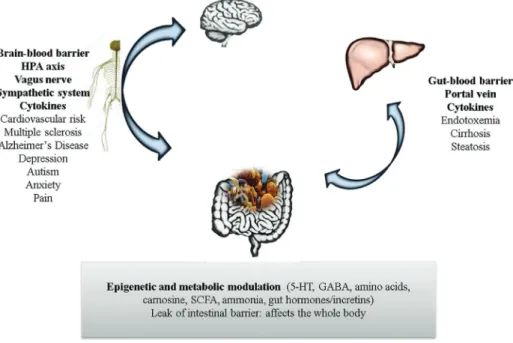
The features of IEMs are highly heterogeneous; how-ever, the nervous system central (CNS) and liver, due to their high metabolic rate, are particularly susceptible to the effects of any metabolic defect (Sahooet al., 2012). These organs are also closely related to microbiome activity, and a summary of on this matter can be found in Figure 1.
The microbiome has wide-ranging influence on the CNS, with probable effects on metabolism (Fuet al., 2015; Montagner et al., 2016), coordination (Sampson et al., 2016), mood (Slykermanet al., 2017), behavior (Tillischet al., 2013), cognition (Steenbergenet al., 2015), tempera-ture control (Chevalieret al., 2015), and sensation (Chiuet al., 2013). This influence may begin before birth, via the maternal microbiome (Rautavaet al., 2012), and may be perpetuated throughout life, playing essential roles in the development of the blood–brain barrier (Braniste et al., 2014), maturation of the immune system (Chung et al., 2012), and also myelination of the prefrontal cortex (Hoban
et al., 2016). Communication between the microbiome and
the CNS is two-way, occurring both through metabolites and toxins produced by the bacterial community on the one hand, and via the immune, metabolic, nervous, and endo-crine systems on the other (Powellet al., 2017). Over the years, disruption of the microbiome-brain-gut axis has been associated with various diseases. A breach in system ho-meostasis may occur at any point along this axis. Stressful situations affecting the brain, for instance, may affect the gut microbiome via the hypothalamic-pituitary-adrenal (HPA) axis, with repercussions for immune cell activity and bowel function (Moloneyet al., 2014). Bacterial com-ponents, in turn, can stimulate secretion of proinflamma-tory cytokines from epithelial cells, dendritic cells, and macrophages. Knowingly, several neuropsychiatric disor-ders, including depression, anxiety, schizophrenia, and au-tism spectrum disorders, are associated with elevated circulating levels of proinflammatory cytokines (Liuet al., 2015a; Petraet al., 2015). In addition to these pathways, ce-rebral function can also be modulated by microbial metabo-lites capable of crossing the blood–brain barrier (Li and Zhou, 2016). Pierre and Pellerin (2005) reported that monocarboxylate transporters (MCTs), which transport lactate, pyruvate, ketone bodies, and other SCFAs, are widely expressed in cerebral tissue, and especially so in the cortex, hippocampus, striatum, and cerebellum (Pierre and Pellerin, 2005). In rats, G protein-coupled receptors (GPCRs) activated by propionic acid (PPA) are also highly expressed in brain tissue (Boniniet al., 1997). Antibiotic therapy, which is commonly used in the treatment of some IEMs, depletes the microbiome and can affect levels of neuromodulatory substances (tryptophan, monoamines, and neuropeptides), thus influencing anxiety and cognition patterns (Desbonnetet al., 2015).
Microbiome
and
IEM:
an
interplay
519
Table 1- Inborn errors of metabolism addressed in this review, main phenotypic features, and overview of management.
EIM (Substrate accumulated)
Affected protein/gene Main clinical features Long-term management Reference
Phenylketonuria (Phenylalanine)
Phenylalanine-4-hydroxylase (PAH) Neurologic impairments, with physical, cognitive, and
behav-ioral consequences, even in well-controlled PKU
Restriction of dietary phenylalanine. Phe-free medical formula.
Sapropterin (BH4) supplementation in
respon-sive patients.
Large neutral amino acids (LNAA)
Regier and Greene, 2000; OMIM #261600
Tyrosinemia type I (Tyrosine, maleyla-cetoacetate, fumarylac etoacetate, and succinylacetone)
Fumarylacetoacetate hydrolase (FAH) Hepatomegaly, acute liver failure, cirrhosis and hepatocellular
carcinoma
Episodic paralysis and episodic peripheral neuropathy Renal Fanconi syndrome, renal
fail-ure, glomerulosclerosis, nephromegaly, nephrocalcinosis Gastrointestinal bleeding, paralytic ileus
Pancreatic islet-cell hypertrophy, splenomegaly Rickets, chronic weakness
Dietary management with reduced intake of phenylalanine and tyrosine;
Nitisinone Liver transplantation
Das, 2017; Sniderman et al., 2006; OMIM #276700
Urea cycle disorders (Ammonia)
Carbamoylphosphate synthetase I
(CPS1); Ornithine transcarbamylase
defi-ciency (OTC); Argininosuccinate
Synthase 1 (ASS1); Argininosuccinate
lyase (ASL), Arginase-1 (ARG1);
N-acetylglutamate synthase (NAGS);
Or-nithine transporter (SLC25A15); or citrin
(SLC25A13)
Vomiting, lethargy, and behavioral abnormalities.
Neurologic impairments. Seizures in acute hyperammonemia. Liver impairments
Dietary management with reduced intake of proteins,
Essential amino acids supplementation. Vitamin and mineral supplementation, Medications to increase the nitrogen excre-tion.
Liver transplantation,
Ah Mew et al., 2003; Häberle et al., 2012.
Alkaptonuria (Homo-gentisic acid and its oxidation products)
Homogentisate 1, 2-dioxygenase (HGD) Urine that turns dark on standing, alkalinization, black
ochronotic pigmentation of cartilage and collagenous tis-sues, arthritis (especially in the spine).
Cardiovascular impairments: Aortic and/or mitral valve calcifi-cation, coronary artery calcificalcifi-cation, aortic dilatation. Urolithiasis, ochronotic prostate stones (in males)
Nitisinone * Introne and Gahl, 2003;
Mistry et al., 2013; OMIM #203500
Propionic acidemia (Propionic acid)
Propionyl-CoA carboxylase (PCC) Central nervous system impairments: acute
encephalopathy, lethargy, axial hypotonia, limb
hypertonia, coma, seizure, psychomotor retardation, cerebral at-rophy, dystonia, cerebellar hemorrhage (rare), ischemic stroke in the basal ganglia (rare).
Decreased appetite, feeding difficulties, vomiting, dehydration. Hepatomegaly, pancreatitis.
Pancytopenia, neutropenia, anemia, thrombocytopenia. Cardiomyopathy, tachypnea, apnea.
Osteoporosis, dermatitis acidemica
L-carnitine, Antibiotics, Low-protein diet,
Vitamin and mineral supplementation, Precursor-free amino acid and/or isoleucine/ valine supplementation.
Baumgartner et al., 2014; OMIM
#606054https://www.ncbi .nlm.nih.gov/pmc/arti-cles/PMC4180313/
Methylmalonic Acidemia (Methyl-malonic acid)
Methylmalonyl-CoA mutase (MUT) Central nervous system impairments: lethargy, hypotonia,
de-velopmental delay, coma, severe involvement of globus pallidus, delay in myelination, cerebellar hemorrhage (rare), ischemic stroke in the basal ganglia (rare).} Leukopenia, thrombocytopenia.
Cardiomyopathy, hepatomegaly, pancreatitis, recurrent epi-sodes of vomiting, interstitial nephritis, chronic renal failure
Same as in PA, plus vitamin B12 in respon-sive patients.
As evidence mounts for a systemic effect of the gut microbiome on the host, the liver has also been found to be affected by changes in the microbiome. In addition to its central role in intermediary metabolism (for instance, many enzymes affected by IEM are only expressed in liver) and bile secretion, the liver is the target organ of therapies for metabolic disorders (Brunetti-Pierri and Lee, 2005) and can also be considered a secondary lymphoid organ (Macphersonet al., 2016). Changes in liver physiology are probably caused primarily by DNA methylation processes, covalent histone modifications, and regulation of gene ex-pression by non-coding RNA (ncRNA) (Macphersonet al., 2016). In addition to SCFAs, isothiocyanates and polyphenols are also produced by the microbiome, and all of these compounds have the potential to cause epigenetic changes. As the liver receives blood from the gut through the portal vein, it is susceptible to exposure to microbial by-products that cross the intestinal barrier. In humans and non-human animals alike, whenever liver or bowel disease causes dysfunction of the barrier role played by these or-gans, there is a breakdown in mutualism between the host and the microbiome, which leads to systemic exposure to gut bacteria and increased immune activation (Chassainget al., 2015). In these situations, the liver becomes a primary immune barrier that mediates host–microbiome mutualism (Balmeret al., 2014).
Hepatocytes are sensitive to microbial byproducts, and may trigger an inflammatory immune response with systemic effects: even exposure to low levels of LPS in-duces IFN-goverexpression and IL-10 underexpression in the liver in animal models of obesity, thus predisposing to the development of steatohepatitis (Yanget al., 1997). On the other hand, deletion of the flagellin receptor TLR5 in mouse hepatocytes has been shown to predispose to hepatic steatosis and fibrosis, as well as other features of the meta-bolic syndrome. In this study, antibiotic treatment was able to reverse steatosis and related aspects in TLR5 knockout mice, suggesting that mechanisms for clearance of micro-organisms capable of gut–liver translocation is essential for maintenance of host systemic health, preventing the chronic inflammation induced by microbial pathogens (Etienne-Mesminet al., 2016). Taking into account the im-portant immune role of the liver, it makes sense that most patients with cirrhosis and severe liver failure die of sepsis, not of metabolic derangements (Leber et al., 2009), as many of these infections are caused by oral commensals or gut microbiota (Gustotet al., 2009). The dysbiosis state it-self impulses inflammatory response and has potential for causing disease. The role of the microbiome in liver disor-ders is further supported by the efficiency of treating these conditions with probiotics, prebiotics, and antibiotics. Studying the microbiome, hence, may provide a better un-derstanding of complex diseases and lay the groundwork for new therapies (Tilget al., 2016).
520 Colonettiet al.
EIM (Substrate accumulated) Affected protein/gene Main clinical features Long-term management Reference Hemochromatosis type 1 (Iron) HFE protein, Hemochromatosis gene ( HFE1 ) Heart involvement: cardiomyopathy, congestive heart fail-ure, arrhythmia, cardiomegaly. Liver involvement: cirrhosis, hepatomegaly, hepatocellular car-cinoma. Diabetes mellitus. Arthritis. Hypogonadotropic hypogonadism. The severe effects of the disease usually do not appear until af-ter decades of progressive iron loading Periodic phlebotomy Seckington and Powell, 2000; OMIM #235200
Trimethylaminuria (Amino-trimethyla- mine)
The microbiome and IEMs: the state of the art
The gut microbiome plays roles in amino acid and carbohydrate metabolism, vitamin and cofactor bio-synthesis, and production of SCFAs, in addition to influ-encing the physiology of the liver, brain, and GI tract, all of which are affected by IEMs. In light of the many important activities of this virtual metabolic organ and its vast impact on the host, some studies have considered the microbiome as a factor that interferes with organic homeostasis in the context of IEMs, and have sought to characterize possible interactions, both endogenous (genetic defect) and exoge-nous (treatment/diet), with host metabolic pathways, as well as the probable consequences of the presence or ab-sence of specific bacteria and their metabolites on the hu-man body.
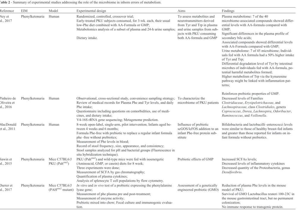
Studies of the association between microbiome and IEMs have focused on aminoacidopathies (such as PKU, tyrosinemia, and alkaptonuria), organic acidemias (methyl-malonic acidemia and propionic acidemia), and hemochro-matosis. The main characteristics of the IEMs addressed in these studies, including their long-term management, are summarized in Table 2. Some possible effects of treatments of IEM on microbiome are showed in Figure 2.
The majority of studies on microbiome–IEM interac-tions has focused on PKU. One of the most thorough among such studies compared the microbiome of eight pa-tients with PKU to that of 10 healthy individuals by
analy-sis of the 16S rRNA gene. In this study, Pinheiro de Oli-veiraet al.(2016) demonstrated reduced abundance of bac-teria in the families Clostridiaceae, Erysipelotrichaceae, and Lachnospiraceae, class Clostridiales, and genera
Coprococcus, Dorea, Lachnospira, Odoribacter,
Ruminococcus, andVeillonella in patients with PKU, as
well as an increase in Prevotella, Akkermansia, and Peptostreptococcaceae populations. Their metabolic pre-diction was associated both with starch and glucose metab-olism and with AA metabmetab-olism (Pinheiro de Oliveiraet al., 2016). The authors raised the hypothesis that bacterial en-richment related to LPS biosynthesis, as observed in pa-tients with PKU, might be associated with peripheral inflammation, as indicated by the proinflammatory circu-lating cytokine profile of these patients (Coakley et al., 2014). In the same study, the authors found a correlation between microbiotic profile and circulating levels of phenylalanine (Phe), which might indicate a relationship between these patients’ microbiome, their treatment re-sponse, and their phenotype.
Focusing on the potential impacts of prebiotic treat-ment in individuals with PKU, a study reported by Mac-Donald et al. (2011) analyzed the effects of prebiotic oligosaccharides (scGOS/lcFOS) as an adjunct to the meta-bolic formula that forms the mainstay of PKU manage-ment. As breastfeeding is highly restricted in children with PKU, the authors theorized that a lack of the oligosaccha-rides present in breast milk might be associated with
in-Microbiome and IEM: an interplay 521
522
Colonetti
et
al.
Table 2- Summary of experimental studies addressing the role of the microbiome in inborn errors of metabolism.
Reference EIM Model Experimental design Aims Findings
Ney et al., 2017
Phenylketonuria Human Randomized, controlled, crossover trial;
Early-treated PKU subjects consumed, for 3-wk. each, their usual low-Phe diet combined with AA-Formula or GMP;
Metabolomics analysis of a subset of plasma and 24-h urine samples;
Dietary intake.
To assess metabolites and neurotransmitters derived from Tyr and Trp in plasma and urine samples from sub-jects with PKU consuming both AA-formula and GMP
Plasma metabolome: 7 of the 40
microbiome-associated compounds showed differ-ential levels with AA-formula compared with GMP;
Significant differences in the plasma profile of secondary bile acids;
Associated compounds showed differential levels with AA-Formula compared with GMP; Urine metabolome: 7 of 45 microbiome; Individ-uals fed with AA formula had a 50% higher intake of Tyr and Trp;
Differential degradation level of Tyr by intestinal microbes of individuals fed with AA-formula, po-tential harmful metabolites formed;
Higher metabolism of Trp via the kynurenine pathway might be linked with inflammation pat-terns;
Reinforces prebiotic properties of GMP. Pinheiro de
Oliveira et al., 2016
Phenylketonuria Human Observational, cross-sectional study, convenience sampling strategy;
Review of medical records for Plasma Phe and Tyr levels, and daily Phe intake;
Questionnaire including questions on comorbidities, use of medi-cines, and dietary intake;
V4-16S rRNA gene sequencing; Metagenome prediction.
To characterize the microbiome of PKU patients
Decreased levels of Families
Clostridiaceae,Erysipelotrichaceae, and
Lachnospiraceae, classClostridiales, genera
Coprococcus,Dorea,Lachnospira,Odoribacter,
Ruminococcus, andVeillonella.
MacDonald et al., 2011
Phenylketonuria Human 8-week open-label, single-arm, pilot intervention; Infants aged
be-tween 4 weeks and 6 months;
Formula Phe-free with prebiotic to replace a regular infant formula phe -free without prebiotics;
Measurement of Phe levels in blood;
Record of stool frequency, size, appearance, and consistency; Stool samples analyzed for pH and bacterial groups (Fluorescence in situ hybridization technique).
Influence of prebiotic scGOS/lcFOS addition to an infant Phe-free protein sub-stitute
Bifidobacteria and lactobacilli–enterococci levels were similar to those of healthy breast-fed infants and greater than those reported for infants on in-fant formula without prebiotics.
Sawin et al., 2015
Phenylketonuria Mice C57BL6/J
PKU (Pahenu2)
PKU (Pahenu2) and wild-type mice were fed with isoenergetic
(Aminoacid, GMP, or casein) diets for 8 week; Three experiments were done;
Measurement of SCFA by gas chromatography; Quantification of plasma cytokines;
Analysis of splenocyte T cell populations by flow cytometry.
Prebiotic effects of GMP Increased SCFAs levels;
Decreased levels of inflammatory cytokines Decreased quantity of the Proteobacteria, genus
Desulfovibrio.
Durrer et al., 2017
Phenylketonuria Mice C57BL6/J
(PAHenu2mutant)
In vitroandin vivotest of a probiotic expressing the phenylalanine lyase gene;
Measurement of phe plasma pre and post-treatment; Measurement of enzyme activity;
Prebiotic mixed into chow; Fecal culture and immunogenic evalua-tion.
Assessment of a genetically engineered probiotic (GMO)
Reduction of plasma Phe levels in the mouse model of PKU;
Survival of GMO Lactobacillus reuteri 100-23C in the mouse gastrointestinal tract, but no permanent colonization;
Microbiome
and
IEM:
an
interplay
523
Reference EIM Model Experimental design Aims Findings
Gertsman et al., 2015
Alkaptonuria Tyrosinemia
Human Collection of samples from patients with alkaptonuria before and
af-ter treatment with NTBC plus samples of Tyrosinemia types I, I and transient patients;
Analysis of the sera y tQ-TOF LC/ MS metabolomic platform; Untargeted metabolomics strategy;
In vitroexperiments with cultures of human cells and intestinal flora cultures to identify the nature of the link between 4-HPP and the ele-vated indoles.
Evaluate the metabolic ef-fects of nitisinone
Increased levels of I3CHO, in patients treated with nitinisone.
Frye et al., 2016
Propionic acidemia (PA)
Human lym-phoblastoid cell lines (LCLs)
Measurement of mitochondrial function in ASD and sex-age-matched control LCLs;
Incubation with PPA and reactive oxygen species.
Effects of PPA in an unfa-vorable redox
microenvironment
PPA can have both beneficial and toxic effects on mitochondrial function, depending on concentra-tion, exposure duraconcentra-tion, and microenvironment re-dox state.
Buhnik-Ro-senblau et al., 2012
Hemochromatosis type 1
Mouse Comparison between wild-type and genetically deficient mouse;
Culture followed by tRFLP and 16S rRNA gene sequencing.
Effects of iron metabolism (Irp2-/-and Hfe-/-genes) on microbiome
Irp2
-/-Increased levels ofL. intestinaliscompared to
Hfe-/-mice andL. murinuscompared to both
Hfe-/-and WT mice;
Hfe
-/-Increased levels ofEnterococcus faecium;
Increased levels ofL. johnsoniito both Hfe-/-and
Irp2-/-mice compared to WT.
PKU: phenylketonúria; Phe: phenylananine; Tyr: tyrosine; Trp: tryptophan; scGOS/lcFOS: neutral short chain galactooligosaccharides and long chain fructooligosaccharides; AA: aminoacids; GMP: glycromacropepide; SCFAs: short chain fatty Acids; GMO: genetically modified organism; I3CHO: indole-3-carboxaldehyde (exclusively produced by microbiota); PA: propionic academia; LCLs: Human lymphoblastoid cell lines; PPA: propionic acid; Irp2: iron regulatory protein 2 gene; Hfe: hemochromatosis protein gene. *The identified compounds are either exclusively synthesized or contributed by intestinal bacteria, as well as by human metabolism.
creased fecal pH and reduced bifidobacterial populations, thus predisposing the patient to infections. Administration of probiotics might mitigate this problem. The experiment assessed the dominant bacterial groups and found that the administered prebiotic oligosaccharides were able to main-tain bifidobacteria levels and low fecal pH, without altering circulating levels of Phe. Despite the small sample size and lack of statistical power, these findings suggest that supple-menting metabolic formula with prebiotics might be an in-teresting strategy in PKU, as the levels of Bifidobacteria and Lactobacilli–Enterococci at the end of the study were similar to those found in healthy children and higher than those reported in children who took the formula alone, without prebiotics. In the only patient who was previously receiving a diet without prebiotics, there was also a reduc-tion in pathogens such as C. perfringensandC. difficile (group Clostridium histolyticum/lituseburense), E. coli,
Shigella, Salmonella, and Klebsiella (subgroup
Enterobacteriaceae) (MacDonaldet al., 2011).
Also regarding prebiotics, recent years have been promising in terms of the use of glycomacropeptide (GMP) as a substitute for Phe-free AA formula in patients with PKU. GMP is highly glycosylated and, when pure, consti-tutes a natural protein source that lacks the AAs (Phe, tyro-sine (Tyr), tryptophan (Trp), histidine, cysteine, arginine) involved in some IEMs, including PKU (Neelima et al., 2013). For now, human trials are seeking to ascertain the ef-ficiency of GMP as a partial (50% formula, 50% GMP) or total replacement for the Phe-free AA formula. In trials, the use of GMP had no significant impact on circulating Phe levels and was preferred by patients over the formula, as GMP is more palatable and, according to patients, provides
greater satiety than a formula-based diet alone (Neyet al., 2016; Zakiet al., 2016). This could make GMP an option to increase treatment adherence.
When the urine and plasma metabolome of the indi-viduals with PKU were compared within the groups fed with AA-formula or GMP, differences were found between the metabolite profile linked to the microbes. There were no differences between fasting plasma concentrations of the Tyr and Trp, but individuals fed with AA formula had a 50% higher intake of Tyr and Trp. This can be explained as a result of higher degradation by the intestinal microbes, raising the levels of microbiome-derived compounds from Tyr. Some of these compounds are potentially harmful. There was no differential degradation of Trp, but the me-tabolism of Trp via the kynurenine pathway was evidenced by higher levels of metabolites linked to this pathway and might be linked with inflammation patterns. Change in plasma profile of secondary bile acids, but not primary bile acids, supports the statement that there are alterations in the gut microbiome with ingestion of AA-formula and GMP, and reinforces the prebiotic proprieties of the GMP (Neyet al., 2017).
Although the effect of GMP on the human gut microbiome has yet to be studied, in mice, GMP was asso-ciated with control of Th2-type immune responses, in-creasedLactobacillusandBifidobacteriumpopulations in as little as three days after treatment (Jiménezet al., 2016), elevated levels of SCFAs and reduced levels of proinflam-matory cytokines, and reduced Proteobacteria counts (ge-nusDesulfovibrio) without affecting circulating Phe levels (Sawinet al., 2015). The genusDesulfovibriois associated with production of hydrogen sulfate, a cytotoxic compound
524 Colonettiet al.
found at higher levels in patients with ulcerative colitis (Rowanet al., 2010).
Regarding disorders of tyrosine metabolism, Gerts-manet al. (2015) described the metabolic effect of niti-sinone (NTBC or 2-(2-nitro-4-fluoromethylbenzoyl)-1,3-cyclohexanedione) in patients with alkaptonuria. Analysis of their metabolic profile showed that indole levels were in-creased in treated patients as compared with controls. Indoles play a key role in signaling pathways (as building blocks for melanin and serotonin) and intercellular commu-nication, facilitate quorum sensing, and have been uniquely associated with dietary intake and microbial metabolism of tryptophan. Among the indoles found to be increased, indole-3-carboxaldehyde (I3CHO) is produced exclusively by the microbiota, while the other two are produced by hu-man cells (Gertshu-manet al., 2015). The authors stressed that the reduced form of I3CHO, indole-3-carbinol, a com-pound also found in cruciferous vegetables, is associated with the prevention of several neoplasms.
Animal experiments also suggest that genetic defects in the host may alter the composition of the gut microbiota, leading to dysbiosis due to a buildup of substances in the cells or lumen of the bowel (Buhnik-Rosenblau et al., 2012). This effect has been observed in hemochromatosis. Hemochromatosis is a disease caused by excess iron ab-sorption by gut cells, which leads to iron overload. This usually becomes clinically detectable in adulthood and is damaging to many organs, including the liver, pancreas (causing diabetes), heart, and skin (Babitt and Lin, 2011). Mutations in the HFE gene account for the majority of cases of hereditary hemochromatosis, especially in individ-uals of Northern European descent (Barton, 2013). In a study of mice with mutations in two genes that encode pro-teins involved in regulation of iron homeostasis (HFE-/-and Irp2-/-), Buhnik-Rosenblauet al.(2012) found abnormali-ties particularly in resident populations of lactic-acid bacte-ria, both in Irp2-mutant and in HFE-mutant mice as compared to controls.
The gut microbiome produces several metabolites, including PPA, a SCFA implicated in several diseases. In autistic populations, the level of the phylumFirmicutesis increased and was largely attributable toClostridiaclass
withRuminococcaceaeandLachnospiraceaefamilies. The
differences in Clostridia species in children with autism spectrum disorder include greater abundance of Clostridiumclusters I, II, XI andC. bolteae(Finegoldet al., 2002; Songet al., 2004; Parrachoet al., 2005; Williamset al., 2011; Strati et al., 2017). SeveralRuminococcaceae
andLachnospiraceae are known butyrate producers and
may thus influence SCFA levels (Louiset al., 2010). So, the treatment with antibiotics can affect producers of SCFA. Some patients’ symptoms improve transiently when antibiotics are administered (Sandler et al., 2000; Shaw 2010). Curiously, a similar effect is seen in patients with propionic acidemia, who can experience the same
neuro-developmental complications seen in autism (Witterset al., 2016). Among the various roles played by PPA, it was re-cently reported to act as a modulator of mitochondrial func-tion. In a study of autism and control cell lines, the effects of PPA depended not only on the concentration of the acid, but also on the level of reactive oxygen species (ROS) pres-ent, as ROS influence mitochondrial ability to use PPA as an energy source. Thus, PPA could have beneficial effects in individuals without mitochondrial dysfunction, and harmful effects in individuals with an unfavorable meta-bolic status and elevated levels of ROS (Fryeet al., 2016). In methylmalonic acidemia, which shares several symp-toms and management strategies with propionic acidemia, vitamin B12(cobalamin) is also used as treatment in
respon-sive patients, in addition to antibiotics. This vitamin is syn-thesized by some gut bacteria, and is also a regulator of microbiome composition and function (Baumgartneret al., 2014; Degnanet al., 2014).
The microbiome can also be considered an exogenous source of tetrahydrobiopterin (BH4), another important
me-tabolite of gut bacteria. BH4is a key cofactor for several
regulatory enzymes, as Phenylalanine-4-hydroxylase, which catalyzes the conversion of L-phenylalanine to L-ty-rosine. The BH4has also been shown to improve working
memory and cerebral activation (Christet al., 2013). In ro-dents, BH4production is age-dependent and is related to the
presence of Actinobacteria in the bowel, especially
Adlercreutzia equolifaciens and Microbacterium
schleiferi. These same species have been identified in the human gut microbiome (Beliket al., 2017). Very little is known about the determinants of responsiveness to BH4
therapy and its effects on cerebral activity and cognition, but these effects are known to be multifactorial, as they vary across individuals with the same genotype (Pérezet al., 2005). The discovery that BH4is naturally produced by
gut microbiota has implications for translational medicine, as this cofactor is used in the treatment of some patients with PKU.
The long-term perspective is that elucidation of the metabolic role of the microbiota and identification of which species play these roles will pave the way for manipulating the microbiome, so that pathways beneficial to the host are stimulated, while those harmful to the host are inhibited. In this line, some authors have raised the hypothesis of using methanogenic bacteria normally present in the human bowel to control metabolites such as trimethylamine (TMA), bypassing the normal route of trimethylamine N-oxide (TMAO) production as an intermediate for CH4to an
alternative pathway (Brugèreet al., 2014). In the liver, defi-ciency in the pathway of TMA conversion into TMAO leads to trimethylaminuria, an IEM that causes strong body odor, impairing the patients’ quality of life and interper-sonal relations (Mackayet al., 2011). Diets rich in com-pounds such as phosphatidylcholine, choline, betaine, and L-carnitine generate TMA via the gut microbiota, which is
then converted in TMAO by the liver. High levels of TMAO are associated with increased risk of cardiovascular disease in the general population (Wanget al., 2011; Koeth et al., 2013; Gregory et al., 2015; Liu et al., 2015b). Making the transition from theory into practice, administra-tion of the probioticLactobacillus reuteri,engineered to express a phenylalanine lyase gene from the cyanobacteria Anabaena variabilis, successfully treated mice with PKU. Blood levels of Phe declined after the fourth day of treat-ment and remained low throughout the experitreat-ment, with no permanent colonization of the gut (Durreret al., 2017), sug-gesting potential for modified probiotics in the treatment of IEMs.
The creation of genetically modified probiotics de-sign especially to normalize defective metabolic pathways in the host is only one of the many potential advantages of microbiome research. IEMs are characterized by substan-tial variability in presentation, and genotype alone cannot explain patients’ clinical pictures. The microbiome may contribute significantly to factors such as tolerance to cer-tain nutrients and responsiveness to cofactors (and to treat-ment itself). Studying the microbiomes of patients with IEMs may provide valuable tools for clinical practice, both advancing our understanding of phenotypes and facilitating the development of new biomarkers and therapies.
Main questions about microbioma and IEM and
how to address them
There are some important issues involved in the study of the human microbiome in IEM. First of all, most of the diseases that compound the IEM class are rare, and usually there are subclasses within the same IEM. This is the reason why the studies normally have a small number of partici-pants. Second, the microbiome is mainly influenced by diet, and diet overload or restriction is one of most common treatments for IEM. This is one of reasons that make obtain-ing an adequate control group very difficult. Third, this class of diseases is derived of a metabolic genetic defect, and defects in a metabolic gene also affect the microbiome. So, if a dysbiotic state is observed in this group of patients will it reflect the genetic or the diet effect? Taken together, all the facts above make it very hard to obtain a homoge-neous and statistically valid group of untreated patients and make difficult the comparison pre and post-treatment to verify if the altered microbiome is mainly affected by ge-netic or diet effects. Additional difficulty is added by the fact that several metabolic diseases, if untreated, can lead to severe impacts through life, so IEM patients should start to be treated as soon as possible.
Despite the difficulties, studying the patterns of the microbiome in groups of treated patients offers the possi-bility to evaluate the real impact of the genetic defect and diet on the microbiome. Patients need lifelong treatment, and the intragroup study of phenotype, microbiome and
diet can be elucidative for some ancient questions that re-main unknown. PKU patients, for instance, were studied in light of the microbiome by Pinheiro de Oliveira et al. (2016) (see Table 2). Even though not capable of answering the question if alteration comes from diet or genetics, a microbiome alteration correlated with Phe blood levels was observed. This is exciting data, due to the fact that it can help explain why some patients are more tolerant to Phe than others, despite having the same genotypes.
In an IEM, the genetic defect and the diet factors co-exist, so the measure of macro- and micronutrients ingested is required. Diet has a strong impact on the microbiome, and in spite of patients having similar lines of treatment all over the world, the source of fibers, carbohydrates and pro-teins can vary geographically and/or culturally. For this reason, microbiome studies should not combine patients of geographically distinct regions or culture to raise the num-ber of participants. Rather, these studies must be done lo-cally and then, if methodologilo-cally possible, make compar-isons that take into account the dietetic/cultural/geographic factors.
As detailed above, there are several other factors that can influence and be influenced by the microbiome. Impor-tant data as sex, age, body mass index, type of birth deliv-ery, breast feeding (duration and transition to solid food), antibiotic and other drug usage, vitamin supplementation, as well as physical exercise, and other diseases (physical and/or mental) must be collected and also analyzed. All subjects included in studies that aim to characterize the microbiome of certain IEMs should be three years or older to avoid the period of drastic changes in microbiome com-position due to the typical change in diet during this period. Given that the microbiome varies according to the stage of life and sex, and certain cultures can also exert some influ-ence, the best way to avoid interference of age and sex is the sex-age-matched strategy.
Another useful strategy is based on experimental studies using animal models. This strategy is very impor-tant since animal models have less genetic variation and are maintained in a highly controlled environment (that in-cludes diet and/or a germ-free environment). Also, a high the number of subjects can be easily obtained in such re-search. This is the better model for initial tests of geneti-cally engineered probiotics and correlations with diseases caused by the genetic defect in the absence or presence of the treatment. This kind of study, besides not being capable of fully reproducing the human reality, can work to gener-ate hypotheses and help to provide better strgener-ategies and comprehension of studies done in humans.
With the development of NGS tools, procedures are no longer the main limitation for human microbiome stud-ies. Microbiome data is currently obtained by three differ-ent approaches: 1) by 16S rRNA gene partial sequencing, 2) by whole DNA shotgun metagenomic sequencing, or 3) by metatranscriptomics (mRNA-seq), to access the active
gene expression pattern For instance, the 16S rRNA gene sequencing method is largely used and has been the first choice method among researchers. Reasons for choosing this approach include the availability of a comprehensive database and scalability. Moreover, studies based in meta-transcriptomics require a better control for sample collec-tion to RNA/metabolites processing. Metagenomics, meta-transcriptomics and all other “omics”, and the associated bioinformatics techniques are allowing comparative analy-ses in an unprecedented way. All of these tools allow for testing a recent hypothesis related to the presence of a com-mon set of microbial taxa universally present in healthy in-dividuals (Turnbaughet al., 2007), also known as microbial core. However large variations in the taxonomic composi-tion observed in the human microbiome rapidly refute such a hypothesis (Bäckhedet al., 2012). Due to the well-known microbial functional redundancy in nature, an alternative hypothesis is the presence of a functional core represented by a set of metabolic functions that are performed by the microbiome within a particular habitat, but are not neces-sarily provided by the same organisms in different people (Shafquatet al., 2014). Still, studies devoted to better un-derstand how deeply the microbiome can affect an organ-ism with critical metabolic pathways that are naturally al-tered, are just in the early stages. Multidisciplinary efforts need to be done to aggregate modern techniques of se-quencing and identification of metabolites that can lead to the phenotype or drug effect in question. Microbial se-quencing alone will not be capable of explaining the pheno-type, but is a fundamental tool in the understanding of the process. Additional techniques based on metabolomics analysis and RNA-seq, as well as gathering information about the immune system and SCFA levels can offer funda-mental pieces of information in the process.
Conclusions
Studies on the microbiome in IEMs are scarce. The effects of the genetic defect itself and of treatment in IEMs, especially in the long term, have yet to be fully understood. As IEMs are commonly managed through dietary interven-tion (nutrient overload and/or restricinterven-tion), dysbiosis is a possibility. This dysbiotic status would alter the patients’ already compromised metabolic state even further, induc-ing or worseninduc-ing abnormalities in secondary metabolic pathways, and thus contributing to phenotypic manifesta-tions, especially liver and brain involvement. Dysbiosis can be treated with antibiotic therapy, dietary prebiotics, or fe-cal transplant, alone or in combination. The administration of probiotics engineered to at least partly meet the meta-bolic needs of the IEM-affected host has practically unex-plored therapeutic potential and may constitute an intervention that is simple to administer, yet has a major impact on the patients’ lives. Collectively, microbiome re-search in patients with IEMs can not only contribute signif-icantly to our understanding of the pathophysiology of
these diseases and to the development of new biomarkers and therapies, but also help to improve the long-term qual-ity of life in affected patients.
Acknowledgments
The authors acknowledge the financial support of CAPES, FAPERGS and FIPE-HCPA.
References
Alfadhel M, Benmeakel M, Hossain MA, Al Mutairi F, Al Othaim A, Alfares AA, Al Balwi M, Alzaben A and Eyaid W (2016) Thirteen year retrospective review of the spectrum of inborn errors of metabolism presenting in a tertiary center in Saudi Arabia. Orphanet J Rare Dis 11:126.
Argmann CA, Houten SM, Zhu J and Schadt EE (2016) A next generation multiscale view of inborn errors of metabolism. Cell Metab 23:13–26.
Ashton JJ, Colquhoun CM, Cleary DW, Coelho T, Haggarty R, Mulder I, Batra A, Afzal NA, Beattie RM, Scott KP,et al.
(2017) 16S sequencing and functional analysis of the fecal microbiome during treatment of newly diagnosed pediatric inflammatory bowel disease. Medicine (Baltimore) 96:e7347.
Babitt JL and Lin HY (2011) The molecular pathogenesis of he-reditary hemochromatosis. Semin Liver Dis 31:280–292. Bäckhed F, Fraser CM, Ringel Y, Sanders ME, Sartor RB,
Sher-man PM, Versalovic J, Young V and Finlay BB (2012) De-fining a healthy human gut microbiome: Current concepts, future directions, and clinical applications. Cell Host Mi-crobe 12:611–622.
Balmer ML, Slack E, de Gottardi A, Lawson MAE, Hapfelmeier S, Miele L, Grieco A, Van Vlierberghe H, Fahrner R, Patuto N,et al.(2014) The liver may act as a firewall mediating mutualism between the host and its gut commensal micro-biota. Sci Transl Med 6:237ra66.
Barton JC (2013) Hemochromatosis and iron overload: From bench to clinic. Am J Med Sci 346:403–412.
Baumgartner MR, Hörster F, Dionisi-Vici C, Haliloglu G, Karall D, Chapman KA, Huemer M, Hochuli M, Assoun M, Ballhausen D,et al.(2014) Proposed guidelines for the diag-nosis and management of methylmalonic and propionic acidemia. Orphanet J Rare Dis 9:130.
Belik J, Shifrin Y, Arning E, Bottiglieri T, Pan J, Daigneault MC and Allen-Vercoe E (2017) Intestinal microbiota as a tetrahydrobiopterin exogenous source in hph-1 mice. Sci Rep 7:39854.
Bilder DA, Kobori JA, Cohen-Pfeffer JL, Johnson EM, Jurecki ER and Grant ML (2017) Neuropsychiatric comorbidities in adults with phenylketonuria: A retrospective cohort study. Mol Genet Metab 121:1–8.
Boelens JJ, Orchard PJ and Wynn RF (2014) Transplantation in inborn errors of metabolism: current considerations and fu-ture perspectives. Br J Haematol 167:293–303.
Bonini JA, Anderson SM and Steiner DF (1997) Molecular clon-ing and tissue expression of a novel orphan G protein-coupled receptor from rat lung. Biochem Biophys Res Commun 234:190–193.
Boyer SW, Barclay LJ and Burrage LC (2015) Inherited meta-bolic disorders: Aspects of chronic nutrition management.
Nutr Clin Pract Off Publ Am Soc Parenter Enter Nutr 30:502–510.
Braniste V, Al-Asmakh M, Kowal C, Anuar F, Abbaspour A, Tóth M, Korecka A, Bakocevic N, Ng LG, Guan NL,et al.
(2014) The gut microbiota influences blood-brain barrier permeability in mice. Sci Transl Med 6:263ra158.
Brugère JF, Borrel G, Gaci N, Tottey W, O’Toole PW and Malpuech-Brugère C (2014) Archaebiotics: Proposed thera-peutic use of archaea to prevent trimethylaminuria and car-diovascular disease. Gut Microbes 5:5–10.
Brunetti-Pierri N and Lee B (2005) Gene therapy for inborn errors of liver metabolism. Mol Genet Metab 86:13–24.
Buhnik-Rosenblau K, Moshe-Belizowski S, Danin-Poleg Y and Meyron-Holtz EG (2012) Genetic modification of iron me-tabolism in mice affects the gut microbiota. Biometals Int J Role Met Ions Biol Biochem Med 25:883–892.
Cani PD, Possemiers S, Van de Wiele T, Guiot Y, Everard A, Rottier O, Geurts L, Naslain D, Neyrinck A, Lambert DM,et al.(2009) Changes in gut microbiota control inflammation in obese mice through a mechanism involving GLP-2-driven improvement of gut permeability. Gut 58:1091–1103. Chassaing B, Koren O, Goodrich JK, Poole AC, Srinivasan S, Ley
RE and Gewirtz AT (2015) Dietary emulsifiers impact the mouse gut microbiota promoting colitis and metabolic syn-drome. Nature 519:92–96.
Chevalier C, Stojanovic O, Colin DJ, Suarez-Zamorano N, Taral-lo V, Veyrat-Durebex C, Rigo D, Fabbiano S, Stevanovic A, Hagemann S,et al.(2015) Gut Microbiota Orchestrates En-ergy Homeostasis during Cold. Cell 163:1360–1374. Chinsky JM, Singh R, Ficicioglu C, van Karnebeek CDM,
Grompe M, Mitchell G, Waisbren SE, Gucsavas-Calikoglu M, Wasserstein MP, Coakley K,et al.(2017) Diagnosis and treatment of tyrosinemia type I: a US and Canadian consen-sus group review and recommendations. Genet Med 19:1380
Chiu IM, Heesters BA, Ghasemlou N, Von Hehn CA, Zhao F, Tran J, Wainger B, Strominger A, Muralidharan S, Horswill AR, et al. (2013) Bacteria activate sensory neurons that modulate pain and inflammation. Nature 501:52–57. Cho I and Blaser MJ (2012) The Human Microbiome: at the
inter-face of health and disease. Nat Rev Genet 13:260–270. Christ SE, Moffitt AJ, Peck D and White DA (2013) The effects of
tetrahydrobiopterin (BH4) treatment on brain function in in-dividuals with phenylketonuria. NeuroImage Clin 3:539–547.
Chung H, Pamp SJ, Hill JA, Surana NK, Edelman SM, Troy EB, Reading NC, Villablanca EJ, Wang S, Mora JR,et al.(2012) Gut immune maturation depends on colonization with a host-specific microbiota. Cell 149:1578–1593.
Coakley K, Douglas T and Singh R (2014) Phenylketonuria is a condition of inflammation associated with high BMI and low bone turnover (1034.2) FASEB J 28:1034.2.
Coleman OI and Nunes T (2016) Role of the microbiota in colorectal cancer: Updates on microbial associations and therapeutic implications. BioResearch Open Access 5:279–288.
Cotillard A, Kennedy SP, Kong LC, Prifti E, Pons N, Le Chatelier E, Almeida M, Quinquis B, Levenez F, Galleron N,et al.
(2013) Dietary intervention impact on gut microbial gene richness. Nature 500:585–588.
Crenn P and Maillot F (2007) Principes et contraintes des régimes au cours des maladies neurométaboliques chez l’adulte. Rev Neurol (Paris) 163:936–941.
Cummings JH, Pomare EW, Branch WJ, Naylor CP and Macfar-lane GT (1987) Short chain fatty acids in human large intes-tine, portal, hepatic and venous blood. Gut 28:1221–1227. Dabrowska K and Witkiewicz W (2016) Correlations of host
ge-netics and gut microbiome composition. Front Microbiol 2016:1357
Das AM (2017) Clinical utility of nitisinone for the treatment of hereditary tyrosinemia type-1 (HT-1). Appl Clin Genet 10:43–48.
David LA, Maurice CF, Carmody RN, Gootenberg DB, Button JE, Wolfe BE, Ling AV, Devlin AS, Varma Y, Fischbach MA,et al.(2014) Diet rapidly and reproducibly alters the human gut microbiome. Nature 505:559–563.
De Angelis M, Francavilla R, Piccolo M, De Giacomo A and Gobbetti M (2015) Autism spectrum disorders and intestinal microbiota. Gut Microbes 6:207–213.
De Filippo C, Cavalieri D, Di Paola M, Ramazzotti M, Poullet JB, Massart S, Collini S, Pieraccini G and Lionetti P (2010) Im-pact of diet in shaping gut microbiota revealed by a compar-ative study in children from Europe and rural Africa. Proc Natl Acad Sci U S A 107:14691–14696.
Degnan PH, Taga ME and Goodman AL (2014) Vitamin B12 as a modulator of gut microbial ecology. Cell Metab 20:769–778.
Desbonnet L, Clarke G, Traplin A, O’Sullivan O, Crispie F, Moloney RD, Cotter PD, Dinan TG and Cryan JF (2015) Gut microbiota depletion from early adolescence in mice: Implications for brain and behaviour. Brain Behav Immun 48:165–173.
Donaldson GP, Lee SM and Mazmanian SK (2016) Gut biogeo-graphy of the bacterial microbiota. Nat Rev Microbiol 14:20–32.
Durrer KE, Allen MS and Hunt von Herbing I (2017) Genetically engineered probiotic for the treatment of phenylketonuria (PKU); assessment of a novel treatment in vitro and in the PAHenu2 mouse model of PKU. PLoS One 12:e0176286. Etienne-Mesmin L, Vijay-Kumar M, Gewirtz AT and Chassaing
B (2016) Hepatocyte Toll-like Receptor 5 promotes bacte-rial clearance and protects mice against high-fat diet-indu-ced liver disease. Cell Mol Gastroenterol Hepatol 2:584–604.
Ezgu F (2016) Inborn Errors of Metabolism. Adv Clin Chem 73:195–250.
Finegold SM, Molitoris D, Song Y, Liu C, Vaisanen ML, Bolte E, McTeague M, Sandler R, Wexler H, Marlowe EM, et al.
(2002) Gastrointestinal microflora studies in late-onset au-tism. Clin Infect Dis 35:S6–S16.
Frye RE, Rose S, Chacko J, Wynne R, Bennuri SC, Slattery JC, Tippett M, Delhey L, Melnyk S, Kahler SG,et al.(2016) Modulation of mitochondrial function by the microbiome metabolite propionic acid in autism and control cell lines. Transl Psychiatry 6:e927.
Fu J, Bonder MJ, Cenit MC, Tigchelaar EF, Maatman A, Dekens JAM, Brandsma E, Marczynska J, Imhann F, Weersma RK,
et al.(2015) The gut microbiome contributes to a substantial proportion of the variation in blood lipids. Circ Res 117:817–824.
Geirnaert A, Calatayud M, Grootaert C, Laukens D, Devriese S, Smagghe G, Vos MD, Boon N and de Wiele TV (2017) Bu-tyrate-producing bacteria supplementedin vitroto Crohn’s disease patient microbiota increased butyrate production and enhanced intestinal epithelial barrier integrity. Sci Rep 7:11450.
Gertsman I, Gangoiti JA, Nyhan WL and Barshop BA (2015) Per-turbations of tyrosine metabolism promote the indolepy-ruvate pathway via tryptophan in host and microbiome. Mol Genet Metab 114:431–437.
Gill SR, Pop M, DeBoy RT, Eckburg PB, Turnbaugh PJ, Samuel BS, Gordon JI, Relman DA, Fraser-Liggett CM and Nelson KE (2006) Metagenomic analysis of the human distal gut microbiome. Science 312:1355–1359.
Goodrich JK, Davenport ER, Waters JL, Clark AG and Ley RE (2016) Cross-species comparisons of host genetic associa-tions with the microbiome. Science 352:532–535.
Gregory JC, Buffa JA, Org E, Wang Z, Levison BS, Zhu W, Wag-ner MA, Bennett BJ, Li L, DiDonato JA,et al.(2015) Trans-mission of atherosclerosis susceptibility with gut microbial transplantation. J Biol Chem 290:5647–5660.
Grenham S, Clarke G, Cryan JF and Dinan TG (2011) Brain–gut–microbe communication in health and disease. Front Physiol 2011:94.
Gustot T, Durand F, Lebrec D, Vincent JL and Moreau R (2009) Severe sepsis in cirrhosis. Hepatol Baltim Md 50:2022–2033.
Häberle J, Boddaert N, Burlina A, Chakrapani A, Dixon M, Huemer M, Karall D, Martinelli D, Crespo PS, Santer R,et al.(2012) Suggested guidelines for the diagnosis and man-agement of urea cycle disorders. Orphanet J Rare Dis 7:32. Han A, Sung YB, Chung SY and Kwon MS (2014) Possible
addi-tional antidepressant-like mechanism of sodium butyrate: targeting the hippocampus. Neuropharmacology 81:292–302.
Henao-Mejia J, Elinav E, Jin C, Hao L, Mehal WZ, Strowig T, Thaiss CA, Kau AL, Eisenbarth SC, Jurczak MJ, et al.
(2012) Inflammasome-mediated dysbiosis regulates pro-gression of NAFLD and obesity. Nature 482:179–185. Hiergeist A, Gläsner J, Reischl U and Gessner A (2015) Analyses
of intestinal microbiota: Culture vs. sequencing. ILAR J 56:228–240.
Hoban AE, Stilling RM, Ryan FJ, Shanahan F, Dinan TG, Claes-son MJ, Clarke G and Cryan JF (2016) Regulation of prefrontal cortex myelination by the microbiota. Transl Psy-chiatry 6:e774.
Human Microbiome Project Consortium (2012) Structure, func-tion and diversity of the healthy human microbiome. Nature 486:207–214.
Jacqueline C, Brazier L, Faugère D, Renaud F, Thomas F and Roche B (2017) Can intestinal microbiota be associated with non-intestinal cancers? Sci Rep 7:12722.
Janssen AWF and Kersten S (2015) The role of the gut microbiota in metabolic health. FASEB J 29:3111–3123.
Jiménez M, Cervantes-García D, Muñoz YH, García A, Haro LM and Salinas E (2016) Novel mechanisms underlying the therapeutic effect of glycomacropeptide on allergy: Change in gut microbiota, upregulation of TGF-b, and inhibition of mast cells. Int Arch Allergy Immunol 171:217–226. Kakihana K, Fujioka Y, Suda W, Najima Y, Kuwata G, Sasajima
S, Mimura I, Morita H, Sugiyama D, Nishikawa H,et al.
(2016) Fecal microbiota transplantation for patients with steroid-resistant acute graft-vs. -host disease of the gut. Blood 128:2083–2088.
Kashtanova DA, Popenko AS, Tkacheva ON, Tyakht AB, Alexeev DG and Boytsov SA (2016) Association between the gut microbiota and diet: Fetal life, early childhood, and further life. Nutrition 32:620–627.
Koeth RA, Wang Z, Levison BS, Buffa JA, Org E, Sheehy BT, Britt EB, Fu X, Wu Y, Li L,et al.(2013) Intestinal micro-biota metabolism of L-carnitine, a nutrient in red meat, pro-motes atherosclerosis. Nat Med 19:576–585.
Koh A, De Vadder F, Kovatcheva-Datchary P and Bäckhed F (2016) From dietary fiber to host physiology: Short-chain fatty acids as key bacterial metabolites. Cell 165:1332–1345.
Lanpher B, Brunetti-Pierri N and Lee B (2006) Inborn errors of metabolism: The flux from Mendelian to complex diseases. Nat Rev Genet 7:449–460.
Larsen N, Vogensen FK, Berg FWJ van den, Nielsen DS, An-dreasen AS, Pedersen BK, Al-Soud WA, Sørensen SJ, Han-sen LH and JakobHan-sen M (2010) Gut microbiota in human adults with type 2 diabetes differs from non-diabetic adults. PLoS One 5:e9085.
Le Poul E, Loison C, Struyf S, Springael JY, Lannoy V, Decobecq ME, Brezillon S, Dupriez V, Vassart G, Van Damme J,et al.
(2003) Functional characterization of human receptors for short chain fatty acids and their role in polymorphonuclear cell activation. J Biol Chem 278:25481–25489.
Leber B, Mayrhauser U, Rybczynski M and Stadlbauer V (2009) Innate immune dysfunction in acute and chronic liver dis-ease. Wien Klin Wochenschr 121:732–744.
Lee WS and Sokol RJ (2015) Intestinal microbiota, lipids and the pathogenesis of intestinal failure-associated liver disease. J Pediatr 167:519–526.
Li Q and Zhou JM (2016) The microbiota-gut-brain axis and its potential therapeutic role in autism spectrum disorder. Neu-roscience 324:131–139.
Liu J, Sun J, Wang F, Yu X, Ling Z, Li H, Zhang H, Jin J, Chen W, Pang M, et al. (2015a) Neuroprotective effects of
Clostridium butyricumagainst vascular dementia in mice via metabolic butyrate. BioMed Res Int 2015:412946 Liu TX, Niu HT and Zhang SY (2015b) Intestinal microbiota
me-tabolism and atherosclerosis. Chin Med J 128:2805–2811. Lloyd-Price J, Abu-Ali G and Huttenhower C (2016) The healthy
human microbiome. Genome Med 8:51.
Lopez-Legarrea P, Fuller NR, Zulet MA, Martinez JA and Cater-son ID (2014) The influence of Mediterranean, carbohydrate and high protein diets on gut microbiota composition in the treatment of obesity and associated inflammatory state. Asia Pac J Clin Nutr 23:360–368.
Louis P, Young P, Holtrop G and Flint HJ (2010) Diversity of hu-man colonic butyrate-producing bacteria revealed by analy-sis of the butyryl-CoA:acetate CoA-transferase gene. Envi-ron Microbiol 12:304–314.
MacDonald A, Cochrane B, Wopereis H and Loveridge N (2011) Specific prebiotics in a formula for infants with Phenylke-tonuria. Mol Genet Metab 104(Suppl):S55-S59.
Mackay RJ, McEntyre CJ, Henderson C, Lever M and George PM (2011) Trimethylaminuria: Causes and diagnosis of a so-cially distressing condition. Clin Biochem Rev 32:33–43.