ZOONOSES AND COMMUNICABLE DISEASES
COMMON TO MAN AND ANIMALS
Third Edition
Volume I
Bacterioses and Mycoses
Scientific and Technical Publication No. 580
PAN AMERICAN HEALTH ORGANIZATION Pan American Sanitary Bureau, Regional Office of the
WORLD HEALTH ORGANIZATION 525 Twenty-third Street, N.W. Washington, D.C. 20037 U.S.A.
ISBN 92 75 31580 9
PAHO Cataloguing-in-Publication Pan American Health Organization
Zoonoses and communicable diseases common to man and animals 3rd ed. Washington, D.C.: PAHO, © 2001.
3 vol.—(Scientific and Technical Publication No. 580)
ISBN 92 75 11580 X I. Title II. Series 1. ZOONOSES
2. BACTERIAL INFECTIONS AND MYCOSES 3. COMMUNICABLE DISEASE CONTROL 4. FOOD CONTAMINATION
5. PUBLIC HEALTH VETERINARY 6. DISEASE RESERVOIRS
NLM WC950.P187 2001 En
The Pan American Health Organization welcomes requests for permission to reproduce or translate its publications, in part or in full. Applications and inquiries should be addressed to the Publications Program, Pan American Health Organization, Washington, D.C., U.S.A., which will be glad to provide the latest information on any changes made to the text, plans for new editions, and reprints and translations already available.
© Pan American Health Organization, 2001
Publications of the Pan American Health Organization enjoy copyright protection in accordance with the provisions of Protocol 2 of the Universal Copyright Convention. All rights are reserved.
The designations employed and the presentation of the material in this publication do not imply the expression of any opinion whatsoever on the part of the Secretariat of the Pan American Health Organization concerning the status of any country, ter-ritory, city or area or of its authorities, or concerning the delimitation of its frontiers or boundaries.
CONTENTS
Prologue . . . vii
Preface to the First Edition . . . ix
Preface to the Second Edition . . . xi
Introduction . . . xv
PART I: BACTERIOSES
Actinomycosis . . . 3Aeromoniasis. . . 6
Animal Erysipelas and Human Erysipeloid . . . 14
Anthrax . . . 21
Botulism . . . 28
Brucellosis. . . 40
Campylobacteriosis . . . 67
Cat-scratch Disease . . . 78
Clostridial Food Poisoning. . . 82
Clostridial Wound Infections . . . 87
Colibacillosis . . . 90
Corynebacteriosis . . . 99
Dermatophilosis. . . 103
Diseases Caused by Nontuberculous Mycobacteria . . . 107
Diseases in Man and Animals Caused by Non-O1 Vibrio cholerae. . . 117
Enterocolitic Yersiniosis. . . 122
Enterocolitis Due to Clostridium difficile . . . 132
Food Poisoning Caused by Vibrio parahaemolyticus. . . 138
Glanders . . . 142
Infection Caused by Capnocytophaga canimorsus and C. cynodegmi. . . 146
Leprosy . . . 149
Leptospirosis . . . 157
Listeriosis . . . 168
Lyme Disease . . . 179
Melioidosis . . . 184
Necrobacillosis . . . 190
Nocardiosis . . . 195
Pasteurellosis . . . 199
Plague . . . 207
Pseudotuberculous Yersiniosis . . . 218
Rat-bite Fever . . . 226
Rhodococcosis. . . 229
Salmonellosis. . . 233
Shigellosis . . . 247
Staphylococcal Food Poisoning . . . 251
Streptococcosis . . . 257
Tetanus . . . 265
Tick-borne Relapsing Fever . . . 271
Tularemia . . . 275
Zoonotic Tuberculosis . . . 283
PART II: MYCOSES
Adiaspiromycosis . . . 303Aspergillosis . . . 305
Blastomycosis . . . 311
Candidiasis . . . 315
Coccidioidomycosis. . . 320
Cryptococcosis . . . 326
Dermatophytosis . . . 332
Histoplasmosis. . . 339
Mycetoma . . . 345
Protothecosis . . . 348
Rhinosporidiosis . . . 350
Sporotrichosis . . . 352
Zygomycosis . . . 356
Index . . . 361
LIST OF TABLES AND ILLUSTRATIONS
Bacterioses Tables 1. Foods giving rise to botulism, and number of outbreaks, United States of America, 1899–1977 . . . 312. Number of cases and deaths from human plague in the Americas, 1971–1980 . . . 210
3. Outbreaks of foodborne salmonellosis in selected countries, 1981–1985 . . . 235
4. Distribution of tetanus morbidity according to political division and climate, Argentina, 1967–1977 . . . 267
Figures 1. Animal erysipelas and human erysipeloid (Erysipelothrix rhusiopathiae). Mode of transmission . . . 17
2. Anthrax. Transmission cycle . . . 25
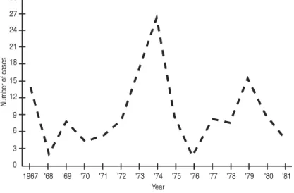
3. Botulism (transmitted by foods). Reported cases and deaths per year, United States of America, 1960–1980 . . . 29
4. Reported cases of botulism per year, Argentina, 1967–1981 . . . 32
5. Bovine brucellosis (Brucella abortus). Mode of transmission . . . 52
7. Caprine and ovine brucellosis (Brucella melitensis).
Mode of transmission . . . 54
8. Campylobacteriosis (Campylobacter jejuni). Mode of transmission. . . 70
9. Campylobacteriosis (Campylobacter fetus). Probable mode of transmission . . . 75
10. Enterocolitic yersiniosis (Yersinia enterocolitica). Supposed mode of transmission . . . 127
11. Glanders. Mode of transmission . . . 144
12. Leptospirosis. Synanthropic transmission cycle . . . 162
13. Melioidosis (Pseudomonas pseudomallei). Mode of transmission . . . 187
14. Number of cases and deaths from human plague worldwide, 1971–1980 . . . 209
15. Plague. Domestic and peridomestic transmission cycle . . . 213
16. Pseudotuberculous yersiniosis (Yersinia pseudotuberculosis). Probable mode of transmission . . . 222
17. Salmonellosis. Mode of transmission (exceptSalmonella typhi and the paratyphoid serotypes) . . . 241
18. Tick-borne relapsing fever (Ornithodorosspp.). Mode of transmission . . . 273
19. Tularemia. Mode of transmission in the Americas . . . 279
20. Tuberculosis (Mycobacterium bovis). Mode of transmission . . . 293
vii
Zoonoses and communicable diseases common to man and animals continue to have high incidence rates and to cause significant morbidity and mortality. Infections and parasitoses of cattle can reduce meat or milk production and can lead to the death or destruction of the animals, all of which diminishes the supply of available food for man. These diseases are also an obstacle for international trade, as well as a serious financial drain for cattle farmers and, more broadly, for a com-munity’s or a country’s economy, which can have wide repercussions for a society’s health.
With the aim of helping to solve these problems, the Pan American Health Organization (PAHO)—an international public health organization that has devoted itself to improving the health and living conditions of the people of the Americas for nearly one hundred years—established the Veterinary Public Health Program. The Program’s overall objective is to collaborate with PAHO’s Member Countries in the development, implementation, and evaluation of policies and programs that lead to food safety and protection and to the prevention, control, or eradication of zoonoses, among them foot-and-mouth disease.
To this end, PAHO’s Veterinary Public Health Program has two specialized regional centers: the Pan American Foot-and-Mouth Disease Center (PANAFTOSA), created in 1951 in Rio de Janeiro, Brazil, and the Pan American Institute for Food Protection and Zoonoses (INPPAZ), established on November 15, 1991 in Buenos Aires, Argentina. INPPAZ’s precursor was the Pan American Zoonoses Center (CEPANZO), which was created through an agreement with the Government of Argentina to help the countries of the Americas combat zoonoses, and which oper-ated from 1956 until 1990.
Since its creation in 1902, PAHO has participated in various technical coopera-tion activities with the countries, among them those related to the surveillance, pre-vention, and control of zoonoses and communicable diseases common to man and animals, which cause high morbidity, disability, and mortality in vulnerable human populations. PAHO has also collaborated in the strengthening of preventive medi-cine and public health through the promotion of veterinary health education in learn-ing, research, and health care centers. An example of this work is the preparation of several publications, among which the two previous Spanish and English editions of Zoonoses and Communicable Diseases Common to Man and Animalsstand out.
Scientific knowledge has progressed since the last edition. Also, the countries of the Americas have modified their livestock production strategies in recent years, which has affected the transmission of zoonotic infections and their distribution. The publication of this third edition is an attempt to address these changes. The third edi-tion is presented in three volumes: the first contains bacterioses and mycoses; the second, chlamydioses, rickettsioses, and viroses; and the third, parasitoses.
viii PROLOGUE
evaluation and in the design of epidemiological surveillance systems for the prevention and timely control of emerging and reemerging zoonoses. In summary, we are confident that this book will contribute to the application of the knowledge and resources of the veterinary sciences for the protection and improvement of public health.
ix
This book considers two groups of communicable diseases: those transmitted from vertebrate animals to man, which are—strictly speaking—zoonoses; and those common to man and animals. In the first group, animals play an essential role in maintaining the infection in nature, and man is only an accidental host. In the sec-ond group, both animals and man generally contract the infection from the same sources, such as soil, water, invertebrate animals, and plants; as a rule, however, animals do not play an essential role in the life cycle of the etiologic agent, but may contribute in varying degrees to the distribution and actual transmission of infections.
No attempt has been made to include all infections and diseases comprised in these two groups. A selection has been made of some 150 that are of principal inter-est, for various reasons, in the field of public health. The number of listed zoonoses is increasing as new biomedical knowledge is acquired. Moreover, as human activ-ity extends into unexplored territories containing natural foci of infection, new zoonotic diseases are continually being recognized. In addition, improved health services and better differential diagnostic methods have distinguished zoonoses pre-viously confused with other, more common diseases. A number of diseases described in this book have only recently been recognized, examples of which include the Argentine and Bolivian hemorrhagic fevers, angiostrongyliasis, rotaviral enteritis, Lassa fever, Marburg disease, and babesiosis.
The principal objective in writing this book was to provide the medical profes-sions a source of information on the zoonoses and communicable diseases common to man and animals. Toward that end, both medical and veterinary aspects, which have traditionally been dealt with separately in different texts, have been combined in a single, comprehensive volume. As a result, physicians, veterinarians, epidemi-ologists, and biologists can all gain an overview of these diseases from one source. This book, like most scientific works, is the product of many books, texts, mono-graphs, and journal articles. Many sources of literature in medicine, veterinary med-icine, virology, bacteriology, mycology, and parasitology were consulted, as were a large number of reports from different biomedical disciplines, in order to provide up-to-date and concise information on each disease. It is expected that any errors or omissions that may have been committed can, with the collaboration of the readers, be corrected in a future edition.
Where possible, explanations were attempted with special emphasis on the Americas, particularly Latin America. An effort was made, one which was not always successful, to collect available information on diseases in this Region. Data on the incidence of many zoonoses are fragmentary and frequently not reliable. It is hoped that the establishment of control programs in various countries will lead to improved epidemiologic surveillance and disease reporting.
More space has been devoted to those zoonoses having greatest impact on public health and on the economy of the countries of the Americas, but information is also included on those regionally less important or exotic diseases.
x PREFACE TO THE FIRST EDITION
given the appropriate ecologic factors for existence of the etiologic agents. Today, public health and animal health administrators, physicians, and veterinarians must be familiar with the geographic distribution and pathologic manifestations of the various infectious agents so that they can recognize and prevent the introduction of exotic diseases.
We, the authors, would like to give special recognition to Dr. Joe R. Held, Assistant Surgeon-General of the United States Public Health Service and Director of the Division of Research Services of the U.S. National Institutes of Health, who gave impetus to the English translation and reviewed the bacterioses sections.
We would also like to express our utmost appreciation to the experts who reviewed various portions of this book and offered their suggestions for improving the text. These include: Dr. Jeffrey F. Williams, Professor in the Department of Microbiology and Public Health, Michigan State University, who reviewed the chapters dealing with parasitic zoonoses; Dr. James Bond, PAHO/WHO Regional Adviser in Viral Diseases, who read the viroses; Dr. Antonio Pío, formerly PAHO/WHO Regional Adviser in Tuberculosis and presently with WHO in Geneva, and Dr. James H. Rust, PAHO/WHO Regional Adviser in Enteric Diseases, both of whom reviewed the bacterioses; and Dr. F. J. López Antuñano, PAHO/WHO Regional Adviser in Parasitic Diseases, who read the metazooses.
We would like to thank Dr. James Cocozza, PAHO/WHO Veterinary Adviser, for his review of the translation and Dr. Judith Navarro, Editor in the Office of Publications of PAHO, for her valuable collaboration in the editorial revision and composition of the book.
xi
The fine reception accorded the Spanish, English, and French versions of this book has motivated us to revise it in order that it still may serve the purpose for which it was written: to provide an up-to-date source of information to the medical profession and allied fields. This book has undoubtedly filled a void, judging by its wide use in schools of public health, medicine, and veterinary medicine, as well as by bureaus of public and animal health.
The present edition has been considerably enlarged. In the seven years since the first edition was published, our knowledge of zoonoses has increased broadly and rapidly, and new zoonotic diseases have emerged. Consequently, most of the dis-cussions have been largely rewritten, and 28 new diseases have been added to the original 148. Some of these new diseases are emerging zoonoses; others are patho-logic entities that have been known for a long time, but for which the epidemiopatho-logic connection between man and animal has been unclear until recently.
The use this book has had outside the Western Hemisphere has caused us to aban-don the previous emphasis on the Americas in favor of a wider scope and geomed-ical view. Moreover, wars and other conflicts have given rise to the migration of populations from one country or continent to another. A patient with a disease heretofore known only in Asia may now turn up in Amsterdam, London, or New York. The physician must be aware of these diseases in order to diagnose and treat them. “Exotic” animal diseases have been introduced from Africa to Europe, the Caribbean, and South America, causing great damage. The veterinary physician must learn to recognize them to be able to prevent and eradicate them before they become entrenched. It must be remembered that parasites, viruses, bacteria, and other agents of zoonotic infection can take up residence in any territory where they find suitable ecologic conditions. Ignorance, economic or personal interests, and human customs and needs also favor the spread of these diseases.
xii PREFACE TO THE SECOND EDITION
noted. Another topic deeply interesting to researchers is the mystery of the radical antigenic changes of type A influenza virus, a cause of explosive pandemics that affect millions of persons around the world. Evidence is mounting that these changes result from recombination with a virus of animal origin (see Influenza). That this should occur is not surprising, given the constant interaction between man and animals. As a rule, zoonoses are transmitted from animal to man, but the reverse may also occur, as is pointed out in the chapters on hepatitis, herpes simplex, and measles. The victims in these cases are nonhuman primates, which may in turn retransmit the infection to man under certain circumstances.
Among emerging zoonoses we cite Lyme disease, which was defined as a clinical entity in 1977; the etiologic agent was found to be a spirochete (isolated in 1982), for which the name Borrelia burgdorferi was recently proposed. Emerging viral zoonoses of note in Latin America are Rocio encephalitis and Oropouche fever; the latter has caused multiple epidemics with thousands of victims in northeast Brazil. Outstanding among new viral disease problems in Africa are the emergence of Ebola disease and the spread of Rift Valley fever virus, which has caused tens of thousands of human cases along with great havoc in the cattle industry of Egypt and has evoked alarm around the world. Similarly, the protozoan Cryptosporidiumis emerging as one of the numerous agents of diarrheal diseases among man and animals, and prob-ably has a worldwide distribution.
As the English edition was being prepared, reports came to light of two animal diseases not previously confirmed in humans. Three cases of human pseudorabies virus infection were recognized between 1983 and 1986 in two men and one woman who had all had close contact with cats and other domestic animals. In 1986, sero-logic testing confirmed infection by Ehrlichia canisin a 51-year-old man who had been suspected of having Rocky Mountain spotted fever. This is the first known occurrence of E. canisinfection in a human. These two diseases bear watching as possible emerging zoonoses.
The space given to each zoonosis is in proportion to its importance. Some diseases that deserve their own monographs were given more detailed treatment, but no attempt was made to cover the topic exhaustively.
We, the authors, would like to give special recognition to Dr. Donald C. Blenden, Professor in the Department of Medicine and Infectious Diseases, School of Medicine, and Head of the Department of Veterinary Microbiology, College of Veterinary Medicine, University of Missouri; and to Dr. Manuel J. Torres, Professor of Epidemiology and Public Health, Department of Veterinary Microbiology, College of Veterinary Medicine, University of Missouri, for their thorough review of and valuable contributions to the English translation of this book.
We would also like to recognize the support received from the Pan American Health Organization (PAHO/WHO), the Pan American Health and Education Foundation (PAHEF), and the Pan American Zoonoses Center in Buenos Aires, Argentina, which enabled us to update this book.
Mr. Carlos Larranaga, Chief of the Audiovisual Unit at the Pan American Zoonosis Center, deserves our special thanks for the book’s artwork, as do Ms. Iris Elliot and Mr. William A. Stapp for providing the translation into English. We would like to express our most sincere gratitude and recognition to Ms. Donna J. Reynolds, editor in the PAHO Editorial Service, for her valuable collaboration in the scientific editorial revision of the book.
xv
INTRODUCTION
This new edition of Zoonoses and Communicable Diseases Common to Man and Animals is published in three volumes: I. Bacterioses and mycoses; II. Chlamydioses and rickettsioses, and viroses; and III. Parasitoses. Each of the five parts corresponds to the location of the etiologic agents in the biological classifica-tion; for practical purposes, chlamydias and rickettsias are grouped together.
In each part, the diseases are listed in alphabetical order to facilitate reader searches. There is also an alphabetical index, which includes synonyms of the dis-eases and the etiologic agents’ names.
In this edition, the numbers and names of the diseases according to the International Statistical Classification of Diseases and Related Health Problems, Tenth Revision (ICD-10), are listed below the disease title. However, some zoonoses are not included in ICD-10 and are difficult to classify within the current scheme.
In addition, for each disease or infection, elements such as synonyms; etiology; geographical distribution; occurrence in man and animals; the disease in man and animals; source of infection and mode of transmission; role of animals in the epi-demiology; diagnosis; and control are addressed. Patient treatment (for man or other species) is beyond the scope of this work; however, recommended medicines are indicated for many diseases, especially where they are applicable to prophylaxis. Special attention is paid to the epidemiological and ecological aspects so that the reader can begin to understand the determining factors of the infection or disease. Some topics include simple illustrations of the etiologic agent’s mode of transmis-sion, showing the animals that maintain the cycle of infection in nature. Similarly, other graphics and tables are included to provide additional information on the geo-graphical distribution or prevalence of certain zoonoses.
The data on the occurrence of the infection in man and animals, along with data on the geographical distribution, may help the reader judge the relative impact that each disease has on public health and the livestock economy in the different regions of the world, given that the importance of different zoonoses varies greatly. For example, foot-and-mouth disease is extremely important from an economic stand-point, but of little importance in terms of public health, if animal protein losses are not considered. In contrast, Argentine and Bolivian hemorrhagic fevers are impor-tant human diseases, but their economic impact is minimal, if treatment costs and loss of man-hours are not taken into account. Many other diseases, such as brucel-losis, leptospirosis, salmonelbrucel-losis, and equine encephalitis, are important from both a public health and an economic standpoint.
ICD-10 A42.9
Synonyms:Actinostreptotrichosis, mandibular cancer, ray fungus disease.
Etiology: Actinomyces israelii is the principal etiologic agent in man, and A. bovisthe main one in animals. A. naeslundi,A. viscosus,A. odontolytical,A. meyeri and Arachnia propionica(A. propionicus) are isolated less often, although A. visco-susplays an important role in canine actinomycosis. Some reports indicate isolation of A. israelii from animals (Georg, 1974) and A. bovisfrom man (Brunner et al., 1973). Actinomyces are higher bacteria with many characteristics of fungi. They are gram-positive, do not produce spores, are non–acid-fast, range from anaerobic to microaerophilic, and are part of the normal flora of the mouth and of women’s gen-ital tract (Burden, 1989).
Geographic Distribution:Worldwide.
Occurrence in Man:Infrequent; however, data are very limited. Fewer than 100
cases of the disease are recorded each year by the Public Health Laboratory Service’s Communicable Disease Surveillance Centre in Great Britain (Burden, 1989). According to older data, 368 cases were recorded in Wales and England over 12 years (1957–1968), with an incidence of 0.665 per million inhabitants, with a higher incidence among industrial workers (Wilson, 1984). In Scotland, the annual incidence was three per million and the rate of attack was 10 times higher in agri-cultural workers than among others.
The historical ratio of two cases in men to one in women is probably no longer valid because of the number of cases of genital actinomycosis in women using intrauterine contraceptive devices (IUDs).
Occurrence in Animals: The frequency of the disease varies widely among
regions and is also influenced by different livestock management practices. The dis-ease usually appears as sporadic cases. Small outbreaks have occurred in some marshy areas of the United States and the former Soviet Union.
The Disease in Man:A. israelii, the main causal agent in man, is a normal com-ponent of the flora of the mouth. As a result of wounds or surgery, it can enter the soft tissues and bones, where it causes a suppurative granulomatous process that opens to the surface through fistulas. Several clinical forms have been identified according to their location: cervicofacial, thoracic, abdominal, and generalized. Cervicofacial, which is the most common (from 50% to more than 70% of cases), is usually caused by a tooth extraction or a jaw injury; it begins with a hard swelling under the mucous membrane of the mouth, beneath the periosteum of the mandible, or in the skin of the neck. At a later stage, softened areas, depressions, and openings to the exterior with a purulent discharge are evident. These secretions usually con-tain the characteristic “sulphur granules,” which are actinomyces colonies. The tho-racic form is generally caused by breathing the etiologic agent into the bronchial tubes where it establishes a chronic bronchopneumonia that affects the lower por-tions of the right lung (Burden, 1989), with symptoms similar to pulmonary tuber-culosis. As the disease progresses, invasion of the thoracic wall and its perforation
by fistulous tracks may occur. The abdominal form usually occurs after surgery and appears as an encapsulated lesion that often becomes localized in the cecum and the appendix, where it produces hard tumors that adhere to the abdominal wall.
The generalized form is infrequent and results from the erosive invasion of blood vessels and lymphatic system, resulting in liver and brain disease.
In recent years, reports of actinomycosis in the genital tract of women using intrauterine contraceptive devices have multiplied, with the rate of infection increas-ing in proportion to the duration of IUD use. In one study (Valicenti et al., 1982), the infection was found in 1.6% of women in the general population of IUD users and in 5.3% of those attending the clinics. Another study of 478 IUD users found a rate of infection of 12.6% based on Papanicolaou (Pap) smears (Koebler et al., 1983). Attempts to isolate the bacteria in Pap smears rarely yield positive results. However,A. israeliiis also isolated from the genital tract of women who do not use IUDs, indicating that actinomyces are part of the normal flora (Burden, 1989). In the vast majority of cases, colonization by actinomyces produces only a superficial or asymptomatic infection.
Treatment consists of prolonged high doses of penicillin (weeks or months). Erythromycin, clindamycin, and tetracycline may also be used. Surgical drainage of abscesses is important. In women with an endometrium colonized by actinomyces, removing the IUD is sometimes enough for the endometrium to return to normal.
The Disease in Animals: A. bovis is the principal agent of actinomycosis in bovines and, occasionally, in other animal species. In bovines, it centers chiefly in the maxillae where it forms a granulomatous mass with necrotic areas that develop into abscesses. These open via fistulous passages and discharge a viscous, odorless, yellow pus. The pus contains small, yellow, sulphur granules, which are rosette-shaped when viewed under a microscope. In some cases chewing becomes very dif-ficult, and the animal stops eating and loses weight.
The cost-benefit ratio must be measured when treating bovine and equine actino-mycosis. Long-standing chronic lesions do not respond readily to treatment. If the lesions are small and circumscribed, they may be removed surgically. In other cases, curettage can be performed on the abscesses and fistulas, which are then packed with gauze saturated with iodine tincture. Medical treatment is the same as for human actinomycosis, preferably using penicillin.
In swine the etiologic agent localizes principally in the sow’s udder, where it gives rise to abscesses and fistulas. Its pathway of penetration is the lesion caused by the teeth of suckling pigs. This infection is attributed to Actinomyces suis, whose tax-onomy is still uncertain.
In dogs, the disease produces cervicofacial abscesses, empyemas accompanied by pleurisy and osteomyelitis, and, more rarely, abdominal abscesses and cutaneous granulomas. The most common agent encountered prior to 1982 was A. viscosus (Hardie and Barsanti, 1982).
Source of Infection and Mode of Transmission:The infection is endogenous.
Actinomyces develop as saprophytes within and around carious teeth, in the mucin on dental enamel and in the tonsillar crypts. In studies carried out in several coun-tries, actinomyces have been found in 40% of excised tonsils and have been iso-lated in 30% to 48% of saliva samples or material from decayed teeth, as well as from the vaginal secretions of 10% of women using IUDs (Benenson, 1992).
Infections and pathological developments are the product of tissue trauma, lesions, or prolonged irritation. It has not been possible to isolate the agent of actinomyco-sis from the environment. It is believed that the causal agent penetrates the tissues of the mouth through lesions caused by foods or foreign objects, or by way of den-tal defects. From the oral cavity, the bacteria can be swallowed or breathed into the bronchial tubes.
Role of Animals in the Epidemiology of the Disease: The species of Actinomycesthat attack man are different from those that affect animals. Rarely is A. israeliifound in animals or A. bovisfound in man. The designation of species prior to 1960 is doubtful (Lerner, 1991) and thus, distinguishing one species from another presents great problems. The infection in animals is not transmitted to man, nor is it transmitted from person to person or animal to animal.
Diagnosis:The clinical picture may be confused with other infections, such as
actinobacillosis, nocardiosis, and staphylococcosis, as well as neoplasia and tuber-culosis. The first step in confirming the diagnosis is to obtain pus, sputum, or tissue samples for microscopic examination and culture, and to inspect them for granules. Filament masses are visible by direct observation. In smears of crushed granules or pus stained by the Gram and Kinyoun methods, gram-positive and non–acid-fast filaments or pleomorphic forms, occasionally with bacillary-sized branching, may be seen (Cottral, 1978). It is possible to identify the species of actinomyces causing the disease only by culturing and typing the isolated microorganism. In testing women who use IUDs, direct immunofluorescence has yielded good results (Valicenti et al., 1982).
Control:Prevention in man consists of proper oral hygiene and care after dental
extractions or other surgery in the oral cavity. No practical means have been estab-lished yet to prevent actinomycosis in animals.
Bibliography
Ajello, L., L.K. Georg, W. Kaplan, L. Kaufman. Laboratory Manual for Medical Mycology. Washington, D.C.: U.S. Government Printing Office; 1963. (Public Health Service
Publication 994).
Benenson, A.S., ed. Control of Communicable Diseases in Man. 15th ed. An official report of the American Public Health Association. Washington, D.C.: American Public Health Association; 1990.
Brunner, D.W., J.H. Gillespie. Hagan’s Infectious Diseases of Domestic Animals.6th ed.
Ithaca: Comstock; 1973.
Burden P. Actinomycosis [editorial]. J Infect19:95–99, 1989.
Cottral, G.E., ed. Manual of Standardized Methods for Veterinary Microbiology. Ithaca: Comstock; 1978.
Dalling, T., A. Robertson, eds. International Encyclopaedia of Veterinary Medicine. Edinburgh: Green; 1966.
Georg, L.K. The agents of human actinomycosis. Cited in: Lerner, P.L. Actinomycesand Arachnia species. In: Mandell, G.L., R.G. Douglas, Jr., J.E. Bennett, eds. Principles and Practice of Infectious Diseases. 3rd ed. New York: Churchill Livingstone, Inc.; 1990.
Hardie, E.M., J.A. Barsanti. Treatment of canine actinomycosis. J Am Vet Assoc
Koebler, C., A. Chatwani, R. Schwartz. Actinomycosis infection associated with intrauter-ine contraceptive devices. Am J Obstet Gynecol145:596–599, 1983.
Lerner, P.L. Actinomycesand Arachnia species. In: Mandell, G.L., R.G. Douglas, Jr., J.E. Bennett, eds. Principles and Practice of Infectious Diseases. 3rd ed. New York: Churchill
Livingstone, Inc.; 1990.
Pier, A.C. The actinomycetes. In: Hubbert, W.T., W.F. McCulloch, P.R. Schnurrenberger, eds. Diseases Transmitted from Animals to Man.6th ed. Springfield: Thomas; 1975.
Valicenti, J.F., Jr., A.A. Pappas, C.D. Graber, H.O. Williamson, N.F. Willis. Detection and prevalence of IUD-associated Actinomyces colonization and related morbidity. A prospective study of 69,925 cervical smears. JAMA247:1149–1152, 1982.
Wilson, G. Actinomycosis, actinobacillosis, and related diseases. In:Smith, G.R., ed. Vol
3: Topley and Wilson’s Principles of Bacteriology, Virology and Immunity. Baltimore:
Williams & Wilkins; 1984.
AEROMONIASIS
ICD-10 AO5.8 other specified bacterial foodborne intoxications
Etiology:The genus Aeromonasis classified within the family Vibrionaceaeand shares some characteristics with members of other genera of this family. However, genetic hybridization studies indicate that the genus Aeromonasis sufficiently dif-ferent to place it in a new family, with the suggested name of Aeromonadaceae. Two groups can be distinguished in the genus Aeromonas. The first group is psy-chrophilic and nonmotile and is represented by Aeromonas salmonicida, an impor-tant pathogen for fish (the agent of furunculosis). It does not affect man because it cannot reproduce at a temperature of 37°C. The second group is mesophilic and motile, and it is this group that causes aeromoniasis, a disease common to man and animals. These aeromonas are gram-negative, straight bacilli ranging from 1 to 3 microns in length. They have a polar flagellum and are oxidase positive and facul-tatively anaerobic. They essentially include the species A. hydrophila,A. sobria, and A. caviae(Janda and Duffey, 1988), to which A. veroniiand A. schubertiwere added later, as well as the genospecies A. jandaeand A. trota. However, only A. hydrophila and A. sobriaare of clinical interest.
More recent hybridization studies show that the A. hydrophilacomplex is geneti-cally very variable. Thirteen different genospecies have been established, but from a practical standpoint the three principal phenospecies are retained. It is possible to identify 95% of isolates on the basis of their biochemical properties (Janda, 1991).
A system of 40 serogroups was established based on the somatic antigens (O) of A. hydrophilaand A. caviae. All the O antisera contain antibodies to the rugose form (R) of the bacillus, and thus the antisera must be absorbed by culturing the R form before being used (Sakazaki and Shimada, 1984). Typing is done by gel protein electrophoresis, isoenzyme analysis, and genetic analysis. Isoenzyme analysis made it possible to identify genospecies through four enzymes. All these methods have
shown that the clinical strains are very diverse and that no single clone is responsi-ble for most of the infections (Von Graevenitz and Altwegg, 1991).
Over the last decade, researchers have tried to define the virulence factors of this genus, both in terms of structural characteristics and the extracellular products they secrete. Considered important among the structural characteristics is a type of pilus, the “flexible” or curvilinear pilus. It is expressed when stimulated by certain envi-ronmental conditions that give the bacteria the ability to colonize. Another structural characteristic that was first discovered in autoagglutinating strains of A. salmonicida is the S layer, which is outside the cell wall. The loss of this layer—which can be seen with an electron microscope—decreases pathogenicity for fish 1,000 to 10,000 times. A similar layer was later discovered in certain strains of A. hydrophilaand A. sobriain infected fish and mammals, but their functional role seems to differ sub-stantially from the same S layer in A. salmonicida(it does not make the surface of the bacteria hydrophobic).
The substances externally secreted by aeromonas include beta-hemolysin that is produced by certain strains of A. hydrophilaand A. sobria. It has been determined that this hemolysin has enterotoxigenic effects on lactating mice and ligated ileal loops of rabbits. Purified beta-hemolysin inoculated intravenously into mice is lethal at a dose of 0.06 µg. The cytotonic enterotoxin that causes an accumulation of fluid in the ligated ileal loop of the rabbit, as well as other effects, has also been described. Between 5% and 20% of the strains produce a toxin that cross reacts with the cholera toxin in the ELISA test (Janda, 1991).
Based on tests conducted in mice and fish (the latter are much more susceptible), it can be concluded that A. hydrophila and A. sobria are more virulent than A. caviae. In addition, there is a great difference in the virulence of the strains within each species (Janda, 1991). These variations cannot be attributed to a single viru-lence factor. In addition, it was not possible to detect a common mechanism in the pathogenic capacity of Aeromonasspp. in humans or in animals.
An enzyme (acetylcholinesterase) isolated from fish infected by A. hydrophila proved to be highly active against the central nervous system. The toxin was lethal for fish at a dose of 0.05 µg/g of bodyweight; no lesions were observed in the tis-sues. The same toxin was obtained from six different strains (Nieto et al., 1991).
A comparison was made of 11 environmental strains and 9 human strains. All the environmental strains and four of the human strains proved to be pathogenic for trout, at a dose of 3 x 10⁷ colony forming units (CFU). Only the human strains caused death or lesions through intramuscular inoculation of mice. The virulent strains produced more hemolysis and cytotoxins in cultures at 37°C than at 28°C (Mateos et al., 1993).
Geographic Distribution:The motile aeromonas appear worldwide. Their
prin-cipal reservoir is in river and estuary waters, as well as in salt water where it meets fresh water. Population density is lower in highly saline waters and waters with lim-ited dissolved oxygen. It has sometimes been possible to isolate Aeromonasfrom chlorinated water, including muncipal water supplies. These bacteria are more pro-lific in summer than in winter (Stelma, 1989).
Occurrence in Man:Aeromoniasis generally occurs sporadically. There is no
that suggest the possibility of outbreaks are those described in 1982 and 1983. In late 1982, some 472 cases of gastroenteritis associated with the consumption of raw oysters occurred in Louisiana (USA). One year later, another outbreak affected seven people in Florida. This was also attributed to raw oysters that came from Louisiana. Pathogenicity tests were performed on 23 of the 28 strains identified as A. hydrophila; 70% tested positive in at least one of the virulence tests (Abeyta et al., 1986). There may have been other outbreaks that were not recognized because food and patient stools were not examined for detection and identification of A. hydrophila(Stelma, 1989).
Occurrence in Animals:A. hydrophilais a recognized pathogen in fish, amphib-ians, and reptiles. The disease may occur individually or epidemically, particularly in fish-farming pools. The agent affects many fish species, particularly fresh water species. Its economic impact varies, but can be severe (Stoskopf, 1993). Aeromoniasis due to A. hydrophila also causes significant illness in colonies of amphibians and reptiles bred for experimental purposes.
The Disease in Man:For some time the aeromonas were considered
opportunis-tic bacteria. Clinical and epidemiological information amassed in recent years seems to confirm that A. hydrophilaand A. sobriaare the primary human pathogens, par-ticularly as agents of enteritis in children.
The disease appears in two forms: enteric and extraenteric. Studies on the patho-genic role of Aeromonasspp. in gastroenteritis have been conducted in Australia, the United States, England, Thailand, and, more recently, in Rosario, Argentina (Notario et al., 1993). Patients with and without diarrhea have been compared, with the lat-ter group consisting of patients suffering from other diseases or healthy individuals. In Argentina, 8 strains (2%) were isolated from 400 fecal samples and from a colon biopsy in children with diarrhea, and no strains were isolated from 230 children without diarrhea. In the United States, the agent was found in 1.1% of the cases and in none of the controls (Agger et al., 1985). The tests in the other countries also iso-lated A. hydrophilaand A. sobria with greater frequency and in greater numbers from diarrheal feces than from nondiarrheal feces.
Enteritis due to Aeromonasspp. occurs more frequently in summer and predomi-nantly in children from 6 months to 5 years of age. The clinical symptoms include profuse diarrhea, slight fever, and abdominal pains; vomiting is occasionally seen in patients under 2 years of age. Cases of gastroenteritis with blood and mucus in the feces have also been described. The disease is generally benign in children and lasts only a few days. Gastroenteritis is much less frequent in adults, but can occur with diarrhea of longer duration (from 10 days to several weeks or months), weight loss, and dehydration. The predominant species are A. hydrophilaand A. sobria, but A. caviaehas also been implicated in some cases (Janda and Duffey, 1988).
The extra-intestinal clinical form can affect different organs and tissues. One very common form of contamination is through wounds and various traumas. The wound generally becomes infected through contact with river water, ponds, or other water reservoirs. The most common clinical expression is cellulitis. The patient recovers completely in such cases.
Some 20 cases have been described of infection caused by medicinal leeches (Hirudo medicinalis) used to treat postoperative venous congestion after grafts or replantations. The leeches inject a very powerful anticoagulant, causing the
congested area to bleed for one to two hours (or longer) and preventing loss of the graft. Leeches may harbor A. hydrophilain their digestive tract and suckers and transmit the bacteria to the patient. These infections are usually limited to contami-nation of the wound, but can cause extensive tissue loss and septicemia (Lineaweaver et al., 1992).
Untreated cellulitis can become complicated by myonecrosis and require amputa-tion of a limb. If there is bacteremia, the infecamputa-tion may ultimately be fatal. Septicemia occurs primarily in immunodeficient patients and rarely in immuno-competent patients. The clinical manifestations are similar to septicemia caused by other gram-negative bacteria and consist of fever and hypotension. Mortality is high in these cases (Janda and Duffey, 1988). Other clinical forms are rare.
Gastroenteritis in children is a self-limiting disease and does not require treat-ment, except in prolonged cases. All other forms should be treated with antibiotics, such as gentamicin, amikacin, chloramphenicol, and cyprofloxacin. All strains of A. hydrophilaand A. sobriaare resistant to ampicillin (Gutierrez et al., 1993),A. trota is not.
The Disease in Animals: Aeromoniasis is primarily a disease that affects
fish, amphibians, and reptiles. The disease is rare in wild or domestic mammals and birds.
FISH:A. hydrophilais the agent of bacterial hemorrhagic septicemia in fish. All
species of fresh water fish are considered susceptible to this disease. The clinical picture is very varied and sometimes other pathogens are isolated that can confuse the diagnosis and signs of the disease. In the very acute form of the disease, death may occur without warning signs. In other cases, scales are lost and localizaed hem-orrhages appear in the gills, mouth, and base of the fins. Ulcers in the skin, exoph-thalmia, and abdomen-distending ascites may also be found. Renal and hepatic lesions are seen in very prolonged cases (Stoskopf, 1993). The disease occurs spo-radically or in outbreaks. Mortality is variable but can be high.
Intensive fish farming can create conditions that favor infection, such as overpop-ulation and adverse environmental factors (increase in organic material and decrease in dissolved oxygen). These factors reduce the resistance of the fish and favor the pathogenic action of A. hydrophila and other bacteria. Pseudomonas spp. often accompanies A. hydrophilain ulcerous lesions in the skin of fish (erythrodermatitis, fin disease). In northern Greece, where great losses of carp (Cyprinus carpio) occurred in ponds due to a disease characterized primarily by cutaneous ulcers, both A. hydrophilaand various species of Pseudomonaswere isolated. It was possible to reproduce the disease experimentally through subcutaneous inoculation of A. hydrophila without the simultaneous presence of other bacteria (Sioutas et al., 1991). Previously, there was an outbreak in Argentina of fin disease in young black catfish (Rhamdia sapo). Both A. hydrophilaand Pseudomonas aeruginosawere iso-lated from fin lesions. When the disease was reproduced experimentally, there was not much difference between the fish inoculated with A. hydrophilaalone and those inoculated with both bacteria (Angelini and Seigneur, 1988).
necrotic lesions. Enteritis and hepatic necrosis are constant lesions (Soliman et al., 1989).
Aeromoniasis in fish can be treated with antibiotics.
AMPHIBIANS: Frogs used for experimental purposes—whether in laboratory
colonies or under natural conditions—die from a disease called “red leg” that causes cutaneous ulceration and septicemia. The Louisiana frog (Rana catesbeiana) suf-fered various epizootics in 1971 and 1972. Of 4,000 tadpoles separated from their natural habitat and kept under laboratory conditions, 70% died during metamorpho-sis and 20% died after completing it.
Of the wild frogs brought to the laboratory, 10% became ill and died during the first year. The tadpoles born in the laboratory that became ill during metamorphosis demonstrated lassitude, edema, and hemorrhage in the tail; accumulation of bloody lymph around the leg muscles; and small ulcers on the operculum and the skin of the abdomen. Death occurred 24 hours after onset of the disease. The disease pro-gressed slowly in adults; it sometimes lasted up to six months and ended in death. Sick frogs had petechial or diffuse hemorrhages on the skin of their entire bodies. The lymphatic sacks were full of a bloody serous fluid and intramuscular hemor-rhages were found on the hind legs and on the periosteum (Glorioso et al., 1974).
“Red leg” disease in Xenopus leavis (a frog of African origin of the family Pipidae) was described in Cuba, the United States, Great Britain, and South Africa. In Cuba, the outbreak of the disease occurred three weeks after the frogs were trans-ferred from the laboratory (where they were kept at 22°C) to ambient temperature in order to acclimate them. The disease lasted for about 48 days and the principal symptoms were lethargy, anorexia, petechiae, and edema. Autopsy revealed subcu-taneous edemas, hemorrhages, and ascites. Aeromonas hydrophilawas isolated from 14 of the 50 frogs (Bravo Fariñas et al., 1989). According to the authors, the disease was unleashed by environmental changes, infrequent changes of water, and traumas, as well as other factors.
In Johor, Malaysia, where there is a small frog-breeding industry, an outbreak occurred that affected 80% of the animals in a population of 10,000. The disease was characterized by ulcers and petechial hemorrhages on the skin and opaque corneas, but no visceral lesions. In a second outbreak, the disease followed a more chronic course, with symptoms such as ascites, visceral tumefaction, and nervous disorders (Rafidah et al., 1990).
The indicated treatment is antibiotics to which A. hydrophilais susceptible.
REPTILES:In a variety of lizards and snakes, infection due to Aeromonasis asso-ciated with ulcerous stomatitis. The lesions may result in septicemia, with hemor-rhages and areas with ecchymoses on the integument. The animals are anorexic and suffer deterioration in their general health. One complication is pneumonia. At autopsy, exudates are found in the lungs and secondary air passages. The viscera and gastrointestinal tract show pronounced congestion with hemorrhagic areas. Treatment consists of removing the necrotic tissue from the mouth, followed by irri-gation with 10% hydrogen peroxide. The use of such antibiotics as chloramphenicol and gentamicin is indicated (Jacobson, 1984).
OTHER ANIMALS:A case was described in Nigeria of aeromoniasis in a caracal
lynx (Felis caracal) at a zoo. The animal was found with profuse diarrhea, anorexia,
and depression. Despite antidiarrheal treatment, it died in a month. The lesions sug-gested that the cause of death was acute septicemia. A. hydrophilawas isolated from the animal’s internal organs (Ocholi and Spencer, 1989). Similar cases had appeared in young ferrets at a research institute in Japan. The agent isolated was identified as A. sobria (Hiruma et al., 1986). A case of polyarthritis in a 3-day-old calf was described in Australia. A. hydrophilawas isolated from the synovial fluid (Love and Love, 1984). In Germany, a septicemic condition attributed to A. hydrophila has been described in turkeys at 3 to 16 weeks of life, with morbidity of 10% and mor-tality of 1%. Cases have also been recorded in canaries and in a toucan suffering from enteritis; A. hydrophilawas isolated from the viscera. A. hydrophila was lated in a routine postmortem examination of 15 wild, farm, and pet birds. The iso-lates were taken primarily in the cold months (Shane et al., 1984). A pure culture of A. hydrophilawas isolated from a parrot (Amazona versicolor) with bilateral con-junctivitis (García et al., 1992). In all cases, the stressful conditions that contributed to the development of the disease were emphasized.
Source of Infection and Mode of Transmission:The primary reservoir of A. hydrophilaand A. sobriais fresh water in rivers, ponds, and lakes. It is also found in estuaries and in low-salinity salt water. Even treated municipal water supplies can contain Aeromonas. In a French hospital, intestinal and extraintestinal aeromoniasis in 12 patients was attributed to the drinking water (Picard and Goullet, 1987).
Due to the increased numbers of motile Aeromonas in the water supply in The Netherlands, health authorities established maximum indicative values for the den-sity of these bacteria in drinking water. These values are 20 CFU/100 ml for the drinking water in water treatment plants and 200 CFU/100 ml for water being dis-tributed (Van der Kooij, 1988).
Motile Aeromonashave not caused outbreaks with multiple cases (Altwegg et al., 1991). It is difficult to understand why, since the bacteria are widely distributed in nature, water, animal feces, and foods of animal origin, and since they also multiply at refrigeration temperatures.
The distribution of the agents in water reaches its highest level during the warm months, as does the disease. The situation seems to be different in tropical countries. In India, the most frequent isolates from river water occur in late winter, declining in summer and the monsoon season (Pathak et al., 1988). These authors believe that fish are an independent or additional reservoir, since Aeromonas can be isolated from them independent of the bacteria’s density in river water.
Water contaminated by virulent strains of A. hydrophilaor A. sobriais the source of infection for man and other animals. Domestic animals, especially cattle and pigs, eliminate in their feces a large amount of Aeromonasthat are probably of aquatic origin. There are indications that, in addition to water, other contaminated foods, such as oysters and shrimp, may be a source of infection for man. A case of enteri-tis caused by eating a shrimp cocktail occurred in Switzerland in a healthy 38-year-old. Only A. hydrophilaand no other pathogen was isolated from the patient’s stool. The strain isolated from the shrimp was biochemically identical and had the same ribosomal DNA sequence (Altwegg et al., 1991).
under-lying disease, trauma, or other stress factors. Wounds become infected upon contact with water. Medicinal leeches can infect the wound they produce with the aeromonas they harbor in their digestive tract and suckers. The most serious form of the disease, septicemia and its various organic complications, occurs in immunode-ficient individuals and the route of infection is usually extraintestinal.
Fish, amphibians, and reptiles—especially in intensive breeding programs—are infected through the mouth. The factors that contribute to infection are stress from overpopulation, temperature changes, lack of hygiene, and inadequate feeding.
Role of Animals in the Epidemiology of the Disease:Aeromoniasis is primarily
a disease common to man and animals. Fish may act as a reservoir in addition to water. Other animals contribute to contamination of the environment with their feces.
Diagnosis:Diagnosis can be obtained by isolating and identifying the species of
the etiologic agent. As a selective medium, Rimler-Shotts agar can be used; it con-tains citrate, novobiocin, and sodium deoxycholate as selective agents, and lysine, ornithine, and maltose as differential agents. Another commonly used medium is agar with ampicillin and sodium deoxycholate as selective agents and trehalose as a differential agent (García-López et al., 1993).
Control:Until more is known about the disease’s epidemiology and the factors
that determine its virulence, the consumption of raw foods of animal origin should be avoided.
Aeromonas are sensitive to heat, and pasteurization is an effective means for destroying them in milk.
The measure introduced by health authorities in The Netherlands of setting a max-imum indicative value for the density of aeromonas in the water in water treatment plants and in the water distribution network should be considered by other countries when warranted by the number of human cases.
Wounds should be cleaned and disinfected to prevent contamination.
In cases of replantation surgery that require the application of medical leeches, it is recommended that the patient be given antibiotics to which A. hydrophilaand A. sobria are sensitive a few days prior to surgery, so as to eliminate them from the digestive tract of the leeches.
Preventing aeromoniasis in aquatic and semi-aquatic animals in intensive breed-ing programs requires avoidbreed-ing overpopulation, changbreed-ing the water, and maintain-ing proper temperature and feedmaintain-ing regimes. Work is bemaintain-ing done to develop vaccines for fish. Tests indicate that they can provide good protection (Plumb, 1984; Lamers et al., 1985; Ruangpan et al., 1986).
Bibliography
Abeyta, C., C.A. Kaysner, M.A. Wekell,et al. Recovery of Aeromonas hydrophilafrom oysters implicated in an outbreak of foodborne illness. J Food Protect 49:643–644, 1986.
Agger, W.A., J.D. McCormick, M.J. Gurwith. Clinical and microbiological features of
Aeromonas hydrophilaassociated diarrhea. J Clin Microbiol21:909–913, 1985.
Altwegg, M., G. Martinetti Lucchini, J. Lüthy-Hottenstein, M. Rohr-Bach. Aeromonas
-associated gastroenteritis after consumption of contaminated shrimp. Europ J Clin Microbiol Infect Dis10:44–45, 1991.
Angelini, N.M., G.N. Seigneur. Enfermedad de las aletas de Rhamdia sapo. Aislamiento de
los agentes etiológicos e infección experimental. Rev Argent Microbiol20:37–48, 1988. Bravo Fariñas, L., R.J. Monté Boada, R. Cuellar Pérez, S.C. Dumas Valdiviezo. Aeromonas hydrophila, infección en Xenopus leavis. Rev Cubana Med Trop41:208–213, 1989.
García, M.E., A. Domenech, L. Dominguez,et al. Aeromonas hydrophilain a pet parrot
(Amazona versicolor). Avian Dis36:1110–1111, 1992.
García-López, M.L., A. Otero, M.C. García-Fernández, J.A. Santos. Incidencia, compor-tamiento y control de Aeromonas hydrophilaen productos cárnicos y lácteos. Microbiología
9:49–56, 1993.
Glorioso, J.C., R.L. Amborski, G.F. Amborski, D.D. Culley. Microbiological studies on septicemic bullfrogs (Rana catesbeiana). Am J Vet Res35:1241–1245, 1974.
Gutiérrez, J., M.C. Nogales, M.C. Aretio, E. Martín. Patrón de sensibilidad de las
Aeromonasspp. productoras de infecciones extraintestinales. An Med Interna10:65–67, 1993.
Hiruma, M., K. Ike, T. Kume. Focal hepatic necrosis in young ferrets infected with
Aeromonasspp. Jpn J Vet Sci48:159–162, 1986.
Jacobson, E.R. Biology and diseases of reptiles. In: Fox, J.G., B.J. Cohen, F.M. Loew, eds.
Laboratory Animal Medicine. Orlando: Academic Press; 1984.
Janda, J.M. Recent advances in the study of taxonomy, pathogenicity, and infectious syn-dromes associated with the genus Aeromonas. Clin Microbiol Rev4:397–410, 1991.
Janda, J.M., P.S. Duffey. Mesophilic aeromonads in human disease: Current taxonomy, lab-oratory identification, and infectious disease spectrum. Rev Infect Dis10:980–997, 1988.
Lamers, C.H., M.J. De Haas, W.B. Muiswinkel. Humoral response and memory formation in carp after infection of Aeromonas hydrophilabacterin. Dev Comp Immunol9:65–75, 1985. Lineaweaver, W.C., M.K. Hill, G.M. Buncke,et al. Aeromonas hydrophilainfections follow-ing use of medicinal leeches in replantation and flap surgery. Ann Plast Surg29:238–244, 1992.
Love, R.J., D.N. Love. Aeromonas hydrophilaisolated from polyarthritis in a calf. Aust Vet J61:65, 1984.
Mateos, O., J. Anguita, G. Navarro, C. Paniagua. Influence of growth temperature on the production of extracellular virulence factors and pathogenicity of environmental and human strains of Aeromonas hydrophila. J Appl Bacteriol74:111–118, 1993.
Nieto, T.P., Y. Santos, L.A. Rodríguez, A.E. Ellis. An extracellular acetylcholinesterase produced by Aeromonas hydrophila is a major toxin for fish. Microbiol Pathogenesis
11:101–110, 1991.
Notario, R., E. Careno, N. Borda,et al. Aeromonasspp. en niños con síndrome diarreico
agudo. Infect Microbiol Clin5:85–89, 1993.
Ocholi, R.A., T.H. Spencer. Isolation of Aeromonas hydrophilafrom a captive caracal lynx (Felis caracal). J Wildl Dis25:122–123, 1989.
Pathak, S.P., J.W. Bhattache, N. Kalra, S. Chandra. Seasonal distribution of Aeromonas hydrophilain river water and isolation from river fish. J Appl Bacteriol65:347–352, 1988.
Picard, B., Goullet P. Epidemiological complexity of hospital aeromonas infections revealed by electrophoretic typing of esterases. Epidemiol Infect98:5–14, 1987.
Plumb, J.A. Immunisation des poissons d’eau chaude contre cinq agents pathogenes impar-tants. Symposium sur la vaccination des poissons. Paris, Office Internationale des Epizooties (OIE), 20–22 février 1984.
Rafidah, J., B.L. Ong, S. Saroja. Outbreak of “red leg”—An Aeromonas hydrophila infec-tion in frogs. J Vet Malaysia2:139–142, 1990.
Ruangpan, L., I. Kitao, I. Yoshida. Protective efficacy of Aeromonas hydrophilavaccines in
nile tilapia. Vet Immunol Immunopathol12:345–350, 1986.
Sakazaki, R., T. Shimada. O-serogrouping scheme for mesophilic Aeromonasstrains. Jpn J Med Sci Biol37:247–255, 1984.
Shane, S.M., K.S. Harrington, M.S. Montrose, R.G. Roebuck. The occurrence of
Sioutas, S., R.W. Hoffmann, C. Pfeil-Putzien, T. Scherl. Carp erythrodermatitis (CE) due to an Aeromonas hydrophilainfection. J Vet Med B38:186–194, 1991.
Soliman, M.K., M. el S. Easa, M. Faisal,et al. Motile Aeromonasinfection of striped (grey)
mullet,Mugil cephalus. Antonie Van Leeuwenhoek56:323–335, 1989.
Stelma, G.N. Aeromonas hydrophila. In: Doyle, M.T., ed. Foodborne Bacterial Pathogens.
New York: Marcel Dekker; 1989.
Stoskopf, M.K. Bacterial diseases of goldfish, koi and carp. In: Stoskopf, M.K., ed. Fish Medicine. Philadelphia: W.B. Saunders; 1993.
Van der Kooij, D. Properties of aeromonads and their occurrence and hygienic significance in drinking water. Zbl Bakteriol Mikrobiol Hyg 187B:1–17, 1988.
Von Graevenitz, A., M. Altwegg. Aeromonas and Plesiomonas. In: Balows, A., K.L. Herrmann, H.D. Isenberg, H.J. Shadomy, eds. Manual of Clinical Microbiology. 5th ed.
Washington, D.C.: American Society for Microbiology; 1991.
ANIMAL ERYSIPELAS AND HUMAN ERYSIPELOID
ICD-10 A26.0 cutaneous erysipeloid
Synonyms:Rosenbach’s erysipeloid, erythema migrans, erysipelotrichosis, rose
disease (in swine).
Etiology: The etiologic agent is Erysipelothrix rhusiopathiae(E. insidiosa), a gram-positive (with uneven coloration), facultatively aerobic or anaerobic, non-motile bacillus 0.6 to 2.5 microns long that does not produce spores. When found in the rugose phase it tends to form filaments. It is resistant to environmental factors, and survives 5 days in water and 15 days in mud (Jones, 1986). The number of serotypes is increasing: in 1987, 23 (from 1 to 23) had been recognized, with sub-serotypes 1a, 1b and 2a, 2b (Norrung et al., 1987), and by 1991, there were already 26 serotypes (Norrung and Molin, 1991). Serotyping is important in epidemiology and immunization.
A second species, E. tonsillarum, was isolated from the tonsils of apparently healthy swine (Takahashi et al., 1987).
The classification and nomenclature of the genus Erysipelothrix is still under investigation. DNA:DNA hybridization studies have shown that one group of E. rhu-siopathiae serotypes is genetically more related to this species, while another is genetically more related to E. tonsillarum. Two serotypes, 13 and 18, possibly belong to a new species, given their low level of hybridization with both species (Takahashi et al., 1992).
Geographic Distribution: The etiologic agent is distributed on all continents
among many species of domestic and wild mammals and birds. It has also been iso-lated from aquatic animals, such as dolphins, American alligators and crocodiles, and sea lions.
Occurrence in Man:Human erysipeloid is for the most part an occupational
dis-ease affecting workers in slaughterhouses and commercial fowl-processing plants, fishermen and fish-industry workers, and those who handle meat (particularly pork) and seafood products. It is not a notifiable disease and little is known of its inci-dence. In the former Soviet Union, nearly 3,000 cases were reported between 1956 and 1958 in 13 slaughterhouses in the Ukraine, and 154 cases were reported in the Tula region in 1959. From 1961 to 1970, the U.S. Centers for Disease Control and Prevention confirmed the diagnosis of 15 cases in the US. A few isolated cases have occurred in Latin America. Some epidemic outbreaks have occurred in the former Soviet Union, in the United States, and on the southern Baltic coast (see section on source of infection and mode of transmission).
Occurrence in Animals:The disease in swine (rose disease, swine erysipelas) is
important in Asia, Canada, Europe, Mexico, and the United States. It has also been seen in Brazil, Chile, Guatemala, Guyana, Jamaica, Peru, and Suriname, but the incidence is low in these countries. However, the disease seems to be increasing in importance in Chile (Skoknic et al., 1981). Polyarthritis in sheep due to E. rhu-siopathiaehas been described in many sheep-breeding areas of the world.
The Disease in Man:The cutaneous form is known by the name erysipeloid to
distinguish it from erysipelas caused by a hemolytic streptococcus. The incubation period ranges from one to seven days. Erysipeloid localizes primarily in the hands and fingers and consists of an erythematous, edematous skin lesion with violet col-oration around a wound (the inoculation point) that may be a simple abrasion. Arthritis in the finger joints occurs with some frequency. The patient experiences a burning sensation, a pulsating pain, and at times an intense pruritus.
The course of the disease is usually benign and the patient recovers in two to four weeks. If the infection becomes generalized, septicemia and endocarditis may cause death. In the US, most cases reported in recent years have been the septicemic form generally associated with endocarditis (McClain, 1991). An analysis of 49 cases of systemic infection occurring over a 15-year period (Gorby and Peacock, 1988) found that E. rhusiopathiaehas a peculiar tropism toward the aortic valve. In 40% of the cases, there was a concomitant cutaneous erysipeloid lesion and fatality was 38%. In slightly more than 40%, there was a history of prior valvular disease. Only 17% had a history that could be characterized as involving a compromised immune system. The principal symptoms were fever (92%), splenomegaly (36%), and hematuria (24%).
Nelson (1955) did not record any cases of endocarditis among 500 cases of erysipeloid in the US, which would indicate that the systemic disease is rather rare. The first case of endocarditis in Brazil was described by Rocha et al.(1989). The disease began with an erysipeloid and progressed to septicemia and endocarditis. The patient was an alcoholic with a prior history of aortic insufficiency, who had pricked himself with a fishbone.
The preferred treatment is penicillin, to which E. rhusiopathiaeis very sensitive. Treatment with cephalosporins can be substituted for patients who are allergic to penicillin (McClain, 1991).
The Disease in Animals:Many species of domestic and wild mammals and birds
SWINE:Swine erysipelas is an economically important disease in many countries.
In several central European countries, swine can only be raised profitably where sys-tematic vaccination is practiced. Morbidity and mortality vary a great deal from one region to another, perhaps due to differences in the virulence of the etiologic agent. At present, acute forms are infrequent in western Europe and in North America.
The incubation period lasts from one to seven days. There are three main clinical forms: acute (septicemia), subacute (urticaria), and chronic (arthritis, lymphadeni-tis, and endocarditis). These forms may coexist in a herd or appear separately. The acute form begins suddenly with a high fever. Some animals suffer from prostration, anorexia, and vomiting, while others continue to feed despite the high fever. In some animals, reddish purple spots appear on the skin, particularly in the ears. There is splenomegaly and swelling of the lymph nodes. In the final phase of septicemic erysipelas, dyspnea and diarrhea are the most obvious symptoms. The disease has a rapid course and mortality is usually very high (Timoney et al., 1988). The subacute form is characterized by urticaria, which initially appears as reddish or purple rhom-boid-shaped spots on the skin. These spots are found particularly on the abdomen, the inside of the thighs, the neck, and the ears. The plaques later become necrotic, dry up, and fall off.
The chronic form is characterized by arthritis. At first, the joints swell and move-ment is painful; later, the lesion may develop into ankylosis. Losses from arthritis are considerable because the animals’ development and weight gain are affected and because they may be confiscated from the abattoirs. The chronic form may also appear as endocarditis, with progressive emaciation or sudden death. Lymphadenitis is another manifestation of the chronic form (Timoney et al., 1988; Blood and Radostits, 1989).
Among the isolates of E. rhusiopathiae obtained from swine with clinical erysipelas, serotypes 1 (subtypes 1a and 1b) and 2 predominate. Subtype 1a is usually isolated from the septicemic form, serotype 2 from the urticarial and arthritic form, and serotypes 1 and 2 from endocarditis. A study conducted in Japan typed 300 isolates from swine with erysipelas. Most belonged to serotypes 1a, 1b, or 2. Serotype 1a was also isolated in 9.7% of arthritis and lymphadenitis cases. Only 6.7% belonged to other serotypes: 3, 5, 6, 8, 11, 21, and N (could not be typed), isolated from the chronic form of erysipelas. These latter strains were analyzed experimentally for their pathogenicity in swine and were found to produce the urticarial form.
The strains of serotype 1a isolated from swine with arthritis or lymphadenitis pro-duced various symptoms: generalized urticaria with depression and anorexia in some animals, localized urticaria lesions in other animals, and no symptoms in the remaining animals (Takahashi, 1987).
Acute cases can be treated with simultaneous administration of penicillin and antiserum.
SHEEP AND CATTLE:E. rhusiopathiaecauses arthritis in lambs, usually after tail docking or sometimes as a result of an umbilical infection. The infection becomes established about two weeks after tail docking or birth, and the main symptoms are difficulty in movement and stunted growth. Recovery is slow.
In Argentina, Brazil, Chile, Great Britain, and New Zealand, a cutaneous infec-tion caused by E. rhusiopathiaehas been observed on the hooves of sheep a few
days after they have undergone a benzene hexachloride dip. The lesions consist of laminitis and the animals experience difficulty moving. The disease lasts about two weeks. As with human erysipeloid, the infection gains entry through small skin abrasions. It can be prevented by adding a disinfectant such as a 0.03% solution of cupric sulfate to the dip. Serotype 1b was the most common of the isolates found in Australia, not only in swine but in domestic and wild sheep and fowl as well. Serotypes 1a and 2 were less frequent in sheep (Eamens et al., 1988).
Other forms of erysipelas in sheep are valvular endocarditis, septicemia, and pneumonia (Griffiths et al., 1991).
Arthritis has been observed in calves, and the agent has been isolated from the tonsils of healthy adult cows.
FOWL:A septicemic disease caused by E. rhusiopathiaeoccurs in many species of domestic and wild fowl; turkeys are the most frequently affected. Symptoms include general weakness, diarrhea, cyanosis, and a reddish-purple swollen comb. The disease tends to attack males in particular. Mortality can vary between 2.5% and 25%. The lesions consist of large hemorrhages and petechiae of the pectoral and leg muscles, serous membranes, intestine, and gizzard. The spleen and liver are enlarged. Symptoms and lesions are similar in chickens, ducks, and pheasants.
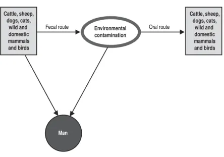
Source of Infection and Mode of Transmission (Figure 1): Many animal
species harbor E. rhusiopathiae. The principal reservoir seems to be swine; the eti-ologic agent has been isolated from the tonsils of up to 30% of apparently healthy swine. In a study carried out in Chile, the agent was isolated from tonsil samples of 53.5% of 400 swine in a slaughterhouse (Skoknic et al., 1981). E. rhusiopathiaewas isolated from 25.6% of soil samples where pigs live and from their feces (Wood and
Figure 1. Animal erysipelas and human erysipeloid (Erysipelothrix rhusiopathiae). Mode of transmission.
Infected animals (boar)
Susceptible animals
(boar) Environmental
contamination (pasture, water)
Ingestion, through the skin
Man
Through the skin b
y handling
animals and animal products
,