ANATOMY AND HISTOLOGY OF SECONDARY SEXUAL CHARACTERS,
GONADS
AND LIVER OF THE ROCK-POOL BLENNY, PARABLENNIUS
SANGUINOLENTUS PARVICORNIS, (PISCES: BLENNIIDAE) OF THE AZORES
RICARDO S E R ~ O SANTOS
ARQUIP$AG()
SANTOS, RCARDO SERRAO 1995. Anatomy and histology of secondary sexual characters, gonads and liver of the rock-pool blenny, Parablennius sanguinolentus parvicornis (Pisces: Blenniidae) of the Azores. Arquipe'lago. Life and Marine Sciences 13A: 21-38. Angra do Heroismo. ISSN 0870-6581.The main histologic aspects of the reproductive organs of Parablennius sanguinolentus parvicornis, a rocky intertidal pool fish of the Azores with alternative life history tactics, are described and illustrated. Eight stages of m y t e development (plus atretic ones) are considered. The development of the ovaries is classified into 6 stages. Ovaries may contain, according to female size, from 4000 up to 41000 mytes in the month which preceeds the reproductive season. Gonad maturation of the males is assigned to 8 stages. Seasonal variations of gonad histology, of both males and females, are illustrated. Histology of the livers shows that they play an important role in the storage of energy. Lipidic reserves are used while the breeding season progresses.
SANTOS, RICARDO SERRAO 1995. Anatomia e histologia dos caracteres sexuais secundArios, g6nadas e figado do caboz-das-pqas, Parablennius sanguinolentus parvicornis, (Pisces: Blenniidae) dos Aqores. Arquipe'lago. CiCncias Biol6gicas e Marinhas 13A: 21-38. Angra do Heroismo. ISSN 0870-6581.
Neste artigo descrevo os brgZos reprodutores de Parablennius sanguinolentus parvicornis, peixe que habita as pqas rochosas do estrato entre-maris dos Aqores. Esta espicie apresenta ticticas alternativas de acasalamento. SZo considerados e descritos oito estidios de desenvolvimento dos 6citos (mais um estidio degenerativo). 0 desenvovimento dos ov5rios 6 classificado em seis estidios. 0 s ovkios, no m&s anterior ao inicio da postura, cont&m, conforme o tamanho das fsmeas, entre 4000 e 41000 &citos em diferentes estidios de desenvolvimento. 0 process0 de maturaqZo das g6nadas dos machos t classificado em oito estidios. SZo ilustradas, atravis de preparaqBes histol6gicas, as variaqaes sazonais do desenvolvimento das ghadas. A histologia hepitica mostra que o figado desempenha um importante papel como reservat6rio energktico. As reservas lipidicas, acumuladas durante o Inverno e Primavera sZo gastas durante a curta estaqZo de reproduqZo.
Ricardo Serrcio Santos, Universidade dos Aqores, Departamento de Oceanografa e Pescas, PT-9900 Horta, A~ores, Portugal.
-
Unidade de Estudos Ocefinicos IMAR, PT- 9900 Horta, Acores, Portugal.INTRODUCTION the studies were concentrated on populations
The reproductive biology and phenology of from the British Isles, Mediterranean and several species of blennies and their close Adriatic Seas.
relatives of the north-eastern Atlantic and the Blennies, together with gobies, comprise the Mediterranean and adjacent seas, have been majority of the resident (sensu GIBSON 1969) fish studied from a variety of viewpoints (see GIBSON species in the shallow water of these regions 1969, 1982; ALMADA & SANTOS 1995). Most of (GIBSON 1969, 1982; ZANDER 1972; ARRUDA
ANATOMY ANO
HI
STOLOGY OF SECONDARY SEXUAL C
H
ARACTERS,
GON
AD
S AND LIVER
OF
THEROCK-POOL BLENNY,
PARABLENNIUSSANGUINOLENTUS PARVICORNIS,
(PISCES: BLENNIIDAE) OF
THE
AZO
RE
S
~CARDOSERRÃOSANTOS
ARQUI
PÉLAGO
IN1RODUCTION
SANTOS, RICARDO SERRÃO 1995. Anatomy and histology of secondary sexual
characters, gonads and li ver of the rock-pool blenny, Parablennius
sanguinoLentus parvicomis (Pisces: Blenniidae) of the Azores. Arquipélago. Life
and Marine Sciences 13A: 21-38. Angra do Heroísmo. ISSN 0870-6581.
The main histologic aspects of the reproductive organs of Parablennius sanguinolentus
parvicornis, a rocky intertidal pool fish of the Azores with altemative life history tactics,
are described and illustrated. Eight stages of oocyte development (plus atretic ones) are considered. The development of the ovaries is classified into 6 stages. Ovaries may contain, according to female size, from 4000 up to 41000 oocytes in the month which preceeds the reproductive season. Gonad maturation of the males is assigned to 8 stages. Seasonal variations of gonad histology, of both males and females, are illustrated. Histology of the Iivers shows that they play an important role in the storage of energy. Lipidic reserves are used while the breeding season progresses.
SANTOS, RICARDO SERRÃO 1995. Anatomia e histologia dos caracteres sexuais
secundários, gónadas e fígado do caboz-das-poças, Parablennius sanguinolentus
parvicomis, (Pisces: Blenniidae) dos Açores. Arquipélago. Ciências Biológicas e
Marinhas 13A: 21-38. Angra do Heroísmo. ISSN 0870-6581.
Neste artigo descrevo os órgãos reprodutores de Parablennius sanguinolentus parvicornis,
peixe que habita as poças rochosas do estrato entre-marés dos Açores. Esta espécie apresenta tácticas alternativas de acasalamento. São considerados e descritos oito estádios de desenvolvimento dos oócitos (mais um estádio degenerativo). O desenvovimento dos ovários é classificado em seis estádios. Os ovários, no mês anterior ao início da postura, contêm. conforme o tamanho das fêmeas, entre 4000 e 41000 oócitos em diferentes estádios de desenvolvimento. O processo de maturação das gónadas dos machos é classificado em oito estádios. São ilustradas, através de preparações histológicas, as variações sazonais do desenvolvimento das gónadas. A histologia hepática mostra que o fígado desempenha um importante papel como reservatório energético. As reservas lípídicas, acumuladas durante o Inverno e Primavera são gastas durante a curta estação de reprodução.
Ricardo Serrão Santos, Universidade dos Açores, Departamento de Oceanografia e
Pescas, PT-9900 Horta, Açores, Portugal. - Unidade de Estudos Oceânicos [MAR,
PT-9900 Horta, Açores, Portugal.
The reproductive biology and phenology of several species of blennies and their dose re1atives oi' the north-eastern Atlantic and the Mediterranean and adjacent seas, have been studied from a variety of viewpoints (see GIBSON
1969, 1982; ALMADA & SANTOS 1995). Most of
the studies were concentrated on populations
from the British Isles, Mediterranean and Adriatic Seas.
B1ennies, together with gobies, comprise the
majority of the resident (sensu GIBSON 1969) fish
species in the shallow water of these regions (GIBSON 1969, 1982; ZANDER 1972; ARRUDA 21
1990; SANTOS et al. 1994). They all lay demersal eggs and provide parental care to the developing embryos. Their general biology was reviewed at length by GIBSON (1969, 1982, 1986).
In general, the blennies live more than two years and occasionally up to ten or thirteen years (e.g. Lipophrys pholis: QASIM 1956b; DUNNE 1977). Most of them are iteroparous; sexual activity extending for two or more years. In. temperate zones the reproductive season lasts for two or more months, with several spawnings (QASIM 1956a, 1956b; SHACKLEY & KING 1977a; SANTOS 1992). At lower latitudes females may spawn all the year round (NURSALL 1977, 1981; JOHANNES 1978). Eggs are attached to the substratum by adhesive discs (e.g. SANTOS 1989) or threads.
The testes of blennies and gobies are unique among teleost for possessing a special gland. In blennies the testicular gland has cells containing lipids (PATZNER & SEIWALD 1987c; SEIWKD & PATZNER 1987). The spermatids mature in the testicular gland where they receive nutrients (LAHNSTEINER & PATZNER 1990% 1990b; LAHNSTEINER et al. 1990). The testicular gland is also an important source of mucus secretion, which is added to the spermatozoids. At the end of the spawning season it is involved in the phagocytosis of remaining spermatozoa (LAHNSTEINER & PATZNER 1990b; LAHNSTEINER et al. 1990).
There are often structures used to enhance behaviour associated with reproduction. Colour patterns of males frequently change during the reproductive phase (temporary dichromatism), with development of strikingly conspicuous colour marks (head masks) (GIBSON 1969). Most species of blennies possess special structures in the head region such as tentacles and crests. Crests are usually present in breeding males, or, if present in both sexes, they are specially developed in males during the reproductive season (QASIM 1956a; PAPACONSTANTINOU 1979; PATZNER et al. 1986; PATZNER & SEIWALD
1987b; ALMADA 1989). Male anal glands are distinctive secondary sexual characters in many species (Scartella cristata by: SMITH 1974;
Parablennius gattorugine: KOTRSCHAL &
GOLDSCHMID 1983; P. pilicornis: DENOIX 1984;
P. ruber: SANTOS 1987; Salaria pavo: PATZNER
& SEIWALD 1987a, 1987b, and see also ZANDER 1975). Their function is still obscure.
Parablennius sareguinolentus parvicomis
(Valenciennes in Cuvier & Valenciennes 1836) is the dominant resident rock-pool intertidal species in the Azores (SANTOS et al. 1994). It is one of the few species of blennies (the other being
Salaria pavo, ALMADA et al. 1994, 1995) which
is known to have developed alternative mating tactics, in which the large males act as parentals, while the small males stay as satellites of the parental territories (SANTOS 1985; SANTOS &
ALMA 1988). The demography and growth, and the biometry of reproductive phenology of this species was studied in detail (SANTOS et al. 1995, 1996). Agressions in the context of reproductive phenology, both in males and in females, were analysed by SANTOS & NASH (1996).
In this paper we provide a description of gonad development in the general context of the phenology of P. s. parvicomis, with
a
view to enhance detail to the biometric phenological analysis presented by SANTOS et al. (1996). The histology of the gonads and liver are examined in relation to the process of maturation and reproductive effort. From this the pathways of energy allocation and transfer can be elucidated. It is well known that some of the lipids deposited in the oocytes during maturation do not come directly from ingested food, but are transferred from lipid stores in the liver and muscles (ZAHND1959; LARSON 1974; HTUN-HAN 1978; CRUPKIN et al., 1988). It has been shown in Lipophrys
pholis (SHACKLEY & KING 1977b, 1978, 1979)
that the yolk incorporates exogenous protein during oocyte development, which was synthesized in the liver.
In the present work other morphological and histological aspects associated with reproduction have also been considered, such as the development of the anal glands and testicular structures. Some of these subjects are poorly understood for many other blennies.
MATERIAL AND METHODS
1. Sampling
A total of 2,580 fishes were sampled monthly between January 1987 and December 1988 with and without the anaesthetic ChinaldinB (Merck) in rock pools at Feteira on the south coast of Faial Island, Azores. From these, the gonads of ca. 200 males and females, and the livers of ca. 100 males and females were prepared for histology. Total length and weight of the body, gonads and livers were obtained from recently dead fishes (always killed with an overdose of the anaesthetic Quinaldine). Width (diameter) of male anal glands (first and second transformed rays of anal fin -see Fig. 1) were measured with a caliper.
2. Preservation and preparation of material Gonads and liver were preserved for histological studies and further examination. One or both of the gonads and the whole liver or a part of it, depending on its size, were preserved in aqueous Bouin's solution (COSTA & CHAVES 1943) for a period of between two and four days. Subsequently they were prepared for histological examination (following the standard histological series, adapted from COSTA & CHAVES 1943), by being stained with the eosine/haematoxiline method (after COSTA & CHAVES 1943).
Development of the gonads and the livers were described from histological preparations and relative weights. Diameters of oocytes at different stages as well as other structures (e.g. the thickness of adherent discs) were measured with an accuracy of +1 pm. Diameter of fat vacuoles of the liver were measured in some histological preparations.
Sub-samples of the gonads of known weight (WSG) of 33 pre-spawning mature females were preserved in Gilson solution to enable separation of the oocytes for counting and measuring (KARTAS & QUIGNARD 1984). Since Gilson
solution was observed to reduce egg diameter, 50 eggs, from two different females, were measured in fresh condition and remeasured after immersion. The mean reduction (mean f
standard deviation) of the egg diameter was 21 f
5%.
Estimates of the total number of oocytes per female were made using a sub-sampling technique. All the oocytes were put into a small receptacle which was divided into 200 quadrats and twenty of these quadrats (NQs) were randomly selected. All the oocytes they contained (NEQs) were counted and measured. Oocytes were measured to +1 pm accuracy using a Mitutoyor profile projector (PJ- 100). Total number of oocytes in the sample (NES) was estimated as NES= NEQs/NQs x 200. Total number of oocytes per female (TFE) was estimated as WE= GW/WSG x NES, where GW is gonad weght.
RESULTS
1. Genera! morphology 1.1. Female reproductive organs
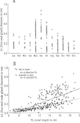
The female gonad is composed of two sausage- shaped ovaries, that extend along the body cavity in a dorsal position above the intestines and below the bladder (Fig. 2). Colouration varies from transparent to milky white in immature females to purple in ovaries with fully developed oocytes. Separate oviducts emerge from each gonad. These connect anterior to the single genital opening which is horseshoe shaped and posterior to the anus.
The maximum diameter of mature oocytes was 0.9 mm to 1.1 mm. In a single ovary there were oocytes in several stages of development. The adherent disc, a structure that enables the egg to stay fixed to a surface after spawning, was distinguishable during early egg development being fully developed by the end of oocyte maturation.
': . .<;.::''. ) .'.. : . . . . . . . . .. Liver ... :,. . al glands . , , , . . . . . . . . . . .. . .. , , ,... :.::>: T.
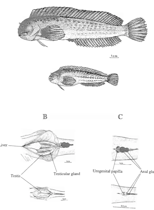
Fig. 1. A- Parental (above) and satellite (below) males of P. s. pamicornis. B- ventral internal view of the males (parental above, satellite below) to show the gonads and liver. C- Ventral external view of the males (parental above, satellite below) to show the urino-genital papilla, the anus and the anal glands.
Urogenital papilla
Anus
anal fin ray
Ovaries
/
Fig. 2. A- Female of Parablennius sanguinolentus parvicornis. B- Ventral external view of the female, showing the urino-genital papilla, the anus and the beginning af the anal fin. C- Ventral internal view of the female to show the liver and gonads.
1.2. Male reproductive organs and secondary sexual characteristics.
Males can be easily distinguished by external characters such as the shape and structure of genital papillae. The males have three openings on the urinogenital papillae, all posterior to the anus. Their arrangement is transverse to the body (Fig. 1). This is apparent very early being present just after metamorphosis. Small individuals need
to be examined with a hand lens to confirm the shape of the genital papillae.
Larger males, in reproductive condition, have other obvious characters that make the distinction from females easy. They tend to be much darker, even black, during the territoriaYparenta1 phase. The profile of the head is more rounded and proeminent in males, probably due to the storage of fat tissues. Males also display two distinctive anal glands on the first and second anal fin rays.
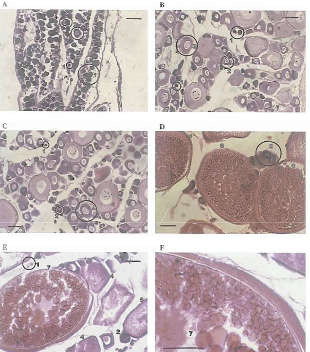
Development of anal glands is ontogenetic and seasonal (Fig. 3). Small individuals have very small glands, which are difficult to distinguish (Fig. 1). They are more apparent during the summer due to a darker colouration. In bigger animals glands are highly reduced after the reproductive season but undergo rapid growth in May.
" I "
Jan Feb Mar Apr May Jun Jul Aug Sep Oct Nov Dee
8 10 12 14 16 18 20
TL (total length in cm)
Fig. 3.A- Monthly variations of the width diameter of the first anal gland of the males. B- Relationship of the width diameter of the first anal gland of the males and their total length, r2 (May to August)=0.72; (September to April)=0.44.
The testes of P. s. panticornis (Fig. 1) are similar to those of other Blenniidae composed of paired elongated bodies situated below the kidney and consisting of tubules of the unrestricted spermatogonial type (GRIER et al. 1980; GRIER
1981; PATZNER & SEIWALD 1987a). When mature their colour is milky white. The vas deferens emerge posteriorly from both testes.
Like in all other Blenniidae, the testes have other specialized structures. The most obvious is the testicular gland, which is situated ventrally in the testes. Its colour is rnilkish-rose. The vas deferens are connected with the testicular gland and conduct the sperm to the genital openings.
2. Development 2.1. Ovaries
The eight stages of development of the oocytes are described with detail in Table 1, and illustrated in Figure 4. The six stages of macroscopic gonad maturation, which are directly related with gonadosomatic indices (see SANTOS et al., in press) are described in Table 2. Oocyte stages compared with the ovary condition are shown in Table 3.
2.2. Testes and accessory organs
Male gonads of P. s. sanguinolenrus are composed of two main, and very distinctive components: the testis and the testicular gland (Fig. 1 and Fig. 5). The testicular gland is situated ventrally in relation to the testis. It has connections to the tubules and the vas deferens (see Fig. 5). It is composed of tubuli, which are separated by cell membranes. In ripe gonads, tubules are easily distinguishable in the testicular gland as filled with sperm (see Fig. 5). The vas deferens are located at the end of the testicular gland. Blood vessels and blood cells are easily distinguishable (see Fig. 5).
Both small (i.e. non-parental males, including satellites) and parental males have well developed gonads during the months of reproduction. Mature spermatozoa are present in the vas deferens before spawning in April and May). This indicates that male gametes mature before female gametes. After the spawning season, in September and October, mature spermatozoa still remain in the vas deferens. Six stages of gonad development were characterized (Table 4).
The size of the testis, relative to the testicular gland, was much bigger in small mahre non- parental males (e.g. satellites) (meanestd. in May with n= 16: 6.80k1.1) than in older parental males (May, n= 14: 1.84k1.27). The testis shrink substantially after the reproductive season. The proportional relationship between testicular glandltestis clear distinct before and after the reproductive season (May, n= 33: 0.41k0.24 and in September, n= 14: 2.46k0.41).
3. Preliminary estimates of "fecundity" The number of oocytes in the ovary varied from just under 4,000 in a female 8.50 cm total length, to over 41,000 in a female 17 cm total length (mean+s.d.= l3,420+5,117). In May, only a
small proportion of oocytes belonged to the mature size classes. Being a multiple spawner, it is likely that only a small proportion of the oocytes in the ovary are released at any one time. Therefore, in a multiple spawning species such as this, absolute fecundity in terms of eggs released, is difficult to estimate (KARTAS &
QUIGNARD, 1984). It was found that the number of eggs
(I?)
was best correlated with total weight2
(TW) (F= 3606.4+399.176 'IW, r = 0.56, p< 0.001, n= 33) rather than with gonad weight
2
(GW) (F= 8102.63+3036.243 GW, r = 0.32, p< 0.001, n= 33) or total length (TL) (F= -18091.7+
2
2549.504 TL, r = 0.46; p< 0.001; n= 33). The relationship between number of eggs and total length is best expressed by the fuction: F=63.096
~ ~ 2 . 1 2 9 (r2=0.53, p<o.OOl).
Table 1
Scale of oocyte development of P. s. parvicornis.
Stage Name and Size Description
I Oogonia (up to 30 pm) I1 Primary oocyte (30 to
100 pm).
111 Oocytes between 100 to 150 pm.
IV Oocyte diameter about 230 pm
V Oocyte diameter about 300 pm
VI Oocyte diameter about 500 pm.
VII Oocyte diameter about 700 pm.
VIII Oocyte diameter up to
Present singly or in small nests in the germinal epithelium. Nucleus large. No nucleoli visible. Cytoplasm dark.
Oocytes already surrounded by a thin layer of follicular epithelium. Nucleus located in the middle of the cell and surrounded by nucleoli. Cytoplasm dark.
Easily distinguishable in histological preparations since the cytoplasm is much lighter. Nucleus, located in the middle of the cell, surrounded by several nucleoli at the periphery. Development of the adherent disc recognizable at one pole of the oocyte forming a layer, 10 pm thick.
Cytoplasm appears much lighter and a few small vacuoles distinguishable. Yolk development begins. Nucleoli dispersed all over the nucleus. The thickness of the germinal chorion around 3 pm. Thickness of the adherent disc around 17 pm. Cytoplasm nearly filled with vacuoles, except for a ring circling the nucleus. Nucleoli
' spread all over the nucleus. The adherent disc is 27 pm thick.
Oocytes characterized by the presence of yoUc droplets associated with vacuoles. The cytoplasm completely filled with yolk granules. Nucleoli concentrated in the middle of the nucleus in most cases. Thickness of adherent disc now up to 30 microns, while that of the chorion is 10 pm.
Ripe oocytes ready to be released. Cytoplasm completely filled with large yolk
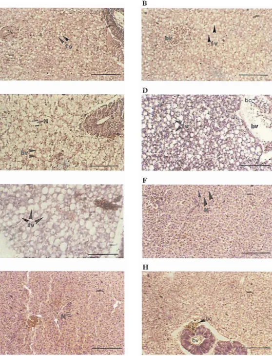
Fig. 4. Histology of female gonads: stages of m y t e development, from 1 to 7, are illustrated (see Table I for descriptive details). A- histology of a gonad from a female with 5.27 cm TL, from March. B- histology of a gonad from a female with 12.80 cm TL, from March. C- histology of a gonad from a female with 14.60 cm TL, from February. D- histology of a gonad from a female with 13.94 cm TL, from June. E and F- histology of a gonad from afemale with 13.00 cm TL, from June (scale: 100 pm).
Ti' Ti'
Fig. 5. Histology of male gonads: A- male with 15.90 cm TL from January. B- male with 15.38 cm TL from March. C- male with 9.01 cm TL from April. D- male with 16.84 cm TL from April. E- male with 9.00 cm from May. F- male with 14.55 cm
TI,
from May. G- male with 9.36 cm TL from July. H- male with 16.50 cm TL from July. I- male with 11.85 cm TL from August. K and L- male with 15.36cm TL from September. M- male with 16.40cm TL from October. N- male with 11.97cm TL from November. 0- male with 17.39 cm from December. S and T- seminiferous tubuli. (bv) blood vessel. T- testis. T and G- testicular gland. V and D- vas deferens (sp) spermatocytes (scale: 200 pm).Table 2
Scale of ovary condition of P. s. pawicornis, with reference to oocyte development and gonadosomatic
index.
Stage Name Description
I Immature Ovaries are very small. Weight less than 0.06g. representing 0.07 to 0.9 % of total body weight and 0.09 to 0.8% of the eviscerated weight. Ovaries form a thin transparent to yellowish ribbon. Oocytes mainly in stage I and 11.
I1 Maturing Ovaries slightly swollen and creamylyellowish. Oocytes now just visible macroscopically. Some ovaries do not complete development to mature stags. Weight not exceed 0.8g representing a maximum of 8% of total body weight. Oocytes present at stages of development I to V.
111 Ripening. Ovaries very large, occupy a large proportion of abdominal cavity. Oocytes easily visible macroscopically. Ovaries light yellow. Weight between 0.2g and 3.2g representing up to 8% of total body weight. Oocytes from stage I to VI.
IV Ripelpre- Ovaries distended and occupying almost the whole abdominal cavity. Oocytes easily visible spawning. macroscopically. Ovaries purplish. Weight may reach 6.5g representing up to 15% of total body weight and 18% of the eviscerated weight. Ripelpre-spawning females found at the end of May and beginning of June. Ovaries have oocytes in all developmental stages
V Ripelspawning Ovaries may be less distended and less purple due to recent release of mature oocytes. Weight varies between 0.7g and 4.2g representing up to 10% of total body weight and 13% of eviscerated weight. Ripelspawning ovaries found from June until August. All developmental stages of oocytes present. VI Post-spawning Ovaries shrunken and coloured cream to darkcream. Non-released eggs and atretic eggs can be seen
macroscopically. Weight from 0.04g to l g representing up to 2% of total body weight. Oocytes from stage 1 to stage III.
Table 3
Summary of oocyte stages and ovary condition in different months of the year
Month Oocytes stages Ovary condition
January February March April May June July August September October November December I to III (+ atretic) I to
Iv
I to IV I tov
I to VI I tovm
I to WI I to VIII I to 11, VIII (+ atretic) I to 111, VIII (+ atretic) I to I11 (+ atretic) I tom
(+ atretic) I and I1 I and I1 I and I1 I and I1 I to I11 I tov
I, 11,111, V and VI I, I1 and VI I, I1 and VI I,II
and VI I and I1 I and I1 4. Liver 4.1. General descriptionThe liver is one of the largest organs located in the abdominal cavity. It is found just behind the transverse septum on the first half of the abdominal cavity (Fig. 2 and 3). Its colour may vary from dark cream to brown. As an organ with an important role in the storage of energy the liver exhibits considerable variation in its weight and structure that cannot be simply related with individual growth. These variations are strongly seasonal.
4.2. Histology
4.2.1. Seasonal variations in the structure of the female liver
In February the cells were filled with fat vacuoles of small and medium size (Fig. 6/A) in most of the individuals, but some of the bigger animals had large fat vacuoles (Fig. 6/B
Table 4
Scale of male gonad development in P. s. parvicomis, with reference to gonadosomatic index.
Stage Maturation Description
Immature Testes very small and transparent. Immature testes found in 0' individuals until January. Mean gonadosomatic index 0.14%
Maturing Gonads much larger accounting from 0.5% to 3.4% of total body weight. Maturation begins in JanuaryIFebmary. In fishes maturing for their second or third time testicular gland is consistent and cells look more compact than in younger fish. Testicular gland proportionally bigger than testis in large animals. In smaller males testicular gland only develops to a small size. From January until April the enlargement of the testis occurs. In April, the tubules may or may not be completely filled. Small cavities may be present in the centre. Not all stages of spermatogenesis will have been completed.
111 Ripe By the end of May maturation appears to be complete. Gonad milky white in appearance and testicular gland slightly pink. Testes full of cells in different stages of spermatogenesis. Seminiferous tubules with spermatozoa distinct in the testicular gland. Spermatozoa found in vas deferens. Testes reach maximum size in relation to the testicular gland. Gonadosomatic indices reach their maximum value.
Spawning Spawning occurs in June, July and early August. The testis full of ripe spermatozoa. Cells in all other stages of development are present. Spermatozoa mainly seen in the testicular gland. In most cases tubuli empty in the middle indicating that spermatozoa have been released into the sperm ducts. Testicular gland showing a loose formation, with cavities, in August.
Post-spawning Gonads much shrunken and greyish. Testicular gland prominent. The testis look like a fine band over the gland. Spermatozoa remaining in the testis and in the testicular gland. Condition maintained until November. Testicular gland proportionally wider than the testis. Gonadosomatic indices at their minimum in adults.
Recovering Recoven, of the gonad initiated bv December. when s~ermatoaenesis occurs again.
and Table 5). In May, at the onset of the the liver (Fig. 7lA). These fat vacuoles were reproductive season, the livers were completely very enlarged in May (Fig. 7/B) and still filled with fat vacuoles. These were large in present in June (Fig. 7lC). In July the liver bigger females (Fig. 6/C), and of intermediate showed a compact structure (Fig. 7/D), which
size in smaller females (Fig. 6D). remained at least until October.
After the reproductive season, in August, the liver was much more compact and the fat
vacuoles were rare, if present at all. The Table 5
nucleus of the cells were much closer, and were
A descriptive quantitative scale of liver very distinct (Fig. 6E). The liver seemed to
structures for both males and females. rest in this condition until October (Fig. 61F).
The storage of l i ~ i d s u amears to be reinitiated
. .
(measurements in pm)in November (Fig. 6/G), but some of the livers diameter of lipid distance n
were still of the compact type at this time. vacuoles between nuclei
compact --- 4.58
+
1.93 254.2.2. Seasonal variations in the structure of small fat vacuoles 2 5 6.44k3.12 32
the male liver
medium fat 2 1 0 10.00
+_ 1.67 37
The pattern of seasonal variations of the
structire of the males liver seems very similar large fat 2 20 - - -
-
-- - -
20 to that of the females. In March fat vacuoles fillFig. 6. Histology of female liver. A- female of 7.79 cm TL from February. B- female of 12.10 cm from February. C- female of 15.83 cm TL from March. D and E female of 11.75 cm TL from October. F female of 17.40 cm TL from August. G- female of 16.09 cm TL from October. H- female of 11.75 cm
TL
from November (fi) fat vacuoles (bc) blood cells (N) cell nucleus (scale 100 p).Fig. 7. Histology of male liver. A- male of 15.38 cm TL from March. B- male of 14.55 cm TL from May. C- male of 16.38 cm TL from June. D- male of 15.75 cm
TL
from July. E- male of 11.85 cmTL
from August. F- male of 15.36 cmTL
from September. G- male of 10.71 cm TL from October (f.v.) fat vacuoles (B) blood cell (bc) blood cells (N) cell nucleus (scale 100 pm).DISCUSSION
Anal glands are distinctive male sexual traits of many blennid species (e.g. . Salaria pavo,
PATZNER & SEIWALD 1987b; Parablennius ruber, SANTOS 1987; and unpublished observations on P. incognitus and Ophioblennius atlanticus atlanticus of the Azores). Their appearance and shape may be rather different even among species from the same genus (GOLDSCHMID et al. 1980), as for instance in P. ruber (SANTOS 1987) and P. gattorugine (KOTRSCHAL & GOLDSCHMID 1983) whose taxonomic distinction only recently was confirmed (ALMEIDA 1979; BATH 1982). The exact function of these organs is still being debated. It has been speculated that they produce pheromones (LOSEY 1969; LAUMEN et al. 1974), or that they produce antibiotic secretions that could be used by the male to protect the eggs against bacterial infections (QASIM 1956a; PETERSON 1984). The size of the glands is reduced out of the reproductive season. Since these glands are only fully developed in parental males during the reproductive season, and not in sneakers and satellites, it is a clear indication that they have a parental or epigamic function. This subject deserves to be further investigated.
Male gonads of blennioids are unique among teleosts in possessing a testicular gland. It stores lipids and spermatozoa (LAHNSTEINER &
PATZNER 1990d; LAHNSTEINER et al. 1990). LAHNSTEINER & PATZNER (1990c, 1990d) proposed that structural differentiation of the male gonads of blennies ("testis" and testicular gland), would have the advantage of shortening the duration of the spermatid cycle in the "testis", since final differentiation and consequent storage occurs in the testicular gland. Spawning in blennies may occur on successive days over a long period of time. The ability to accelerate the initial rate of spermatogenesis, associated with the possibility of spermatozoa storage would be an advantage, under these circumstances. The size and morphology of testicular glands of large and small mature males of P. s. parvicornis are very distinct, as in Tripterygion tripteronotus and
T.
,delaisi (JONGE et al. 1989). Testicular glands are
clearly more developed in parental males. It is probable that this gland has other roles, besides the storage of spermatozoa. This subject is worth to be further investigation.
The histology of female gonads show that young females (size between 7.5 and 10 cm) only produce one batch of eggs during their first year as mature females. The oocytes of some of them reach only intermediate stages of maturation and are probably retained for the next spawning season. Major investment in reproduction begin when females reach their second year (SANTOS et al. 1996). Only a small proportion of the oocytes mature at a time, but this small proportion occupies the majority of the ovary volume. These are distributed by several spawning acts. In Salaria pavo more than 70% of the number of oocytes in the ovary are in the first stage of oogenesis (PATZNER 1985). Ripe oocytes occupy from 66.8% up to 75.3% of the ovary volume (PATZNER 1985). One out of 7 oocytes present in the ovary in the middle of the reproductive season will be retained for the next season. SHACKLEY &
KING (1977a) found that each female produces eight different clutches of oocytes each breeding season. The production of multiple batches of eggs, reaching maturation and being spawned at different times, may present, at least, two types of advantages. One is related to female performance and management of resources, and the other to the survival of the embryos and the larvae. The first benefit is related to hydrodynamic performance. As MILLER (1979), following an hypothesis discussed by WILLIAMS (1959) pointed out: "a direct increase in gonadosomatic index raises problems of hydrodynamic performance and predator attraction, in addition to intrinsic levels of food intake. However, a high reproductive effort may be achieved by summation of repeat (partial, batch, heterochronal) spawning in which successive batches of eggs are produced during a lengthy period of reproduction.". The other benefit is linked to the advantages of polyandry, on the one hand, and to temporal shifting of larvae in an unpredictable habitat, on the other. Distributing the eggs among several males,
reduces the risks of absolute loss of progeny due to wrong choice of the male. This is particularly relevant for a species in which survival of the embryos is directly dependent on the ability of the male to care for them. Distributing eggs for several spawnings during two months also assures that larvae will occur in the plankton at different times. This may contribute to reduced risks of total loss of progeny due to extreme unfavourable biotic andlor abiotic conditions during planktonic life. This is a good strategy in warm temperate and subtropical areas without marked seasonality of production such as the spring bloom, typical of cold temperate areas.
The dynamics of storage and depletion of resources in the liver is another important aspect of the phenology of P. s. pawicornis, which is clearly illustrated by histology. Liver growth is predominantly linked with an increase in cell numbers (LOVE 1970), but it also undergoes changes that can be related to seasonal variations in storing and utilization of fat and glycogen by the individuals. It is known that the liver of the fish stores lipids and glycogen (LOVE 1970; PODROSCHKO et al. 1985). Both males and females seem to rely on lipids stored in the liver after the winter during the reproductive season. It is known that energy stored in ,the liver is re-utilized in muscular activity (LOVE 1970), and effort of reproduction (e.g. in the process of ovary maturation). Females store lipids, which are later transferred to the ovary to be used by the deveIoping embryo before it can feed (LOVE 1970). They transfer energy directly from feeding to the developing oocytes, but also rely on the fat they have stored in the liver. P. s. parvicornis with its well differentiated reproductive season shows the expected seasonal differences in liver size and histology, as well as structural differences related to age. Maturing females (sized from about 9 cm to 12 cm) store lipids in the liver from January until May. The compact structure
of the livers of females sized between 7.5 crn and 10 cm during the months from January to March, and of smaller females until April shows that at these immature sizes do not store fat vacuoles (lipids) in the liver. It is probable that
they invest directly in growth. Their investment in reproduction later on that year will be nil in the smaller size class. In May all females show abundant fat stores in the liver. The size of the fat vacuoles is less in small females than in larger ones. In June some of the big females have lost a great part of their liver fat reserves, certainly transferred to egg yolk, to be used by the embryos during their development. From August to December the liver has lost most of the fat reserves in all size classes. It is most probable that females will be investing directly in body restoration, maintenance and growth during this period. The synthesis of protein yolk is exogenous to the egg. It occurs in the liver from which it is transferred to the developing oocyte SHACKLEY & KING (1978, 1979). These observations confirm the conclusion that the liver is an organ with a role in the storage of energy which is spent during the reproductive season in this species.
ACKOWLEDGEMENTS
The author acknowledge the field support given by Norberto Serpa. I am also grateful for the comments andlor help of Stephen J. Hawkins and Richard D. M. Nash (Port Erin Marine Laboratory), Robin Gibson (Scottish Marine Biological Association), Robert A. Patzner (University of Salzburg), Helen Rost Martins (DOPIUA) and two anonymous referees. The drawings included were made by Les Gallagher. This work was supported by JNICT (CiZncia Program) and Secretaria Regional da Agricultura e Pescas (DRP), to whom I am grateful.
REFERENCES
&ADA, V. 1989. Eco-etologia de Sakaria pavo Risso, 1810 (Pisces: Blenniidae) no Parque Natural da Ria Formosa. Relat6rio. S.N.P.R.C.N., Lisboa. 148pp.
&ADA, V.C. & R.S. SANTOS 1995. Parental care in the rocky intertidal: a case study of adaptation and exaptation in Meditemean and Atlantic blennies. Reviews of Fish Biology and Fisheries 5: 23-37.
ALMADA, V.C., E.J. GONCALVES, A.J. SANTOS & C.
B ~ S T A 1994. Breeding ecology and nest aggregations in a population of Salaria pavo
Teleostei) as revelead by electron microscopy and enzyme histochemistry. Journal of Fish Biology 37: 85-97.
LARSON, G.L. 1974. Liver weight of brook trout in a high-mountain lake at Washington State. Progress on Fish Culture 35: 234- 236.
LAUMEN, J., U. PERN & V. BLUM 1974. Investigations on the function and hormonal regulation of the anal appendices in Blennius pavo (Risso). Journal of Experimental Zoology
190: 47-56.
LOSEY, G.S. 1969. Sexual pheromone in some fishes of the genus Hypsoblennius Gill. Science 153: 181-183.
LOVE, R.M. 1970. The Chemical Biology of Fishes. Academic Press, London. 547pp.
MILLER, P.J. 1979. Adaptiveness and implications of small size in teleosts. Pp. 263-306 in: P. J. Miller (Eds), Fish Phenology: Anabolic Adaptiveness in Teleosts Academic Press, London XV
+
449. NURSALL, J.R. 1977. Territoriality in redlip blennies(Ophioblennius atlaticus
-
Pisces: Blenniidae). Journal of Zoology 182: 205-223.NURSALL, J.R. 1981. The activity budget and use of temtory by a tropical blenniid fish. Zoological Journal of the Linnean Society 72(1): 69-92. PAPACONSTANTINOU, C.A. 1979. Secondary sex
characters of blennioid fishes (Pisces: Blenniidae). Thalassographica 1 (3): 57-75. PATZNER, R.A. 1985. The reproduction of Blennius
pavo (Teleostei, Blenniidae). 111. Fecundity. Zoologischer Anzeiger 214 (112): 1-6.
PATZNER, R.A. & M. SE~WALD 1987a. The reproduction of Blennius pavo Risso (Teleostei, Blenniidae). VI. Testicular cycle. Zoologischer Anzeiger 219 (516): 265-273.
PATZNER. R.A. & M. SEIWALD 1987b. The reproduction of Blennius pavo Risso (Teleostei, Blenniidae). VII. Secondary sexual organs and accessory glands of the testis during the reproductive cycle. Proceedings of the V
Congress of European Ichthyologists, Stockholm, 1985: 293-298.
PATZNER, R.A. & M. SEIWALD 1987c. The reproduction of Blennius pavo Risso (Teleostei, Blenniidae). VIII. The testicular gland - preliminary results. Proceedings of the V Congress of European Ichthyologists, Stockholm, 1985: 299-304.
PATZNER, R.A., M. SEIWALD, M. ADLGASSER & G. KAURIN 1986. The reproduction of Blennius pavo. V. Reproductive behaviour in natural
environment. Zoologischer Anzeiger 216(616): 338-350.
PETERSON, H.P. 1984. Morphologische und histochemische Untersuchungen an den 'Flossendriisen von blenniiden Schleimfischen.
Thesis, Universitat Hamburg. 90pp.
PODROSCHKO, S., R.A. PATZNER & H. ADAM 1985. The reproduction of Blennius pavo (Teleostei, Blenniidae). IV. Seasonal variation in HSI, the liver glycogen value and histological aspects of the liver. Zoologischer Anzeiger 215 (516): 265- 273.
QASIM, S.Z. 1956a. The spawning habitats and embryonic development of the shanny (Blennius pholis L.). Proceedings of the Zoological Society of London 127: 79-93.
QASIM, S.Z. 1956b. Time and duration of the spawning season in some ,marine teleosts in relation to their distribution. Journal du Conseil Permanent International pour lrExploration de la Mer 21: 144-155.
SANTOS, R.S. 1985. Parentais e sattlites: tiicticas alternativas de acasalamento nos machos de Blennius sanguinolentus Pallas (Pisces: Blenniidae). Arquipklago
-
SCrie Ciencias da Natureza 6: 119-146.S N O S , R.S. 1987. Estudos sobre a ecologia e comportamento da fauna litoral dos A~ores: I. Primeiras obsewa$Bes sobre o comportamento territorial e parental dos machos de Parablennius ruber (Pisces: Blenniidae), com uma pequena nota sobre os embriBes. A~oreana 6 (1): 295-320. SANTOS, R.S. 1989. Obsewa$ies sobre os intervalos
de desenvolvimento de Blennius sanguinolentus Pallas (Pisces: Blenniidae). Arquivos do Museu Bocage. Nova SCrie l(19): 293-3 10.
SANTOS, R.S. 1992. Behavioural Ecology, Phenology and Ethology of Parablennius sanguinolentus parvicornis (Valenciennes in Cuvier & Valenciennes 1836) (Pisces: Blenniidae) of the Azores. PhD Thesis. DEEB, University of Liverpool X
+
293.S N O S , R.S. & V. ALMADA 1988. Intraspecific variations in reproductive tactics in males of the rocky intertidal fish Blennius sanguinolentus, in Azores. Pp. 421-447 in: G. Chellazi & M. Vannini (Eds) Behavioral Adaptation to Intertidal Life Plenum Press. New York IX
+
524.SANTOS, R.S. & R.D.M. NASH 1996. Seasonal variations of injuries suffered by individuals of the Azorean rock-pool blenny, Parablennius sanguinolentus parvicornis (Blemiidae). Copeia 1996(1)'216-219.
SANTOS, R.S., R.D.M. NASH & S.J. HAWKINS 1994. Fish assemblages on intertidal shores of the island of Faial, Azores. Arquipe'lago. Life and Marine Sciences 12A: 87-100.
SANTOS, R.S., R.D.M. NASH & S.J. HAWKINS 1995. Age, growth and sex ratio of the Azorean rock-pool bleny, Parablennius sanguinolentus parvicomis, a fish with alternative mating tactics. Journal of the Marine Association of the United Kingdom 75 (3): 75 1-754.
SANTOS, R.S., S.J. HAWKINS & R.D.M. NASH 1996. Reproductive phenology of the Azorean rock-pool blenny (Parablennius sanguinolentus parvicornis) of the Azores, a fish with alternative mating tactics. Journal of Fish Biology: in press
SEIWALD, M. & R.A. PATZNER 1987. The reproduction of Blennius pavo Risso (Teleostei, Blenniidae). VII. The testicular gland
-
preliminary results. Proceedings of the V Congress of European Ichthyologists, Stockolm 1985: 299-304.SHACKLEY, S.E. & P.E. KING 1977a. The reproductive cycle and its control; frequency of spawning and fecundity in Blennius pholis L. Journal of Experimental Marine Biology and Ecology 30: 73- 83.
SHACKLEY, S.E. & P.E. KING 1977b. Oogenesis in a marine teleost, Blennius pholis L. Cell Tissue Research 181: 105-128.
SHACKLEY, S.E. & P.E. K w G 1978. Protein yolk synthesis in Blennius pholis L. Journal of Fish Biology 13: 179-193.
SHACKLEY, S.E. & P.E. KING 1979. Aminoacid incorporation by isolated oocytes of the marine teleost Blennius pholis. Joumal of Fish Biology 14: 475-480.
S m , R. C. 1974. On the biology of Blennius cristatus with special refrence to anal fin morphology. Bulletin of Marine Science 24 (3): 595-605.
WILLIAMS, G.C. 1959. Ovary weights of darters: a test of the alleged association of parental care with reduced fecundity in fishes. Copeia 1959: 18-24.
ZAHND, J.P. 1959. Modifications hCpatiques liCes au
cycle ovarien chez deux poissons ovovivipares: Xiphophoms helleri et Lebistes reticulatus. Archives dlAnatomie Microscopique et de Morphologie Expe'rimentale 48: 23 1-259.
ZANDER, C.D. 1972. Beitrage zur Okologie und Biologie von Blenniidae (Pisces) des Mittelmeeres. Helgolander Wissenschafrliche Meeresuntersungen 23: 193-231.
ZANDER, C.D. 1975. Secondary sex characteristics of Blennioid fishes (Perciformes). Pubblicazioni della Stazione Zoologica di Napoli 39: 7 17-727. Accepted 17 April 1995.