Distribution of immune response cells in the pelvic urethra
and the prepuce of rams
1Jorge Acosta-Dibarrat2*, Alejandro Buendía-Jiménez.3, Edgardo Soriano-Vargas2, Roberto Montes de Oca-Jiménez2 and Jorge Tórtora-Pérez3
ABSTRACT.- Acosta-Dibarrat J., Buendía-Jimenez. A., Soriano-Vargas E., Oca-Jiménez R.M. & Tórtora-Pérez J. 2014. Distribution of immune response cells in the pelvic urethra and the prepuce of rams.Pesquisa Veterinária Brasileira 34(3):270-276. Centro de Investigación y Estudios Avanzados en Salud Animal, Facultad de Medicina Veterinaria y Zootecnia, Univer-sidad Autónoma del Estado de México, Km 15.5 Carretera Toluca-Atlacomulco, C.P. 50100, Toluca, Estado de Mexico, México. E-mail: [email protected]
The pathogens of the reproductive system in the male can penetrate and establish by as-cending route, from to the prepuce to the urethra, accessory glands, epididymis and testicles. The aim of this paper is determine the distribution and number of cells involved in the im-mune response in prepuce and pelvic urethra of rams, without apparent clinical alterations in testicle, epididymis and prepuce. The distribution of some of the cells involved in the immune response at the level of the prepuce and the pelvic urethra was quantified in four one-year-old rams seronegative for B. ovis and A. seminis and without apparent lesions in the testicles, the epididymis, and the prepuce. At the moment of slaughter, samples were taken from the prepu-tial fornix and the pelvic urethra and placed in 10% formalin and under freezing conditions. CD4, CD8, WC1, CD45RO, CD14 and CD1b cells were demonstrated by immunohistochemis-try, and immunoglobulin-containing cells (ICC) of the IgA, IgG and IgM classes were demons-trated by immunofluorescence. The labeled cells present in the mucosa of both organs were counted with an image analyzer. The total number of cells was compared between both tis-sues and differentially between the epithelium and the connective tissue of the mucosa. Sig-nificant differences were found in the total number of CD4, CD45RO, and WC1 lymphocytes, in CD14 macrophages, and CD1b dendritic cells, with mean values being greater in the fornix than in the urethra (p<0.05) in all cases. Only dendritic cells were found in the prepuce. No differences were found in the number of CD8 lymphocytes between both organs. The ratio between each cell type in the connective and the intraepithelial tissues and between organs was 10/1 for CD4 in the fornix (p<0.05), against 7/1 in the urethra (p<0.05), while CD8 had a 1/1 distribution in both mucosae. The WC1 ratio was 5/1 in both mucosae (p<0.05). CD45RO labeling was 19/1 in the prepuce (p<0.05) and 1/1 in the urethra. IgA-containing cells did not show differences in the total number of cells in both tissues. In the urethra, no IgG-containing cells were observed and IgM-containing cells were scarce; in contrast, both cell types were present in the prepuce, in amounts greater than in the urethra (p<0.05). IgA-, IgG-, and IgM--containing cells were located in both organs in the mucosal connective tissue. The presence of antigen-presenting cells, macrophages, and dendritic cells, as well as of lymphocytes CD4, CD8 TCR γδ (WC1), IgA-, IgG and IgM positive cells, and CD45RO cells suggests that both mu -cosae may behave as inductive and effector sites for the mucosal immune response.
INDEX TERMS: Rams, prepuce, pelvic urethra, immune response cells.
1 Received on September 24, 2013.
Accepted for publication on January 21, 2014.
2 Centro de Investigación y Estudios Avanzados en Salud Animal,
Fa-cultad de Medicina Veterinaria y Zootecnia, Universidad Autónoma del Estado de México, Km 15.5 Carretera Toluca-Atlacomulco, C.P. 50100,
Toluca, Estado de México, México. *Corresponding author: jpacosta00@ hotmail.com
3Facultad de Estudios Superiores Cuautitlán, Universidad Nacional
RESUMO.- [Distribuição de células da resposta imune na uretra pélvica e em prepúcio de carneiros.] Os pató-genos do aparelho reprodutor do macho podem penetrar e se estabelecer por via ascendente, a partir do prepúcio à uretra, glándulas anexas, epidídimo e testículos. Neste tra-balho foi quantificada a distribuição de algumas das células envolvidas na resposta imune, em nível de prepúcio e uretra pélvica, em quatro carneiros de um ano de idade, sem lesões aparentes no testículo, no epidídimo e no prepúcio. No mo-mento da eutanásia foram coletadas amostras do prepúcio e da uretra pélvica, as quais foram conservadas em forma-lina 10% e congeladas. De acordó com as análises de imu-nohistoquímica foram observadas células CD4, CD8, WC1, CD45RO, CD14 e CD1b; e por imunoflorescência foram ob -servadas células que continham imunoglobulinas (CCI) das clases IgA, IgG e IgM. Com um analisador de imagens foi feita a contagem das células marcadas, presentes na mucosa de ambos os orgãos. Foi realizada a comparação entre o total de células entre ambos os tecidos e de forma diferencial en-tre os tecidos epitelial e conectivo da mucosa. Foram encon-tradas diferenças significativas no número total de linfóci -tos CD4, CD45RO e WC1, no número de macrófagos CD14 e células dendríticas CD1b, com valores de médias que foram maiores em todas as amostras coletadas do prepucio, em re-lação as amostras coletadas da uretra (p<0.05). Células den -dríticas só foram encontradas no prepúcio. Não foram en -contradas diferenças significativas no número de linfocitos CD8 entre ambos os orgãos. A relação entre cada tipo celular no tecido conectivo e intra-epitelial e entre os diferentes ór-gãos, resultou para CD4 10/1 no prepúcio (p<0.05), contra 7/1 na uretra (p<0.05), entretanto os CD8 se distribuíram 1/1 em ambas as mucosas, não sendo significativa as dife -renças. Os WC1 foram observados na relação 5/1 em ambas as mucosas (p<0.05). A célula CD45RO, no prepucio, foi ob-servada de 19/1(p<0.05) e na uretra de 1/1, não sendo um resultado significativo. As CC-IgA não mostraram diferença significativa no total de células em ambos os tecidos. Na uretra não foram observadas as CC-IgG, e as CC-IgM foram escassas; em contrapartida, ambos os tipos celulares foram observadas no prepucio, em quantidades menores que na uretra (p<0.05). As CC-IgA, IgG e IgM foram observadas em ambos os tecidos conectivos da mucosa. A presença de célu -las apresentadoras de antígenos, macrófagoss e célu-las den-dríticas, assim como de linfócitos CD4, CD8. TCR γδ (WC1), CC-IgA, IgG e IgM e células CD45RO, determinam que ambas as mucosas podem se comportar como locais de indução e promoção da resposta imune das mucosas.
TERMOS DE INDEXAÇÃO: Carneiros, prepúcio, uretra pélvica, cé-lulas da resposta imune.
INTRODUCTION
Immune response cells in the male genital tract have to car-ry out recognition and response functions against potential pathogens, and they must also tolerate germ cells that ex-pose extraneous antigens to the immune system (Ander-son & Pudney 1999). In contrast to the female reproductive apparatus, in which immune response cells have already been studied regarding their distribution and
characteris-tics, those of the male reproductive tract have been little explored, even in humans (Moldoveanu et al. 2005).
In rams, Brucella ovis, one of the most widely studied diseases, can be experimentally reproduced by inoculating the bacterium by conjunctival, oral, intrapreputial or intra-venous route (Plant et al. 1986). In contrast, in the case of A. seminis, the evidence points to the retrograde entry of the pathogen through the urethra, since it is frequently present in the preputial mucosa of puberal animals (Jansen 1983, Acosta-Dibarrat et al. 2006, 2007), while Tritrichomona fo-etus in cattle is a venereal infection, in which the first line of defense is the epithelial barrier and the normal flora of both the prepuce and the vagina (Corbeil & BonDurant 2001).
Studies that quantify the presence of immunoglobulin--producing cells (Igs) in the reproductive organs are scarce. It has been demonstrated that B. ovis infection generates a strong IgA response in the secretions of the accessory glands, as well as the presence of IgA-containing cells in the ampulla of the ductus deferens. However, the changes in IgA-containing cells in the bulbourethral glands and the total IgG and IgM concentrations in the secretions were not significant (Foster et al. 1988a).
The immune response against T. foetus in the preputial mucosa and the penis of bulls was described by Campero et al. (1990), indicating mononuclear infiltration in both mucosae, presence of plasma cells, and an epithelium in-filtrated with lymphocytes, similar to that described in the tonsils (Flower et al. 1982). These studies define the pre -putial and penile mucosa as inductive sites of the immune response (Campero et al. 1990).
The presence of lymphoid tissue associated with the reproductive mucosa of males would have antigen proces-sing and immune response to potential pathogens as its main function. The aim of this study was to quantify and determine the distribution of cells involved in the immune response at the level of the prepuce and the pelvic urethra.
MATERIALS AND METHODS
Animals and Sample collection
The animals were kept during the performance of the pre-vious examine to slaughter facilities in the FES Cuautitlán UNAM. Sampling protocols and slaughter were endorsed by the Subcom-mittee Institutional Animal Care Experimentation (SICUAE) of the Faculty of Veterinary Medicine, UNAM. This protocol was appro-ved by UNAM at 20/06/2003.
Paired samples of the prepuce were taken at the level of the fornix and the pelvic urethra from four one-year-old Pelibuey rams seronegative for B. ovis and A. seminis and without apparent lesions in the testicles, the epididymis, and the prepuce at the mo-ment of slaughter. One of the samples was used in routine
histo-pathological studies, it was fixed in 10% buffered formol solution, pH 7.4, embedded in paraffin, and was cut at 5 µm and stained
with hematoxylin and eosin (HE).
The other sample was used to obtain sections for
immuno-histochemical and immunofluorescence studies, it was placed
in Tissue-Tek OCT-based cryopreservative (Sakura Finetec,
Tor-rance, USA) and was frozen in liquid nitrogen, it was first placed
over the vapors for 2 min, and then it was immersed in the liquid, it was immediately withdrawn and was kept at -80°C until
pro-cessing. Four consecutive sections of 4 µm in thickness and ap
samples using a microtome and were mounted on slides treated with poly-L-Lysine (Sigma Chemical Co, St. Louis, USA) and were
fixed with acetone for 10 minutes. One of the sections was used
as negative control and the three remaining sections were used for the various marker antibodies; in all cases, a prepuce section was included as positive control, on which positive labeling for the three immunoglobulin-containing cells (IgCC) and for all mo-noclonal lymphocytes assayed in this study, CD4, CD8, CD45RO, WC1, CD14 and CD1b, had been previously demonstrated.
Inmunofluorescence (IF) for the detection of positive IgA-, IgG-, and IgM-containing cells
Tissue Igs were removed by placing the slides in PBS all night,
nonspecific blockade with 1% BSA was carried out subsequently for 1 hour at room temperature, and three five-minute washings
with PBS were performed. The sections were incubated for one hour at 37°C with anti-IgA, anti-IgG, and anti-ovine IgM primary antibodies prepared in rabbit, diluted in PBS with 1% BSA. The source of monoclonal antibodies and the dilutions used are sho-wn in Table 1. The slides were washed and incubated for one hour at 37°C with TRICT-conjugated anti-rabbit IgG secondary antibo-dy (Sigma-Aldrich, St Louis, USA) in a 1:30 dilution in 10% goat serum and 1% BSA. Finally, they were washed again three times
for five minutes and were mounted in glycerine for observation.
The primary antibody was replaced with PBS in the slide’s con-trol section, and the technique previously described was then fol-lowed. The control section was included in face of the possibility
of nonspecific labeling by the anti-rabbit IgG conjugate.
Immunoperoxidase (IP) for detecting the presence of CD4,
tions were dehydrated by successive passages in alcohols, they were cleared with xylene and were mounted with resin. In the staining of the negative control section of the slides, the prima-ry antibody was replaced with PBS, in order to continue with the technique previously described and to discriminate possible
res-ponses to endogenous peroxidases. Three five-minute washings
with PBS were performed between each step.
Cell count
Prepuce and pelvic urethra cell counts were performed with
both techniques in ten 40 X 10 fields, using an image analysis sof
-tware (Image Pro Plus 4.5; Media Cybernetics, Silver Spring, USA). The cells present in the image projected by the program, with a surface area of 1.81x10 -2 mm2, were counted. Intraepithelial cells
and those present in the mucosal connective tissue were counted, delimiting the area with the help of the software, and the result was expressed as the number of cells per mm2.
Statistical analysis
The mean of total positive IgA-, IgG-, and IgM-containing cells, CD4, CD8, WC1, CD45RO, CD1b and CD14 present in the prepuce and the urethra of the four animals and the number of labeled cells in the mucosal epithelium and connective tissue in the pre-puce and the urethra was compared. The Kruskal-Wallis nonpara-metric method was used for these comparative analyses.
The graphs show the mean ± standard error. The existence of a statistical difference with p<0.05 was considered in all cases. The Kruskal-Wallis and Mann-Whitney tests were run with the SPSS 13 software for Windows (SPSS Corp., Chicago, USA).
RESULTS
In the histological review of the HE-stained sections, no elements suggesting an inflammatory lesion or lesions of any other origin were detected in any of the samples.
The results of the distribution study of the different cell types present in the prepuce and the urethra are summari-zed in Table 2 and Figure 1.
In the slides prepared for the observation of Ig-contai-ning cells by fluorescence, the most abundant labeling cor -responded to positive IgA-containing cells, which did not show significant differences between both tissues. In the urethra, scarce IgM-containing cells were observed, 8.3 ± 32.2, and no IgG-containing cells were found, while in the prepuce, similar amounts of both cell types were found and in a significantly greater number, 140±45.2 and 135±65, than in the urethra (p< 0.05), Table 2.
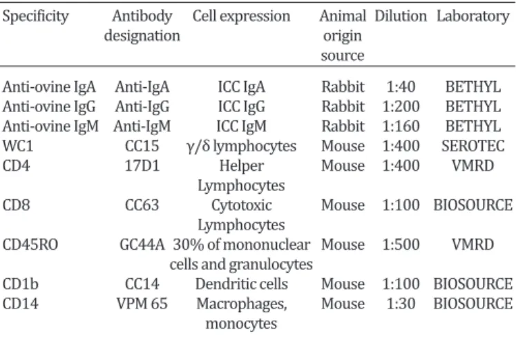
Table 1. Characteristics of the antibodies used in immunofluorescence and immunohistochemistry tests
Specificity Antibody Cell expression Animal Dilution Laboratory
designation origin
source
Anti-ovine IgA Anti-IgA ICC IgA Rabbit 1:40 BETHYL Anti-ovine IgG Anti-IgG ICC IgG Rabbit 1:200 BETHYL Anti-ovine IgM Anti-IgM ICC IgM Rabbit 1:160 BETHYL WC1 CC15 γ/δ lymphocytes Mouse 1:400 SEROTEC CD4 17D1 Helper Mouse 1:400 VMRD
Lymphocytes
CD8 CC63 Cytotoxic Mouse 1:100 BIOSOURCE
Lymphocytes
CD45RO GC44A 30% of mononuclear Mouse 1:500 VMRD cells and granulocytes
CD1b CC14 Dendritic cells Mouse 1:100 BIOSOURCE CD14 VPM 65 Macrophages, Mouse 1:30 BIOSOURCE
monocytes
Table 2. Average of IgA-, IgG-, and IgM- positive cells, CD4, CD8, CD45RO, WC1, CD14 and CD1b in the prepuce and the
pelvic urethra
Prepuce Pelvic urethra
IgA 295.5±67.8 137.3±201
IgG 135.5±65.0 a 0±0
IgM 140.3±45.2 a 8.3±32.2
CD4 850.5±128.5 a 396.1±269
CD8 595.1±117.8 539.3±278
CD45RO 1026±141.0 a 450.8±259
WC1 368.8±152.0 a 20.9±22.8
CD14 450±164.7 a 181.9±55.6
CD1b 38±18.5 a 0±0
a Significance of the comparison between the prepuce and the pelvic ure
-thra p<0.05. CD8, g/d, WC1 lymphocytes, CD45RO cells, macrophages
(CD14), and dendritic cells (CD1b)
sec-Fig.1. Number of IgA-, IgG-, and IgM- positive cells, CD4, CD8, CD45RO, WC1, CD14, and CD1b in the prepuce and the pelvic urethra.
Positive IgA-, IgG-, and IgM-containing cells were seen in the mucosal connective tissue in both organs; IgA-contai-ning cells appeared in groups near the epithelia basement membrane in both mucosae (Fig.2a). In contrast, positive IgG- and IgM-containing cells were located distantly from the epithelium, deeper in the connective tissue (Fig.2b).
Significant differences were found in the total number of CD4, CD45RO, and TCR γδ (WC1) lymphocytes, and in macrophages and dendritic cells between the mucosae of both organs. The mean values found in the prepuce were greater in all cases (p<0.05). No CD1b dendritic cells were found in the pelvic urethra, and they were scarce in the prepuce. No significant differences were found in CD8 cell populations in these tissues.
The comparative results of the count and distribution of intraepithelial cells and of those present in the mucosal con-nective tissues for the different cell types evaluated in the pre-puce and the urethra are summarized in Table 3 and Figure 3.
Fig.3. Number and distribution of IgA-, IgG-, and IgM- positive cells, CD4, CD8, CD45RO, WC1, CD14, and CD1b in the epithe-lium and the submucosa of the prepuce and pelvic urethra.
Table 3. Distribution of CD4, CD8, CD45RO, WC1, CD14, and CD1b in the epithelium and the submucosa of the prepuce
and pelvic urethra
Prepuce Pelvic urethra Intraepithelial Submucosal Intraepithelial Submucosal
CD4 90.9±52.3 759.6±100.2a 49.6±27 346.5±135.5a
CD8 298±74.5 297.3±77.4 228±66.4 311.7±142.2 CD45RO 64,8±34.3 961,6±152.3a 189±72.4 261.6±101.1
WC1 68±25.9 300.8±133.4a 2.8±4 18.1±22.3a
CD14 3.7±1.3 447.2±163.6a 0±0 181.9±80.1a
CD1b 26.2±15.4 12.1±5.4 0±0 0±0
a Intraepithelial and submucosal significance in the prepuce and the pelvic
urethra p<0.05.
The results of the differential count of intraepithelial cells and connective tissue cells show that there were signi-ficant differences in the distribution of CD4, TCR γδ (WC1) lymphocytes and macrophages (CD14) between the pre-puce and the urethra. TCR γδ (WC1) lymphocytes in both
tissues were located intraepithelially or in the connective tissue near the basement membrane of the epithelium. In the urethra, no CD14 positive labeling was found in the epithelium, while in the prepuce, CD14 were scarce in the epithelium and in the connective tissue near the basement
Fig.4. Aspects of the distribution of CD4 and CD8 lymphocytes in the pelvic urethra. (a) Labeling of CD4 lymphocytes. IP, obj.10x. (b) La-beling of CD8 lymphocytes in the urethral mucosa. LaLa-beling is more abundant in the proximity and in the epithelium itself. IP, obj.10x.
Fig.5. Aspects of dendritic cells (CD1b) and CD4, CD8, and WC1 lymphocytes labeling in the prepuce. (a) Dendritic cell (CD1b) near a papilla of the preputial epithelium. IP, obj.40x. (b) CD4 lymphocytes in the preputial mucosa. Some of them are located in the basement membrane of the epithelium. IP, obj.40x.
(c) TCR γδ (WC1) lymphocytes in the preputial mu
membrane and, in contrast, they were quite abundant in the deep connective tissue and in the proximity of mucosal papillae blood vessels. CD14 positive labeling was obser-ved in the endothelia of preputial vessels.
CD8 lymphocytes had a similar distribution in both mu-cosae; intraepithelial CD8 were located more superficially than intraepithelial CD4 (Fig.4). CD45RO lymphocyte dis-tribution in the prepuce showed a clear predominance of intraepithelial cells compared to those present in the con-nective tissue, in contrast, no differences were found in the distribution of these cells in the urethral mucosa. The few CD1b cells observed in the prepuce were found near basal epithelial cells (Fig.5a) or associated to lymphoid nodes, occasionally present in this part of the mucosa. Pictures of CD4, WC1, and CD8 lymphocyte labeling are presented (Fig.5b-e).
For each type of cell evaluated, the relationship betwe-en intraepithelially located cells and cells located in the connective tissue of the mucosa of both organs studied was established. CD4 lymphocytes were found in an average in-traepithelial/connective tissue ratio of 10/1 in the prepuce and 7/1 in the urethra, while CD8 lymphocytes had a 1/1 distribution in both mucosae. WC1 lymphocytes were pre-sent in a 5/1 ratio in both mucosae. The most notable and extreme observation occurred with CD45RO which had an intraepithelial/connective tissue ratio of 19/1 in the pre-puce and a 1/1 ratio in the pelvic urethra. CD14 and CD1b markers did not react in the urethral epithelium.
DISCUSSION
A greater number of positive IgA-containing cells than of IgM and IgG was observed in the prepuce; in contrast, Foster et al. (1988b) reported higher percentages of IgG--positive cells than of IgA- and IgMIgG--positive cells in nor-mal rams free of Brucella ovis and without testicular al-terations. This remarkable difference in the same species may be explained by the animals’ condition and characte-ristics, by the animals’ age, by the presence of pathogens or previous or little apparent inflammatory processes, or may be due to variations in the drawing of samples. The preputial mucosa is structurally more similar to the skin than to a mucosa. However, the presence of IgG-containing cells in the skin may only be expected in dermatitis con-ditions, since the prevailing lymphocytes in the skin are type T lymphocytes (McKeever & Reid 1987, Yirrell et al. 1989, Bos & Kapsenberg 1993). Apparently, the lymphoid tissue and the epithelium itself do not show the same dis-tribution in all preputial portions, and no lymphoid tissue organized in nodules is found in the fornix, as occurs in the mucocutaneous junction (Acosta-Dibarrat et al. 2003); this same variability has been reported in bulls (Flower et al. 1982).
In the urethra, only IgA-containing cells and IgM were found, as has been described in humans (Anderson & Pun-dey 1999, Quayle et al. 1994), however, no positive IgA-con-taining cells are found in the mouse penile urethra.
Regarding the WC1 marker, a part of TCR γδ lym -phocytes express it on their surface; these are relative-ly abundant cells in ruminant blood, especialrelative-ly in young
animals, and may be involved in the rapid release of Th1 cytokines (Pollock & Welsh 2002). TCR γδ constitute a first line of response of T cells in the recognition of cells altered by infection, inflammation, neoplastic changes, intoxica -tion, thermal shock or radiations; therefore, their increase is associated with the occurrence of various inflammatory or infectious diseases (Baldwin et al. 2000). These cells produce keratinocyte growth factor and may be involved in epithelial repair processes (Van der Broek et al. 2005). The prepuce was the site with the greatest expression of WC1 lymphocytes. Studies conducted in ram skin (Van der Broek et al. 2005) using the same markers demonstrated amounts of WC1 similar to those observed in this case. In the normal skin, WC1 cells were more abundant than CD4 and CD8 (Van der Broek et al. 2005), conversely, the num-ber of CD4 and CD8 was higher in the prepuce.
The function of the leukocyte common antigen, CD45, is to modulate the T lymphocyte activation signal transduc-tion. Its CD45RO isoform occurs in CD4 and CD8 T cells and in the memory subclasses (Bembridge et al. 1993). This iso-form is also expressed in monocytes, granulocytes, and in mononuclear cells presenting WC1, TCRγδ, CD4- and CD8-, but it is not expressed by B lymphocytes (Bembridge et al. 1995). This isoform is particularly abundant in the lym-phocytes that reside in the mucosa; in the intestinal lami-na propria they may represent 93%, while they represent 30% of circulating blood lymphocytes (Stephen & Hiroshi 1993). This marker prevailed in both mucosae studied.
A greater amount of WC1 lymphocytes and macro-phages was observed in the preputial mucosa connecti-ve tissue than in that of the urethra, which explains that CD45RO, which marks lymphocytes and macrophages, was also present in a greater relative and absolute number in the prepuce than in the urethra.
The intraepithelial lymphocytes demonstrated in the prepuce and the urethra were mainly CD8 and WC1. The presence of CD4 and CD8 lymphocytes has also been des-cribed in the urethral mucosa in mice (Quayle et al. 1994) and similar observations have been made in human ure-thra, which is consistent with neutrophil and T and B cell infiltration (Anderson & Pudney 1999). As seen in the digestive tract mucosa (Kelsall & Strober 1999), intrae-pithelial lymphocytes in the urethra and the prepuce are mainly CD8. A relatively high number of intraepithelial lymphocytes were WC1 (Tγδ lymphocytes), especially in the prepuce. Intraepithelial lymphocytes are likely to re-cognize a limited variety of microbial and cellular antigens and may be involved in epithelial monitoring and repair processes, rather than in immune response processes (Boismenu 1994).
abs-cence of this marker in the urethral mucosa is an observa-tion consistent with what has been reported for the urethra in humans and mice (Quayle et al. 1994).
The CD14 marker is usually found in the surface of macrophages capable of recognizing LPS of Gram bacte-ria, either alone or associated with proteins (Wright et al. 1990). The observation of endothelial labeling with CD14 in the prepuce may be due to the fact that LPS stimulates its expression in the endothelia, which increases the ac-tivity of endothelial cells in the production of TNF-α and IL-6 (Dai et al. 2002). Quayle et al. (1994), report the pre-sence of numerous macrophages in the urethral mucosa of mouse, similarly to what has been observed in this study for ovines.
CONCLUSION
The presence of antigen-presenting cells, macrophages, and dendritic cells, as well as of CD4, CD8 TCR γδ (WC1) lym -phocytes, IgM-containing cells, IgG, and IgA, and CD45RO cells indicates that the preputial and urethral mucosae of rams have the cell components necessary to act as inducti-ve sites and immune response sites of the mucosae.
Acknowledgements.- This study was supported by the project PAPIIT IN 206101 UNAM and F-PROMEP-38/Rev-03. We are grateful to Aman-da de Souza Aman-da Motta of the Microbiology, Immunology and Parasitology Department, Institute of Basic Health Sciences, Federal University of Rio Grande do Sul for the translation of the abstract to the Portuguese.
REFERENCES
Acosta-Dibarrat J.P., Garrido F.G., Díaz L.A.C. & Tórtora P.J.L. 2003. Análisis histológico y de distribución de linfocitos CD4 y CD8, en linfonódulos asociados a la mucosa prepucial del ovino. Memorias del XII Congreso Nacional Producción Ovina, Tulancingo, Hidalgo, México. (Resumen) Acosta-Dibarrat J., Díaz-Aparicio E., Arellano-Reynoso B.,
Tenorio-Gutié-rrez V. & Tórtora-Pérez J. 2006. Experimental induction of epididymitis in sheep, by intra-urethral inoculation of Actinobacillus seminis: a bac-teriological, serological, and histopathological study. Téc. Pecu. Mex. 44:257-267.
Acosta-Dibarrat J., Díaz-Aparicio E., Tenorio-Gutiérrez V., Suárez-Güemes F. & Tórtora-Pérez J. 2007. Determination of pathological changes in the reproductive tract, IgG, IgM and IgA antibodies in blood, seminal plasma and smegma of rams inoculated with Actinobacillus seminis. J. Anim. Vet. Advances 6:105-113.
Anderson D.J. & Pudney J. 1999. Human Male Genital Tract Immunity and Experimental Models, p.1411-1422. In: Ogra P.L., Mestecky J., Lamm M.E., Strober W., Bienenstock J. & McGhee, J.R. (Eds), Mucosal immunology. 2nd ed. Academic Press, San Diego.
Baldwin C.L., Sathiyaseelan T., Rocchi M. & McKeever D. 2000. Rapid changes occur in the percentage of circulating bovine WC1+ γδTh1 cells. Res. Vet. Sci. 69:176-180.
Bembridge G.P., MacHugh N.D., McKeever D., Awino E., Sopp P., Collins R.A., Gelder K.I. & Howard C.J. 1995. CD45RO expression on bovine T cells: relation to biological function. Immunology. 86:537-544.
Bembridge G.P., Parsons K.R., Sopp P., MacHugh N.D. & Howard C.J. 1993. Comparison of monoclonal antibodies with potential specificity for re -stricted isoforms of the leukocyte common antigen (CD45R). Vet. Immu-nol. Immunopathol. 39:129-136.
Boismenu R. & Havran W.L. 1994. Modulation of epithelial cell growth by intraepithelial gamma delta T cell. Science 266:1253-1255.
Bos J.D. & Kapsenberg M.L. 1993. The skin immune system: progress in cutaneous biology. Immunol. Today 14:75-78.
Campero C.M., Ladds P.W., Hoffmann D. & De’ath G. 1989. Immunoglobu-lin containing cells in normal and inflamed accessory sex glands of bull. Aust. Vet. J. 66:137-140.
Campero C.M., Hirst R.G., Ladds P.W., Vaughan J.A., Emery D.L. & Watson D.L. 1990. Measurement of antibody in serum and genital fluids of bulls by ELISA after vaccination and challenge with Tritrichomonas foetus.
Aust. Vet. J. 67:175-178.
Corbeil L.B. & BonDurant R.H. 2001. Immunity to bovine reproductive in-fection. Vet. Clin. North. Am. Food Anim. Pract. 17:567-583.
Dai L., Gong J., Luo Y. & Liu C. 2002. Expression of CD (14) protein in liver sinusoidal endothelial cells during endotoxemia. Zhonghua Gan Zang Bing Za Zhi. 10:93-95. (Abstract)
Dutia B.M. & Hopkins J. 1991. Analysis of the CD1 cluster in sheep. Vet. Immunol. Immunopathol. 27:189-194.
Flower P.J., Ladds P.W., Thomas A.D. & Watson D.L. 1982. An immunopath-ologic study of de Bovine Prepuce. Vet. Pathol. 20:189-202.
Foster R.A., Ladds P.W., Husband A.J. & Hoffmann D. 1988a. Immunoglobu-lins and immunoglobulin-containing cells in the reproductive tract of rams naturally infected with Brucella ovis. Aust. Vet. J. 65:37-40. Foster R.A., Ladds P.W., Hoffmann D. & Husband A.J. 1988b.
Immunoglobu-lins and immunoglobulin-containing cells in the reproductive tract of normal rams. Aust. Vet. J. 65:16-20.
Jansen B.C. 1983. The epidemiology of bacterial infection of the genitalia in rams. Onderstepoort J. Vet. Res. 50:275-282.
Kelsall B. & Strober W. 1999. Gut-Associated Lymphoid Tissue, p.293-317. In: Ogra P.L., Mestecky J., Lamm M.E., Strober W., Bienenstock J. & McGhee J.R. (Eds), Mucosal immunology. 2nd ed. Academic Press, San
Diego.
McKeever D.J. & Reid H.W. 1987. The response of the supramammary lymph node of the sheep to secondary infection with orf virus. Vet. Microbiol. 14:3-13.
Moldoveanu Z., Huang W.Q., Kulhavy R., Plate M.S. & Mestecky J. 2005. Human Male Genital Tract Secretions: Both Mucosal and Systemic Im-mune Compartments Contribute to the Humoral Immunity. J. Immunol. 175:4127-4136.
Plant J.W., Eamens G.J. & Seaman J.T. 1986. Serological, bacteriological and pathological changes in rams following different routes of exposure to
Brucella ovis. Aust. Vet. J. 63:409-412.
Pollock J.M. & Welsh M.D. 2002. The WC1+ δγ T cell population in cattle: a
possible role in resistance to intracellular infection. Vet. Immunol. Im-munopathol. 89:105-114.
Porcelli S.A., Segelke B.W., Sugita M., Wilson I.A. & Brenner M.B. 1998. The CD1 family of lipid antigen-presenting molecules. Immunol. Today 19:362-368.
Quayle A.J., Pudney J., Muñoz D.E. & Anderson D.J. 1994. Characteriza-tion of T lymphocytes and antigen-presenting cells in the murine male urethra. Biol. Reprod. 51:809-820.
Rhind S.M. 2001. CD1-The pathology Perspective. Vet. Pathol. 38:611-619. Stephen P.J. & Hiroshi K. 1999. Gastrointestinal Lamina Propria T Cells,
p.381-393. In: Ogra P.L., Mestecky J., Lamm, M.E. Strober W. Bienenstock, J. & McGhee J.R. (Eds), Mucosal Immunology. 2nd ed. Academic Press, San
Diego.
Van der Broek A.H.M., Huntley J.F., MacKellar A., Machell J., Taylor M.A. & Miller H.R.P. 2005. Characterization of lesional infiltrates of dendritic cells and T cell subtypes during primary infestation of sheep whit Pso-roptes ovis, the sheep scab mite. Vet. Immunol. Immunophatol. 105:141-150.
Yirrell D.L., Reid H.W., Norval M. & Howie S.E.M. 1989. Immune response of lambs to experimental infection with orf virus. Vet. Immunol. Immu-nopathol. 22:321-332.