www.nature.com/scientificreports
Depth-specific fluctuations of gene
expression and protein abundance
modulate the photophysiology in
the seagrass Posidonia oceanica
Gabriele Procaccini
1, Miriam Ruocco
1, Lázaro Marín-Guirao
1, Emanuela Dattolo
1,
Christophe Brunet
1, Daniela D’Esposito
1, Chiara Lauritano
1, Silvia Mazzuca
2,
Ilia Anna Serra
2, Letizia Bernardo
2, Amalia Piro
2, Sven Beer
3, Mats Björk
4, Martin Gullström
4,
Pimchanok Buapet
4,5, Lina M. Rasmusson
4, Paulo Felisberto
6, Sylvie Gobert
7,
John W. Runcie
8, João Silva
9, Irene Olivé
9, Monya M. Costa
9, Isabel Barrote
9& Rui Santos
9Here we present the results of a multiple organizational level analysis conceived to identify acclimative/ adaptive strategies exhibited by the seagrass Posidonia oceanica to the daily fluctuations in the light environment, at contrasting depths. We assessed changes in photophysiological parameters, leaf respiration, pigments, and protein and mRNA expression levels. The results show that the diel oscillations of P. oceanica photophysiological and respiratory responses were related to transcripts and proteins expression of the genes involved in those processes and that there was a response asynchrony between shallow and deep plants probably caused by the strong differences in the light environment. The photochemical pathway of energy use was more effective in shallow plants due to higher light availability, but these plants needed more investment in photoprotection and photorepair, requiring higher translation and protein synthesis than deep plants. The genetic differentiation between deep and shallow stands suggests the existence of locally adapted genotypes to contrasting light environments. The depth-specific diel rhythms of photosynthetic and respiratory processes, from molecular to physiological levels, must be considered in the management and conservation of these key coastal ecosystems.
Seagrasses are marine angiosperms that have evolved to complete their whole life cycle submerged. They repre-sent one of the most productive components of benthic coastal ecosystems, and in addition to being exposed to daily fluctuations in irradiance have adapted to large spatial (i.e. bathymetric) variations in both irradiance and spectral quality. In the Mediterranean Sea, the endemic seagrass Posidonia oceanica colonizes extensive portions of sea bottom along most of the coastline1. The extensive meadows formed by this species are among the most val-uable marine ecosystems, fulfilling important ecosystem services from carbon sequestration to coastal protection and maintenance of fisheries2–4. Meadow can spread from the surface down to 45 meters depth, where distinct ramets (but possibly the same genets5,6) are exposed to a wide range of light conditions and different photoper-iods, imposing specific adaptive responses. Such extensive P. oceanica meadows offer a valuable opportunity to explore how depth affects light responses and diel rhythms in seagrasses.
The amplitude of physiological changes of the species associated with depth-related variations in light is sim-ilar to the one shown by the congeneric P. sinuosa7 and can be considered low in relation to other seagrasses8,9. These changes include modifications in the plants’ photosynthetic and respiratory rates and in the ability of leaves to harvest light (e.g. refs 10,11) and involve long-lasting adjustments of key metabolic processes including carbon metabolism, stress defense and proteolysis. As a response to low-light conditions, regulatory changes of important 1Stazione Zoologica Anton Dohrn, Naples, Italy. 2Universitá della Calabria, Rende, Italy. 3Tel Aviv University, Tel Aviv,
Israel. 4Stockholm University, Stockholm, Sweden. 5Prince of Songkla University, Songkhla, Thailand. 6LARSyS,
University of Algarve, Faro, Portugal. 7MARE Centre, University de Liege, Liege, Belgium. 8University of Sydney,
Sydney, Australia. 9CCMAR, University of Algarve, Faro, Portugal. Correspondence and requests for materials should
be addressed to G.P. (email: [email protected]) Received: 14 September 2016
Accepted: 16 January 2017 Published: 17 February 2017
OPEN
functional groups of proteins have been reported, including down-regulation of the Ribulose-1,5-Bisphosphate Carboxylase/Oxygenase (RuBisCO) large subunit and modification of the PSI/PSII ratio12. Additionally, diver-gence in the level of expression of photoacclimation- and photoprotection-related genes (i.e. RuBisCO, ferre-doxin, chlorophyll a/b binding proteins, zeaxanthin epoxidase), and antioxidant enzymes, has recently been observed in plants from contrasting depths13,14. High-light adaptation/acclimation in shallow stands include a decrease in the antenna size, and an enhancement of photoprotective mechanisms (i.e. the xanthophyll cycle), as well as an increase in the number of reaction centers14. P. oceanica did not show signs of stress response in deeper water14, suggesting that the plant is well adapted to low-light environments.
Marine macrophytes show circadian rhythms, since much of their biochemistry, physiology, and behavior is temporally organized with respect to the environmental oscillation of day and night15,16. Circadian rhythms control many functions in plants such as development and growth17. The correct matching of the endogenous clock with the day-night cycle directly enhances photosynthesis, increasing productivity and conferring fitness18.
Seagrass responses to daily changes in the light regime have been studied almost exclusively at the photophys-iological level19–26. According to daily variation in irradiance levels, seagrasses, like their terrestrial counterparts, maintain a permanent and dynamic trade-off between photosynthetic efficiency and photoprotection through so-called dynamic photoinhibition27. It is not known whether and how this reversible regulation of photosynthe-sis is driven by internal circadian cycles, allowing the species to anticipate upcoming light patterns (i.e. seasonally or daily), or is regulated by environmental cues. Given the important role temporal rhythms can play in the functioning of seagrass meadows, it is of great importance to explore the basis of their genetic, biochemical and physiological attributes.
Here we present the results of a multidisciplinary study conceived to understand the acclimative and adaptive strategies of P. oceanica photophysiology to the daily fluctuations in the light environment, at contrasting depths. The study was performed in the P. oceanica meadow just outside the Marine Research Station of STARESO, Bay of Calvì (Corsica), and plants were collected during a daily cycle at − 5 m and − 20 m (cf. ref. 28). Irradiance, photophysiological parameters derived from chlorophyll a fluorescence measures, leaf respiratory rates, content of photoprotective xanthophyll pigments, leaf total protein and mRNA levels of selected genes and their relative expression, were evaluated.
The spatial genetic structure along the depth gradient was also assessed. Together with a companion study (in preparation), performed at the same time, which focuses on how plant photosynthetic production is conveyed to higher organizational levels of community and ecosystem production, we provide the first comprehensive, multilevel approach of the functional responses of a seagrass ecosystem to the daily irradiance cycle at different depths. Our results represent an important source of information on the adaptive strategies used by this species, in response to variations in light, and provide a better understanding of the overall functioning of P. oceanica meadows.
Results
Light and temperature.
During the study period, dawn and dusk (as defined by PAR < 1 μ mol m−2 s−1) at the shallow (− 5 m) portion of the STARESO P. oceanica meadow occurred at approximately 07:25 and 19:00 GMT + 2, respectively. At − 20 m the daily photoperiod was shortened by 25–30 min. Maximum PAR values at both depths were recorded around 13:00 with averaged values of 430 and 92 μ mol m−2 s−1 at 5 and 20 m depth, respec-tively (Fig. 1A). During the first sampling after sunrise (i.e. at 09:00), available light levels were 109 ± 32 μ mol m−2 s−1 at 5 m and 24 ± 9 μ mol m−2 s−1 at 20 m depth. The integrated daily photosynthetic photon flux density at − 20 m was on average 24% of the corresponding value at − 5 m. Seawater temperature was similar at the two depths, with daily averaged values of 20.4 ± 0.1 °C and 20.0 ± 0.1 °C for shallow and deep meadow portions, respectively, indicating that the water column was not thermally stratified during the study period. At 5 m depth, the temper-ature ranged from a minimum of 20.0 °C before sunrise to 20.9 °C at 15:00. Maximum and minimum seawater temperature at 20 m depth occurred at the same hours, with values between 19.7 and 20.5 °C.Photosynthesis and photoprotection.
The photochemical efficiency of PSII (Δ F/Fm’) showed a similar daily pattern at the two depths, with similar values at every sampling time (Fig. 1B; Supplementary Table S1). Maximum Δ F/Fm’ values were found before sunrise and progressively decreased until midday, when the lowest values were reached. Later in the day, as irradiance started to decrease, photochemical efficiency progressively increased to equally maximum levels around dusk. The Δ F/Fm’ reduction from sunrise to high-light hours (12:00 to 15:00) was about 80% both at 5 and 20 m depth. The quantum yield of regulated non-photochemical quenching (NPQ) showed an opposite pattern to that observed for Δ F/Fm’, with no significant differences between the two depths (Fig. 1B). The lowest values of NPQ were measured before the sunrise and the highest around midday, with similar values at 5 m (0.403 ± 0.05) and 20 m (0.471 ± 0.01), which were twice the minimum ones.As a consequence of the similar Δ F/Fm’ pattern between plants at the two depths and the higher light avail-ability in the shallow meadow portion, electron transport rates (ETRs) of shallow plants during the light hours were significantly higher than deep plants (Fig. 1C). Their daily integrated photosynthetic electron transport rate was on average 5-fold higher than that at 20 m depth. The ETRs of deeper plants held their maximum values for longer (from 09:00 to 15:00) than shallow plants (from 09:00 to 12:00) (Fig. 1C). This pattern of delayed response of deeper plants was also observed in the expression of genes related to photosynthesis and photoprotection (see below).
No significant differences were found between depths or among times of the day in the leaf concentrations of both chlorophylls and carotenoids (Table 1). The ratio between chlorophyll a and b was around 2. Violaxanthin (V) and zeaxanthin (Z) concentrations were one order of magnitude higher than anteraxanthin (A). On the other hand, the de-epoxidation index was significantly higher at − 5 m than at − 20 m but there were no significant dif-ferences between morning and midday.
www.nature.com/scientificreports/
Respiration.
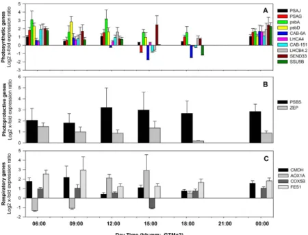
The respiratory activity of P. oceanica leaves was not significantly different between the shallow and deep plants, except at 12:00, where it was higher in the deep site (Fig. 2A). Leaf respiration showed similar diel rhythms at the two depths, with an overall increase at high-light hours and reduced rates at lower irradiances and during the night (Fig. 2A). The response of leaf respiration to light of shallow plants was delayed in respect toFigure 1. Daily cycle of PAR irradiance (A), quantum yield of PSII (circles in B), non-photochemical
quenching downregulation (triangles in (B)) and electron transport rates (C) of shallow (white symbols) and deep (black symbols) P. oceanica plants; and daily variation of mRNA expression of photosynthesis-related genes at 5 m (D) and 20 m (E) depth. Relative expression levels at given time points are calculated over their daily average expression. Dashed lines represent the average expression level of all analyzed genes. Asterisks indicate significant differences among shallow and deep plants at a given sampling time. **p < 0.01; ***p < 0.001. −5 m −20 m 06:00 (n = 5) 12:00 (n = 6) 06:00 (n = 9) 12:00 (n = 9) Chlorophyll a (Chla) 30.3 ± 8.9 46.5 ± 9.5 54.6 ± 5.2 53.3 ± 8.5 Chlorophyll b (Chlb) 16.6 ± 5.6 22.8 ± 4.7 28.6 ± 2.9 26.0 ± 4.3 Total Chlorophyll (ChlT) 46.9 ± 14.4 69.3 ± 14.2 83.2 ± 7.9 79.3 ± 12.8 Chla/Chlb 1.963 ± 0.146 2.087 ± 0.085 1.917 ± 0.054 2.053 ± 0.053 Violaxanthin (V) 35.2 ± 9.9 43.8 ± 3.0 43.3 ± 4.6 45.5 ± 3.0 Anteraxanthin (A) 2.226 ± 0.808 3.963 ± 1.111 2.944 ± 0.866 3.129 ± 0.390 Zeaxanthin (Z) 80.5 ± 28.9 86.7 ± 8.9 34.4 ± 8.7 76.5 ± 21.8 (V + A + Z)/ChlT 117.9 ± 39.1 134.4 ± 6.9 80.6 ± 8.2 125.1 ± 23.7 (A + Z)/(V + A + Z)* 0.686 ± 0.043 0.669 ± 0.076 0.427 ± 0.067 0.546 ± 0.073
Table 1. Posidonia oceanica pigment content at 5 m and 20 m depth at 06:00 and 12:00. Values represent means ± standard error. *Indicates significant differences between depths. Chlorophylls are expressed per square
deeper plants. The leaf respiration of deeper plants peaked from 09:00 to 12:00 whereas in shallow plants the peak was from 12:00 to 15:00 (Fig. 2A). The lowest leaf respiration for both − 5 and − 20 m plants occurred before sun-rise, at 06:00. The shallow plants showed lower respiratory rates than the deep plants during morning samplings (09:00 and 12:00), with significant differences only at 12:00 (Fig. 2A). Leaf respiration was positively correlated with PAR irradiance levels (n = 6, r = 0.85, p = 0.034; Supplementary Table S2) only in deep plants.
Gene expression.
Daily mRNA variation in shallow and deep plants. P. oceanica plants exhibited a dynamicdaily regulation of mRNA levels of selected genes of interest (GOIs). The averaged expression pattern for all ana-lyzed genes indicated a similar daily trend in plants from the shallow and deep meadow portions, but with a gen-eral delay in the deepest stand (Fig. 3A). In shallow plants, GOIs mRNA accumulation started soon after the light onset, peaked around noon (PAR = ~300 μ mol m−2 s−1), and then progressively decreased until dusk, with low values during the night (Fig. 3A). In plants from − 20 m, transcriptional activation of GOIs was delayed by about 3 hours. Activation started at 09:00 and attained maximum expression values at 12:00 and 15:00 under highest PAR values. At 18:30 GOIs were still up-regulated with respect to their daily average expression, in contrast to that observed in shallower plants that were down-regulated at this time (Fig. 3A).
Hierarchical clustering (Fig. 3B) identified two main clusters corresponding to higher and lower expression levels, and further highlighted the time shift between shallow and deep plants. For example, the − 20 m plants at 09:00 (09:00_20 m) belong to the low expression group, while 09:00_5 m falls within the high-expression group and in particular branches with 12:00_20 m. Similarly, plants of 18:30_20 m still cluster with the high-expression group, while 18:30_5 m is included in the low-expression group and clusters with 06:00_20 m.
Most of the photosynthesis-related genes analyzed showed a synchronized diel pattern of expression at each depth (Fig. 1D,E). The expression of some genes was delayed in relation to others, such as LHCA4 and SSU5B at 06:00 and SSU5B, LHCA4 and SEND33 at 15:00, at − 5 m. PSAG at − 20 m was faster to react at 09:00 and at 18:30 (together with SEND33) than the other photosynthetic genes (Fig. 1D). PSAJ and psbA, encoding for subunits of PSI and PSII, respectively, and LHCA4 showed significant correlations with chlorophyll a fluorescence parame-ters. Negative correlations were observed with the photochemical efficiency of plants (Δ F/Fm’; n = 12, p < 0.05) and positive with the non-photochemical quenching (NPQ; n = 12; p < 0.05) and ETRs (n = 12, p < 0.001 and n = 12, p < 0.001, respectively).
A delayed response of photoprotective genes PSBS and ZEP (Fig. 1D,E) in deeper plants in relation to shallow plants was also observed, as in the case of the photosynthetic genes referred above. PSBS expression was positively correlated with PAR (n = 12, r = 0.77, p = 0.003; Supplementary Table S2) and with the dissipative process of non-photochemical quenching NPQ (n = 12, r = 0.85, p < 0.001), at both depths.
The average expression of respiration-related genes showed a similar diel cycle at the two depths, increasing with light to maximum values at 12:00 to 15:00 hours (Fig. 2B,C) and decreasing to lowest levels during the night. AOX1A showed the highest diel variation, especially at 5 m depth. At − 20 m, its expression trend deviated from that of the other respiratory genes, being strongly up-regulated in the early morning (06:00 and 09:00),
Figure 2. Daily cycle of leaf respiration (A) and of mRNA expression of respiration-related genes of shallow
(B) and deep (C) P. oceanica plants. Relative expression levels at given time points are calculated over their daily average expression. Dashed lines represent the average expression level of all analyzed genes. Asterisks indicate significant differences among shallow and deep plants at a given sampling time. *p < 0.05.
www.nature.com/scientificreports/
and down-regulated in the following hours, when CMDH, COX5B and FES1started to be expressed. In shallow plants, the expression peak of this gene occurred later (12:00–15:00) in line with the subsequent rise in respiration (Fig. 2A–C). The level of expression of the AOX1A gene of shallow plants was significantly correlated with light (n = 6, r = 0.89, p = 0.018; Supplementary Table S2) in contrast with the other genes. In deep plants, the genes
CMDH and FES1 were positively correlated with light (n = 6, r = 0.835, p = 0.042 and n = 6, r = 0.83, p = 0.041,
respectively) similarly to the leaf respiratory rates, as described above.
Looking at differences in expression of GOIs within each sampling time, plants from − 5 m showed a stronger induction in respect to − 20 m plants (Fig. 4; Supplementary Table S3). Only after midday and at dusk (15:00 and 18:30) some genes were more expressed in the deep than in the shallow meadow (e.g. CAB-6A and
SSU5B). Transcripts for PSII subunits such as psbA, psbD and PSBS were among those displaying the
strong-est up-regulation in shallow plants, especially during light hours (Fig. 4; Supplementary Table S3). Chlorophyll
a/b binding proteins were also generally more expressed in shallow plants, although significant results were
only found for those associated with PSII (LHC II) (Fig. 4; Supplementary Table S3). The ferredoxin-1 encod-ing gene SEND33 showed large differences in expression levels between plants from the two depths (Fig. 4; Supplementary Table S3). Respiration-related genes, CMDH, COX5B and FES1, were over-expressed in
Figure 3. Average daily expression patterns of all genes of interest in shallow (white circles) and deep (black
circles) P. oceanica plants (A); and Heatmap (B) showing cross-correlation among experimental conditions and similarity of gene expression profiles of the sixteen selected genes analyzed by RT-qPCR. X-axis: columns display the cDNA from the six collection times at − 5 and − 20 m depth, clustered by similarities; y-axis: each row displays the expression level of a given gene in the corresponding cDNA, clustered by similarities across experimental conditions. Color key (white: highest expression strength; red: lowest expression strength).
− 5 m plants at almost all points of time (Fig. 4; Supplementary Table S3). AOX1A exhibited a different trend, being down-regulated at − 5 m in the early morning (06:00 and 09:00), although not significantly (Fig. 4; Supplementary Table S3).
Daily protein variation in shallow and deep plants. Almost 80 protein bands were detected in each gel by
image analysis, 15 of which showed significant variation during the light hours (Supplementary Table S4). Accession, predicted protein sequences and NCBI Blastp against non-redundant protein sequences are reported in Supplementary Table S5. Most of these were structural proteins and enzymes involved in key steps of the light-dependent reactions of photosynthesis (e.g. PSI and PSII subunits, chlorophyll a/b binding proteins, chlo-roplastic ATP synthase), carbon fixation (RuBisCO and RuBisCO activase), and glycolysis (phosphoglycerate kinase, glyceraldehyde 3-phosphate dehydrogenase).
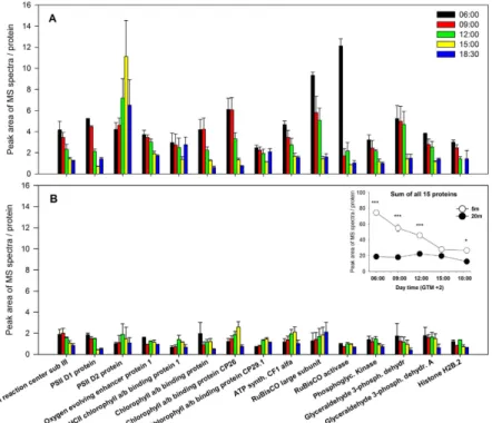
The overall daily pattern of identified proteins revealed strong differences among plants from the two depths (significant D × T interaction for 9 out of the 15 proteins analyzed, Supplementary Table S6). Shallow plants exhibited a significant and progressive reduction in protein abundance, with maximum accumulation before sunrise (06:00) and in early morning (09:00) and lower values during the afternoon and just after sunset (18:30) (Fig. 5). RuBisCO large subunit and RuBisCO activase displayed the highest concentrations at 06:00, but their levels underwent a huge decline just after sunrise. A group of proteins formed by glyceraldehyde 3-phosphate dehydrogenase proteins, Phophoglycerate kinase and oxygen-evolving enhancer protein 1 were very abundant until noon, significantly decreasing thereafter until the sunset. A different trend was observed for the photosys-tem II D2 protein, peaking at 15:00 with 5-fold significantly higher concentrations than morning samples (06:00 and 09:00). Chlorophyll a/b binding protein of LHCII type 1 and the chlorophyll a/b binding protein CP29.1 con-versely showed a significant reduction at 15:00, and similar concentrations for the rest of sampling times (Fig. 5). Deep plants presented lower leaf concentrations of all analyzed proteins than shallow plants and the patterns of variation throughout the day were not as consistent as in shallow plants (Fig. 5). Protein concentration was similar all along the day except for PSII D1 that exhibited significantly lower concentrations at 15:00 and 18:30 with respect to previous hours.
The PSII D2 protein was the only one showing a significant and positive correlation with PAR values (n = 10, r = 0.73, p = 0.015; Supplementary Table S2).
Genetic diversity and structure. Pair-wise comparison of FST estimates between the shallow and deep sites (FST = 0.169) indicated high genetic differentiation related to depth. The shallow site showed higher genotypic richness, percentage of polymorphic loci, total number of alleles and average number of alleles per locus, com-pared to the deep site (Supplementary Table S7). Seventeen private alleles were present at the shallow site, while only two were present at the deep site (Supplementary Table S7). Observed heterozygosity (Ho) was always higher than expected (Supplementary Table S7), with several loci (15 in shallow plants, 14 in deep plants) deviating sig-nificantly from the Hardy-Weinberg equilibrium (indicated with asterisk in Supplementary Table S8). Analyses of Molecular Variance (AMOVA) was performed partitioning the molecular variance into two levels (among depths and within depths), and almost 50% of the total variation was explained by the between-depths component (48%;
Figure 4. Relative quantification of photosynthesis (A), photoprotection (B) and respiratory-related (C)
genes, in shallow P. oceanica plants (− 5 m) at given time points, considering gene expression in deep plants (− 20 m) as control condition. Relative quantification was obtained using REST 200996. Significant results with corresponding P(H1) values are reported in Table S3.
www.nature.com/scientificreports/
Supplementary Table S9). Principal Coordinate Analysis (PCoA), with the first axis explaining 46.42% of the total variance, clearly separates the meadow into two clusters corresponding to the two depths (Fig. 6).
Discussion
Our results revealed for the first time not only that diel oscillation of P. oceanica photophysiological and respira-tory responses were related to transcriptional responses and protein expression of the genes involved in those processes, but also that there was a response asynchrony between shallow and deep plants, probably due to strong differences in the light environment.
The daily patterns of fluorescence-derived parameters indicate a dynamic regulation of the photosynthetic process in P. oceanica19,20. As light increases during the day, plants activate photoprotective mechanisms, decreas-ing photochemical efficiency. This photosynthetic downregulation was similar across depths, and recovered fully by nightfall, suggesting that plants did not experience accumulated photodamage at the light conditions experi-enced in this time of the year. This finding contrasts with results obtained in the seagrass Halophila stipulacea at similar light levels, where photochemical efficiency of shallow plants at 8 m depth did not recover by the end of the day, but showed a gradual increase overnight indicating the longer term activation of recovery processes21. Figure 5. Daily expression pattern of accumulation of 15 proteins of P. oceanica at 5 m (A) and 20 m (B) depth.
Inner panel represents the daily pattern of the sum of all proteins. Asterisks indicate significant differences among shallow and deep plants at a given sampling time. *p < 0.05; **p < 0.01; ***p < 0.001.
Figure 6. PCoA analysis based on the genetic distance matrix, for the two depths (−5 m and −20 m) of the STARESO P. oceanica meadow. The light and dark blue symbols correspond to genotyped individuals from
shallow and deep site, respectively. Percentages of variation explained by the first two axes are 46.42 and 19.51, respectively.
The NPQ induction, the decrease in photochemical efficiency and the upregulation of the photoprotective genes observed both in shallow and deep plants of P. oceanica during high-light hours suggest the triggering of energy dissipation mechanisms such as the xanthophyll cycle29. The de-epoxidation index of P. oceanica was significantly higher in shallow than in deep plants, indicating higher energy dissipation through the xanthophyll cycle. This corresponds to the up-regulation of photoprotective genes in shallow plants, as compared to deep ones, and reveals that P. oceanica has the potential needed for photoprotection in the highly transparent waters of Stareso. However, we did not detect differences in the de-epoxidation index between dawn and midday both in shallow and deep plants, due to high retention of zeaxanthin before sunrise. A possible explanation is the onset of a process called sustained dissipation, which refers to thermal dissipation that is not dependent on the trans-thylakoid Δ pH as for the xanthophyll cycle, and does not relax rapidly in darkness30,31. It involves the con-tinuous engagement of zeaxanthin and its dark retention and is the reason of decreased photosynthesis caused by high stress30–32. Despite this, NPQ increased during the day due to other quenching mechanisms such as those involving PsbS. The accumulated zeaxanthin may actually prompt NPQ induction allowing a faster response to high-light conditions32,33.
Interestingly, the mRNA of the photosystem II subunit S (PSBS) exhibited a daily oscillation highly and positively correlated with the process of non-photochemical quenching at both depths. This points towards the important role of PsbS in activating NPQ mechanisms in seagrasses, as described for terrestrial plants34–37. Photosynthesis is one of the most fundamental processes regulated by circadian clocks at both the metabolic and gene-expression levels38–40. Accordingly, mRNAs of most photosynthesis-related components, including cab genes encoding chlorophyll a/b binding proteins, and subunits of photosystems I and II, followed the daily pat-tern of photophysiological parameters and exhibited a clear diurnal rhythmicity in plants from both depths, with increasing levels slightly before or after sunrise, peak abundance around noon, and decreasing expression at dusk and night. In higher plants, the right timing and amplitude of expression patterns of temporally regulated genes are determined by a combination of endogenous rhythms and external signals, primarily light and tempera-ture, that control transcription41–43. Our data do not allow establishing the existence of endogenous rhythms, but the observed positive correlations with daily irradiance levels for some functional and structural photosynthetic genes indicate the important role of external signals.
In the chloroplasts of terrestrial plants and green algae, the expression of the psbA gene is regulated via a complex network including transcriptional control, regulation of mRNA stability, and the control at the level of translation44. The interdependence between irradiance and the expression of psbA that encodes the PSII D1 core protein observed here, reflects the functioning of the multistep PSII repair cycle process45–48. Increasing damage of the PSII D1 protein with enhanced irradiance forces plants to continuously repair the impaired PSII to main-tain an efficient photosynthetic process44,49. Our analysis revealed a different timing for PSII D1 protein and psbA mRNA accumulation during the day. In shallow plants, maximal abundance of PSII D1 protein was detected in the early morning, and then a progressive decline occured, reflecting its consumption in repairing PSII along the day. Differently, the peak of psbA mRNA accumulation was detected at midday, possibly to sustain the processes of light-induced degradation and de novo biosynthesis of the relative protein in high light, which occurr simulta-neously during PSII repair.
Due to the highly oxidative chemistry of water splitting mediated by the oxygen-evolving complex, the PSII extrinsic protein OEE1 (Oxygen-evolving enhancer protein 1) can be released from its binding site, thereby dis-organizing PSII and impairing the complex50,51. The OEE1 subunit of PSII was also found up-regulated in the shallow P. oceanica plants, likely promoting the protection and recovery of the PSII complex and enhancing the rate of electron transport under high-light conditions. In fact, the expression of the nuclear gene encoding OEE1 is required for high levels of photosynthetic oxygen evolution52 and OEE1 may be involved in the regulation of the PSII53.
In general, the higher transcription rate found in shallow plants of P. oceanica is reflected in the higher accu-mulation of proteins. This is supported, for example, by the significant and strong up-regulation of cab and photosystems-associated transcripts, which corresponds to a higher translation and turnover of the related pro-teins. Particularly, the differential rate in translation of proteins of the PSII complex seems to drive P. oceanica acclimation to the light environment and to be the main metabolic pathway for depth acclimation12–14. Higher translation and protein synthesis rates of shallow plants are also evident by the peaks of RuBisCO and RuBisCO activase proteins just before sunrise. In the presence of light and at an optimal temperature, RuBisCO activase promotes the release of the inhibitor ribulose 1,5-bisphosphate from the catalytic sites of RuBisCO, thus activat-ing the enzyme. This daily pattern is in agreement with the circadian oscillations of RuBisCO activase found in tomato54, apple55, and Arabidopsis56. The maximal abundance of RuBisCO protein in shallow plants was shifted forward about seven hours in respect to the corresponding transcripts (SSU5B), suggesting that translation of RuBisCO subunits occurs during the night so that by sunrise the plants are ready to take maximum advantage of available light for photosynthesis. During the day, RuBisCO level drastically decreased to a minimum at sunset. In the deep site there was a strong down-regulation of P. oceanica SSU5B transcripts during the night that explains the low level of RuBisCO protein at sunrise. The up-regulation of SSU5B during light hours is coherent with the increasing RuBisCO level along the day.
On what concerns the cellular energetic metabolism, it was observed that the level of Glyceraldehyde 3-phosphate dehydrogenase (GADPH), the enzyme that catalyzes the first step of the third stage of glycolysis, was highest at sunrise in both shallow and deep plants, to maintain the energy balance within the cells before photo-synthesis was triggered. The enzyme Phosphoglycerate kinase, which also participates in the maintenance of the organic and inorganic carbon balance, showed the same pattern of variation as GADPH. This enzyme catalyzes the transfer of a phosphate group from 1,3-bisphosphoglycerate in the first ATP-generating step of the glyco-lytic pathway. In the opposite direction, it also generates ADP and 1,1,3-bisphosphoglycerate in gluconeogenesis.
www.nature.com/scientificreports/
These processes modulate the respiratory responses, both at the gene expression and at oxygen consumption levels, which were highest from 12:00 to 15:00 both in shallow and deep plants.
The seagrass Zostera marina also revealed varying respiration rates on a diel basis, even though the peaking times of respiration differed from that of P. oceanica (Rasmusson pers. comm.). The substantial change of the respiratory activity over the day in both seagrass species supports the hypothesis that respiration should not be considered constant when assessing and modeling carbon and oxygen fluxes for vegetated marine habitats. A similar respiration rate between plants at different depths has also been shown for other seagrass species. In
Thalassia hemprichii and in Zostera marina respiration was not significantly different at two different depths,
even though the photosynthetic rate was higher in the shallow plants57,58. In our study, although the diel trend in respiration was comparable among plants from the two depths, a phase shift was present, with shallow plants increasing their leaf respiratory activity later than deep plants. While deep P. oceanica plants presented a peak in respiration around noon, shallow plants had their highest respiratory activity from 15:00 to 18:30. At the two depths, the high photosynthetic production of carbohydrates in high-light hours, could induce an elevated level of respiration, since both processes are tightly interdependent59. At those hours, up-regulation of malate dehy-drogenase (CMDH), an important enzyme of the tricarboxylic acid cycle60 and COX5B and FES1, regulating the cytochrome electron transport61,62, was observed, indicating that the respiratory machinery was working to its full extent. In the shallow plants the up-regulation of AOX1 suggests an over-excitation of the mitochondrial electron transport chain since this gene controls the alternative oxidase that is working as a safety mechanism redirecting the flow of excess electrons63,64.
The present study revealed coherent daily oscillations in P. oceanica at different organizational levels (i.e. gene transcription, protein expression and photosynthesis and respiration responses) whose peaks occurred at differ-ent timings in shallow and deep plants. The acclimation of key biological processes to differdiffer-ent light availability cycles ensures the optimal exploitation of light resources that explains the wide bathymetric distribution of this species. Small depth differences in photoperiod and larger differences in irradiance levels and light spectra are likely to have a profound influence on observed oscillations of gene expression and must be responsible for the asynchrony observed between depths. Changes in light quality and quantity affect the central clock’s synchroni-zation and periodicity of functions65. Different organisms respond differently to changes in irradiance, shortening or lengthening the periodicity66–68. Yet, light spectral composition, which implies cardinal roles for the different classes of photoreceptors, can synchronize diverse metabolisms over the diel phases, influencing the time period and the clock phase66,69,70. The whole issue of circadian regulation of biological function is still unexplored in P.
oceanica, and requires ad hoc experimental conditions.
Our results also showed that the shallow and deep P. oceanica portions of the studied meadow are genetically differentiated, with a higher genetic and genotypic diversity at the shallow site. Genetic differentiation between stands growing at different depth is known in P. oceanica5,71, and it remains to be demonstrated whether this is related to the pressure of contrasting environmental cues that require species acclimation strategies as revealed here. A recent study reported different acclimation strategies and possibly local adaptation in plants at different depth, as response to different temperature72.
Although limited to a single autumn day, this is to date the most complete description of the daily physiologi-cal performance of a seagrass species under natural conditions, and offers for the first time converging multilevel evidence of the existence of diurnal oscillations in key processes governing the functioning of a seagrass meadow along its bathymetric distribution. The photochemistry pathway of energy use was more effective in shallow plants due to higher light availability, but those plants needed more investment in photoprotection and photore-pair, requiring higher translation and protein synthesis than deep plants. The genetic differentiation between deep and shallow stands suggests the existence of locally adapted genotypes to contrasting light conditions. Our find-ings also emphasise the importance of considering daily fluctuations in photosynthetic and respiratory processes (from the molecular to the physiological levels) and the intrinsic differences between shallow and deep plants, in the management and conservation of these key coastal ecosystems.
Methods
Study site and sampling design.
The study site is located in the Revellata Bay, at the west side of the Calvì Bay (Corsica, Mediterranean Sea), where a dense P. oceanica meadow extends from 3 to 37 m depth, in front of the STAtion de REcherches Sous-marines et Océanographiques (STARESO) (8°45E, 42°35N). The area is considered as a Low Nutrient-High Chlorophyll system73, with exceptionally low anthropogenic disturbances. The Calvì Bay serves the status of reference body of water for the northwestern Mediterranean, according to the PREI (Posidoniaoceanica Rapid Easy Index) method applied on existing P. oceanica meadows74–76. P. oceanica samples were col-lected by SCUBA diving six times over a 24 hour period at 06:00, 09:00, 12:00, 15:00, 18:30 and 00:00 (GMT+ 2), at 5 and 20 m depth, in mid-October 2011.
Irradiance and in situ chlorophyll a fluorescence.
Measurements of variable fluorescence were obtained with submersible modulated fluorometers (Shutter Fluorometer, Aquation Pty Ltd, Australia), deployed within the meadow at 5 and 20 m depth, minimizing alterations in the canopy structure. P. oceanica leaves were posi-tioned in the sample holders of the fluorometers so that a portion of leaf halfway along the blade was examined. Accounting for the typical pattern of variation among and within seagrass leaves, as function of tissue age23,77,78, measurements were taken on the same leaf portion (15–25 cm above the leaf ligule) on the first mature leaf of randomly selected shoots. Prior to measurements, epiphytic material was gently removed.The fluorometers were programmed to remain open between measurements to expose the leaf to ambient light, then at each time point the shutter closed over the sample leaf and exposed it to a saturating light pulse followed by a 10 s exposure to far red light (735 nm; to measure Fo’), before applying another saturating pulse measurement. Following these measurements, the shutter then opened and remained so until the next time point.
From these variable fluorescence measurements we calculated the maximum and effective quantum yields of PSII photochemistry (Δ F/Fm’), and the quantum yield of photosynthetic downregulation (NPQ)21,79. Electron trans-port rates (ETR) were calculated according to ref. 80 using ambient PAR values measured at the same location and time. Absorptance values were estimated from leaves collected at the site according to ref. 81, and we assumed equal sharing of photons between photosystems I and II for ETR calculation. See ref. 28 for more details of these measurements. Photosynthetically active radiation (PAR) was measured using the PAR sensor of each Shutter Fluorometer. At each sampling depth and time, four independent measures were taken (n = 4).
Photosynthetic pigments.
Shoot samples for the analysis of pigments were collected only before sunrise (06:00) and at midday (12:00), and brought in darkened containers to the surface. Leaves were cleaned of epi-phytes, rinsed with distilled water and rapidly frozen at − 80 °C. Time from sampling to storage was kept at the minimum (about 5 min). A quantity of 100 mg frozen leaf material was ground in liquid nitrogen in the presence of sodium ascorbate. Pigments were extracted in 5 mL 100% acetone buffered with CaCO382. The extracts were sequentially filtered with LS 5.0 mm membrane filters and hydrophobic PTFE 0.2 mm filters and stored in the dark at − 20 °C prior to analysis. The extraction took place under low-light conditions. Chlorophyll a (Chla) andb (Chlb) were quantified by spectrophotometric absorbance reading, using the equations of ref. 83. Carotenoids
were separated and quantified in an isocratic High Performance Liquid Chromatography (HPLC), as described in ref. 84 after85. Chlorophylls were expressed per square meter of leaf area and all carotenoids were expressed on a total chlorophyll basis. The de-epoxidation index was calculated as (A + Z)/(V + A + Z), where V is violaxanthin, A is anteraxanthin, and Z is zeaxanthin29.
Leaf respiratory rates.
P. oceanica shoots from selected depths and times were immediately brought indarkened containers to the laboratory for respiratory measurements. Epiphyte-free leaf segments of ca. 2 cm2 were incubated in darkness in three 3 ml chambers connected to Clark-type oxygen electrodes (DW1/AD, Hansatech, England). Leaves were allowed to acclimate for 15 minutes until steady state respiration was reached. The tem-perature in the chambers was regulated by water from a temtem-perature-controlled tank, flowing through jackets surrounding the chambers, keeping the temperature at 21 ± 1 °C. Natural seawater from the collection site with a pH ranging from 8.08 to 8.25 and a salinity of 3886 was used and renewed for each measurement.
mRNA and protein expression.
For mRNA and protein expression analysis, entire shoots were col-lected and brought in dark containers from the sampling site to the laboratory (within 5 min), where they were rapidly scraped free of epiphytes, towel-dried and either stored in RNAlater© tissue collection (Ambion, Life Technologies) (samples for RNA extraction, n = 3) or frozen in liquid nitrogen (samples for protein extraction, n = 5). Only the youngest fully mature leaves of the shoot were selected.RNA extraction and RT-qPCR. The RNA extraction procedure is detailed in ref. 28. RNA quantity and purity
were assessed by Nanodrop (ND-1000 UV-Vis spectrophotometer; NanoDrop Technologies), and RNA quality was checked by 1% agarose gel electrophoresis. Total RNA (500 ng) was retro-transcribed in cDNA with the iScript
™
cDNA synthesis kit (Bio-Rad). Sixteen Genes of Interest (GOIs) were selected according to their role in photosynthesis, photoprotection and respiration (cf. ref. 28; Table 2), and their expression profiles were eval-uated by RT-qPCR. For a detailed description of primer design and PCR optimization procedures see refs 87,14. Primer’s sequences, efficiencies (E) and correlation coefficients (R2) can be retrieved from Table 2. PCR efficien-cies for all primer pairs were always > 92% and all R2 were > 0.96. RT-qPCR reactions were conducted in triplicate to capture intra-assay variability and each assay included three no-template negative controls for each primer’s pair28. A 1:100 cDNA template dilution was used, in order to allow almost all gene amplifications to fit in the optimal detection window (from 15 to 25 cycles). The gene encoding for the ribosomal protein L23 (GenBank: GO347779) served as reference gene in our assays, which has been previously tested in different light conditions87. Gene expression data are shown as log2 expression ratio ± SE. To visualize gene expression patterns, a heatmap was generated in R using the heatmap.2 function from the gplots package (http://CRAN.R-project.org/package= gplots).Protein extraction and 1-DE electrophoresis. Total proteins were extracted following the protocol in Spadafora et al.88, which is specifically adapted to marine plants, at all time points except 00:00. As the leaf tissues of marine plants are generally rich in secondary metabolites, such as phenols, disaccharides, and lipids, which interfere with protein extraction and purification88,89, the protocol involves the use of trichloroacetic acid. This strong acid precipitates proteins from the tissue powder and washes out phenols, sugars and other soluble molecules when dissolved in the acetone. For each extraction, 1.4 g of leaves were utilized. Proteins were quantified by the Bradford assay. Protein yield was measured as mg of protein per g fresh tissue weight.
Gel preparation, run and staining were performed as in ref. 90. Each lane of the same SDS-PAGE was divided into six slices from 200 to 10 kDa and manually excised from the gel, cut in small pieces, S-alkylated and digested overnight at 37 °C with trypsin91. Digested peptides were extracted from the gel slices and processed using mass spectrometry (LC-MS/MS) with the LTQ-Orbitrap XL (Thermo Fischer Scientific) as fully described in ref. 90. Spectra acquired by LC-MS/MS were used to identify peptide sequences using PEAKS
™
Studio 6 software (http:// www.bioinfor.com/peaks-inchorus, Bioinformatics Solutions, Inc.) against a customized database built with a collection of protein sequences from multiple databases as described in ref. 92, and from the transcriptome sequences of the NGS Illumina dataset (unpublished data). The differential expression of proteins at different depths and times were evaluated by the labeling-free approach using the peptide ion peak area measurements93. Multiple gel slices from each sample were pairwise combined at corresponding ranges of molecular weight to detect differentially expressed proteins at each time and depth. The peptide ion peak area measurements of MS/www.nature.com/scientificreports/
MS spectra were quantified by PEAKS
™
Q tool for label free quantification. The family of proteins can be identi-fied as long as one protein is present in the sample; families were clustered together to avoid false positives.Statistical analyses.
Results are presented as mean values ± standard error of replicate samples, unless indi-cated otherwise. One-way repeated-measures ANOVA was used to assess the statistical significance of depth (as between- subject factor) on the diel trends of chlorophyll a fluorescence parameters (as within-subject factor). Rm ANOVAs were carried out according to the procedures described in ref. 94. Before running the analyses, the variance-covariance matrices were tested for sphericity using Mauchly’s test, and if the assumption was not met (p < 0.05) the Greenhouse-Geisser (G-G) epsilon adjustment was applied to the degrees of freedom, as it is con-sidered a conservative correction to reduce Type I error94. Student-Newman-Keuls’ post-hoc analysis were done to identify significant differences (p < 0.05) among factor levels.Two-Way ANOVAs were used to test main and interactive effects of depth and time at a significance level of p < 0.05, on pigments, leaf respiration and protein expression. The Student-Newman-Keuls post-hoc test was applied to reveal significant differences between individual means95. Statistical analyses were performed using the statistical package STATISTICA (StatSoft Inc, v. 7.0). For mRNA expression data within each sampling time, significant differences in mRNA levels between shallow and deep plants were assessed using the hypothesis test
P(H1) implemented in REST 200996. This software provides proper error propagation and robust statistical anal-ysis by using a random reallocation algorithm with 10,000 iterations. The direct effect of irradiance on P. oceanica responses measured at different plant levels (i.e. mRNAs, proteins, respiration and photochemistry), was esti-mated through Pearson correlation analyses. Correlations among the different parameters were also performed.
Genetic diversity.
A total of 51 P. oceanica shoots were collected at the two depths, at a reciprocal distance of 5–6 m to minimize the risk of sampling within the same clonal patch. Leaf material (about 5 cm) from individual samples was cleaned from epiphytes and dried in silica gel prior to DNA isolation. Genomic DNA was extracted using the NucleoSpin®
96 Plant II kit (Macherey-Nagel) following the manufacturer’s instructions. All samples were genotyped at 29 microsatellite loci, 13 putatively neutral97,98 and 16 EST-linked99. Microsatellites were ampli-fied in multiplex PCR reactions and scoring was performed following5,99.All genetic diversity indices were calculated both at population and depth levels. Genotypic richness was estimated as R = G − 1/N − 1 where G is the number of distinct genotypes and N is the number of individuals. Calculations were performed using the GenClone software100. All subsequent analyses were performed eliminat-ing all but one individual from replicated genotypes. Heterozygosities and FIS values were calculated eliminating the chloroplastic locus (Poc-trn). The software GenAlEx101 was used to estimate standard population genetic sta-tistics such as the number of alleles (A), number of alleles per locus (A/locus), private alleles (PA), observed (Ho) and unbiased expected heterozygosity (uHe). The exact test of Hardy-Weinberg equilibrium for each locus was performed using the software Arlequin ver. 3.5102. F
IS and FST values were calculated using the software FSTAT103.
Gene name Symbol Acc. no. Biological Process S E R2 Primer sequence 5′- 3′
60 s ribosomal protein L23 L23 GO347779 Translation 168 100% 0.99 87* Photosystem II D2 protein psbD KC954696 Photosynthesis 162 100% 0.98 14* Photosystem II Q(B) protein psbA KC954695 Photosynthesis 137 92% 0.99 14* Photosystem II 22 kDa protein PSBS GO346095.1 Photosynthesis 158 100% 0.99 14* Photosystem I reaction center subunit IX PSAJ GO346974.1 Photosynthesis 160 98% 0.99 14* Photosystem I reaction center subunit V PSAG GO348645.1 Photosynthesis 187 100% 0.99 14* Chlorophyll a/b binding protein 6A CAB-6A GO346691.1 Photosynthesis 154 96% 0.99 14* Chlorophyll a/b binding protein 4 LHCA4 GO347781.1 Photosynthesis 200 100% 0.98 14* Chlorophyll a/b binding protein CP29.2 LHCB4.2 GO346860.1 Photosynthesis 195 100% 0.98 14* Chlorophyll a/b binding protein 151 CAB-151 GO347467.1 Photosynthesis 199 93% 0.99 14* Ferredoxin-1 SEND33 GO348399.1 Electron transport 187 100% 0.98 14* Ribulose-bisphosphate carboxylase small chain 5B SSU5B GO346679.1 Carbon dioxide fixation 169 100% 0.99 14* Zeaxanthin epoxidase ZEP GO348250.1 Xanthophyll cycle 197 100% 0.96 14*
Ubiquinol-cytochrome c reductase iron-sulfur subunit FES1 GO347392.1 Ubiquinol-cytochrome c reductase complex 196 100% 0.99 F: GGTGATCCAAGCAAGAGAGC R: CCACGCCACTTGACTGTCA Cytochrome c oxidase subunit 5B COX5B KC954698 Mitochondrial electron transport chain 181 100% 0.98 F: ACGAGCGGGAGGAGATTG
R: CAGCCAAAACCAAACAACATC Alternative oxidase 1A AOX1A KC954697 Mitochondrial electron transport chain 116 100% 0.99 F: TGCTGCATTGCAAGTCTCTAC R: GTTGTGACACCTCCATGAAGGTC Malate dehydrogenase CMDH GO348392.1 Tricarboxylic acid cycle 235 100% 0.99 F: CCTCATCCTCTCGTCTCCTG
R: GAGGAAGAGCAGCATCAACC
Table 2. Genes of interest and reference gene used in Posidonia oceanica RT-qPCR assays. Gene name and symbol, GenBank Accession Number, biological process, product size (S, base pair), percent efficiency (E), correlation coefficient (R2) and primer sequences are shown. Gene names are given according to Swiss-Prot
P values for pair-wise tests of differentiation were obtained after 1000 permutations. The nominal level for mul-tiple tests was set to 0.001. To assess the component of genetic variation due to habitat contrasts, an Analysis of Molecular Variance (AMOVA) was carried using GenAlEx ver.6.5101. To visualize how the genetic characteris-tics of each individual were organized in a multidimensional space based on allelic data, Principal Coordinates Analysis (PCoA) was performed using the software GenAlEx101.
References
1. Green, E. P. & Short, F. T. World Atlas of Seagrasses. (University of California Press, 2003).
2. Costanza, R. et al. The value of the world’s ecosystem services and natural capital. Nature 387, 253–260 (1997).
3. Costanza, R. et al. Changes in the global value of ecosystem services. Global Environmental Change 26, 152–158, doi: http://dx.doi. org/10.1016/j.gloenvcha.2014.04.002 (2014).
4. Barbier, E. B. et al. The value of estuarine and coastal ecosystem services. Ecological Monographs 81, 169–193, doi: 10.1890/10-1510.1 (2010).
5. Migliaccio, M., Martino, F. D., Silvestre, F. & Procaccini, G. Meadow-scale genetic structure in Posidonia oceanica. Marine Ecology Progress Series 304, 55–65, doi: 10.3354/meps304055 (2005).
6. Arnaud-Haond, S. et al. Implications of extreme life span in clonal organisms: Millenary Clones in Meadows of the Threatened Seagrass Posidonia oceanica. PloS One 7, e30454, doi: 10.1371/journal.pone.0030454 (2012).
7. Collier, C. J., Lavery, P. S., Ralph, P. J. & Masini, R. J. Physiological characteristics of the seagrass Posidonia sinuosa along a depth-related gradient of light availability. Marine Ecology Progress Series 353, 65–79, doi: 10.3354/meps07171 (2008).
8. Olesen, B., Enríquez, S., Duarte, C. M. & Sand-Jensen, K. Depth-acclimation of photosynthesis, morphology and demography of Posidonia oceanica and Cymodocea nodosa in the Spanish Mediterranean Sea. Marine Ecology Progress Series 236, 89–97, doi: 10.3354/meps236089 (2002).
9. Silva, J., Barrote, I., Costa, M. M., Albano, S. & Santos, R. Physiological responses of Zostera marina and Cymodocea nodosa to light-limitation stress. PloS One 8, e81058, doi: 10.1371/journal.pone.0081058 (2013).
10. Alcoverro, T., Cerbiān, E. & Ballesteros, E. The photosynthetic capacity of the seagrass Posidonia oceanica: influence of nitrogen and light. Journal of Experimental Marine Biology and Ecology 261, 107–120, doi: http://dx.doi.org/10.1016/S0022-0981(01)00267-2 (http://dx.doi.org/10.1016/S0022-0981(01)00267-2001).
11. Drew, E. A. Factors affecting photosynthesis and its seasonal variation in the seagrasses Cymodocea nodosa (Ucria) Aschers, and Posidonia oceanica (L.) Delile in the mediterranean. Journal of Experimental Marine Biology and Ecology 31, 173–194, doi: http:// dx.doi.org/10.1016/0022-0981(78)90128-4 (1978).
12. Mazzuca, S. et al. Seagrass light acclimation: 2-DE protein analysis in Posidonia leaves grown in chronic low light conditions. Journal of Experimental Marine Biology and Ecology 374, 113–122, doi: 10.1016/j.jembe.2009.04.010 (2009).
13. Dattolo, E. et al. Acclimation to different depths by the marine angiosperm Posidonia oceanica: transcriptomic and proteomic profiles. Frontiers in Plant Science 4, 195, doi: 10.3389/fpls.2013.00195 (2013).
14. Dattolo, E. et al. Response of the seagrass Posidonia oceanica to different light environments: Insights from a combined molecular and photo-physiological study. Marine Environmental Research 101, 225–236, doi: http://dx.doi.org/10.1016/j. marenvres.2014.07.010 (2014).
15. McClung, C. R. Plant Circadian Rhythms. The Plant Cell Online 18, 792–803, doi: 10.1105/tpc.106.040980 (2006).
16. McClung, C. R. Circadian rhythms in plants. Annual Review of Plant Physiology and Plant Molecular Biology 52, 139–162, doi: 10.1146/annurev.arplant.52.1.139 (2001).
17. Covington, M., Maloof, J., Straume, M., Kay, S. & Harmer, S. Global transcriptome analysis reveals circadian regulation of key pathways in plant growth and development. Genome Biology 9, R130 (2008).
18. Dodd, A. N. et al. Plant circadian clocks increase photosynthesis, growth, survival, and competitive advantage. Science 309, 630–633, doi: 10.1126/science.1115581 (2005).
19. Ralph, P. J., Gademann, R. & Dennison, W. C. In situ seagrass photosynthesis measured using a submersible, pulse-amplitude modulated fluorometer. Marine Biology 132, 367–373, doi: 10.1007/s002270050403 (1998).
20. Silva, J. & Santos, R. Daily variation patterns in seagrass photosynthesis along a vertical gradient. Marine Ecology Progress Series
257, 37–44, doi: 10.3354/meps257037 (2003).
21. Runcie, J. W. et al. Photosynthetic responses of Halophila stipulacea to a light gradient. I. In situ energy partitioning of non-photochemical quenching. Aquatic Biology 7, 143–152, doi: 10.3354/ab00164 (2009).
22. Figueroa, F. L. et al. Effects of solar UV radiation on photosynthesis of the marine angiosperm Posidonia oceanica from southern Spain. Marine Ecology Progress Series 230, 59–70, doi: 10.3354/meps230059 (2002).
23. Enríquez, S., Merino, M. & Iglesias-Prieto, R. Variations in the photosynthetic performance along the leaves of the tropical seagrass Thalassia testudinum. Marine Biology 140, 891–900, doi: 10.1007/s00227-001-0760-y (2002).
24. Campbell, S. J., McKenzie, L. J., Kerville, S. P. & Bité, J. S. Patterns in tropical seagrass photosynthesis in relation to light, depth and habitat. Estuarine, Coastal and Shelf Science 73, 551–562, doi: 10.1016/j.ecss.2007.02.014 (2007).
25. Sharon, Y., Levitan, O., Spungin, D., Berman-Frank, I. & Beer, S. Photoacclimation of the seagrass Halophila stipulacea to the dim irradiance at its 48-meter depth limit. Limnology and Oceanography 56, 357–362, doi: 10.4319/lo.2011.56.1.0357 (2011). 26. Sharon, Y. & Beer, S. Diurnal movements of chloroplasts in Halophila stipulacea and their effect on PAM fluorometric
measurements of photosynthetic rates. Aquatic Botany 88, 273–276, doi: http://dx.doi.org/10.1016/j.aquabot.2007.11.006 (2008). 27. Krause, G. H. & Jahns, P. In Chlorophyll a Fluorescence Vol. 19 Advances in Photosynthesis and Respiration (eds GeorgeChristos,
Papageorgiou & Govindjee) Ch. 18, 463–495 (Springer Netherlands, 2004).
28. Mazzuca, S. et al. Establishing research strategies, methodologies and technologies to link genomics and proteomics to seagrass productivity, community metabolism, and ecosystem carbon fluxes. Frontiers in Plant Science 4, 38, doi: 10.3389/fpls.2013.00038 (2013).
29. Demmig-Adams, B. & Adams, W. W. III The role of xanthophyll cycle carotenoids in the protection of photosynthesis. Trends in Plant Science 1, 21–26, doi: http://dx.doi.org/10.1016/S1360-1385(96)80019-7 (1996).
30. Demmig‐Adams, B. & Adams, W. W. Photoprotection in an ecological context: the remarkable complexity of thermal energy dissipation. New Phytologist 172, 11–21 (2006).
31. Demmig-Adams, B., Ebbert, V., Zarter, C. R. & Adams, W. W. III In Photoprotection, photoinhibition, gene regulation, and environment 39–48 (Springer, 2008).
32. Adams, W. W. III, Zarter, C. R., Mueh, K. E. & Demmig-Adams, B. In Photoprotection, photoinhibition, gene regulation, and environment 49–64 (Springer, 2008).
33. Bonente, G., Howes, B. D., Caffarri, S., Smulevich, G. & Bassi, R. Interactions between the photosystem II subunit PsbS and xanthophylls studied in vivo and in vitro. Journal of Biological Chemistry 283, 8434–8445 (2008).
34. Li, X.-P. et al. A pigment-binding protein essential for regulation of photosynthetic light harvesting. Nature 403, 391–395 (2000). 35. Niyogi, K. K., Li, X.-P., Rosenberg, V. & Jung, H.-S. Is PsbS the site of non-photochemical quenching in photosynthesis? Journal of
www.nature.com/scientificreports/
36. Ikeuchi, M., Uebayashi, N., Sato, F. & Endo, T. Physiological functions of PsbS-dependent and PsbS-independent NPQ under naturally fluctuating light conditions. Plant and Cell Physiology 55, 1286–1295, doi: 10.1093/pcp/pcu069 (2014).
37. Roach, T. & Krieger-Liszkay, A. The role of the PsbS protein in the protection of photosystems I and II against high light in Arabidopsis thaliana. Biochimica et Biophysica Acta (BBA) - Bioenergetics 1817, 2158–2165, doi: http://dx.doi.org/10.1016/j. bbabio.2012.09.011 (2012).
38. Hennessey, T. L. & Field, C. B. Circadian rhythms in photosynthesis: oscillations in carbon assimilation and stomatal conductance under constant conditions. Plant Physiology 96, 831–836, doi: 10.1104/pp.96.3.831 (1991).
39. Goulard, F., Lüning, K. & Jacobsen, S. Circadian rhythm of photosynthesis and concurrent oscillations of transcript abundance of photosynthetic genes in the marine red alga Grateloupia turuturu. European Journal of Phycology 39, 431–437, doi: 10.1080/09670260400009908 (2004).
40. Dodd, A., Kusakina, J., Hall, A., Gould, P. & Hanaoka, M. The circadian regulation of photosynthesis. Photosynthesis Research 119, 181–190, doi: 10.1007/s11120-013-9811-8 (2014).
41. Millar, A. J. & Kay, S. A. Integration of circadian and phototransduction pathways in the network controlling CAB gene transcription in Arabidopsis. Proceedings of the National Academy of Sciences of the United States of America 93, 15491–15496 (1996).
42. Somers, D. E., Devlin, P. F. & Kay, S. A. Phytochromes and cryptochromes in the entrainment of the Arabidopsis circadian clock. Science 282, 1488–1490, doi: 10.1126/science.282.5393.1488 (1998).
43. Li, G. et al. Coordinated transcriptional regulation underlying the circadian clock in Arabidopsis. Nature Cell Biology 13, 616–622, doi: http://www.nature.com/ncb/journal/v13/n5/abs/ncb2219.html#supplementary-information (2011).
44. Mulo, P., Sakurai, I. & Aro, E.-M. Strategies for psbA gene expression in cyanobacteria, green algae and higher plants: From transcription to PSII repair. Biochimica et Biophysica Acta (BBA) - Bioenergetics 1817, 247–257, doi: http://dx.doi.org/10.1016/j. bbabio.2011.04.011 (2012).
45. Aro, E.-M., Virgin, I. & Andersson, B. Photoinhibition of Photosystem II. Inactivation, protein damage and turnover. Biochimica et Biophysica Acta (BBA) - Bioenergetics 1143, 113–134, doi: http://dx.doi.org/10.1016/0005-2728(93)90134-2 (1993).
46. Baena-González, E. & Aro, E.-M. Biogenesis, assembly and turnover of photosystem II units. Philosophical Transactions of the Royal Society B: Biological Sciences 357, 1451–1460, doi: 10.1098/rstb.2002.1141 (2002).
47. Nixon, P. J., Michoux, F., Yu, J., Boehm, M. & Komenda, J. Recent advances in understanding the assembly and repair of photosystem II. Annals of Botany 106, 1–16, doi: 10.1093/aob/mcq059 (2010).
48. Järvi, S., Suorsa, M. & Aro, E.-M. Photosystem II repair in plant chloroplasts — Regulation, assisting proteins and shared components with photosystem II biogenesis. Biochimica et Biophysica Acta (BBA) - Bioenergetics 1847, 900–909, doi: http://dx.doi. org/10.1016/j.bbabio.2015.01.006 (2015).
49. Muneer, S., Park, Y. G., Manivannan, A., Soundararajan, P. & Jeong, B. R. Physiological and proteomic analysis in chloroplasts of Solanum lycopersicum L. under silicon efficiency and salinity stress. International Journal of Molecular Sciences 15, 21803–21824, doi: 10.3390/ijms151221803 (2014).
50. Barber, J. & Andersson, B. Too much of a good thing: light can be bad for photosynthesis. Trends in Biochemical Sciences 17, 61–66, doi: http://dx.doi.org/10.1016/0968-0004(92)90503-2 (1992).
51. Asada, K. The water-water cycle in chloroplasts:: scavenging of active oxygens and dissipation of excess photons. Annual Review of Plant Physiology and Plant Molecular Biology 50, 601–639, doi: 10.1146/annurev.arplant.50.1.601 (1999).
52. Mayfield, S. P., Rahire, M., Frank, G., Zuber, H. & Rochaix, J. D. Expression of the nuclear gene encoding oxygen-evolving enhancer protein 2 is required for high levels of photosynthetic oxygen evolution in Chlamydomonas reinhardtii. Proceedings of the National Academy of Sciences of the United States of America 84, 749–753 (1987).
53. Yang, E. J. et al. Oxygen-evolving enhancer protein 2 is phosphorylated by glycine-rich protein 3/wall-associated kinase 1 in Arabidopsis. Biochemical and Biophysical Research Communications 305, 862–868, doi: http://dx.doi.org/10.1016/S0006-291X(03)00851-9 (2003).
54. Martino-Catt, S. & Ort, D. R. Low temperature interrupts circadian regulation of transcriptional activity in chilling-sensitive plants. Proceedings of the National Academy of Sciences of the United States of America 89, 3731–3735 (1992).
55. Watillon, B., Kettmann, R., Boxus, P. & Burny, A. Developmental and circadian pattern of rubisco activase mRNA accumulation in apple plants. Plant Molecular Biology 23, 501–509, doi: 10.1007/BF00019298 (1993).
56. Liu, Z., Taub, C. C. & McClung, C. R. Identification of an Arabidopsis thaliana ribulose-1,5-bisphosphate carboxylase/oxygenase activase (RCA) minimal promoter regulated by light and the circadian clock. Plant Physiology 112, 43–51 (1996).
57. Dennison, W. C. & Alberte, R. S. Photoadaptation and growth of Zostera marina L. (eelgrass) transplants along a depth gradient. Journal of Experimental Marine Biology and Ecology 98, 265–282, doi: http://dx.doi.org/10.1016/0022-0981(86)90217-0 (1986). 58. Hena, M. A., Misri, B., Sidik, B. J., Hishamuddin, O. & Hidir, H. Photosynthetic and respiration responses of Dugong grass
Thalassia hemprichii (Ehrenb) Aschers. at Teluk Kemang seagrass bed, Malaysia. Pakistan Journal of Biological Sciences 4, 1487–1489 (2001).
59. Atkin, O. K., Evans, J. R., Ball, M. C., Lambers, H. & Pons, T. L. Leaf Respiration of snow gum in the light and dark. Interactions between temperature and irradiance. Plant Physiology 122, 915–924 (2000).
60. Miller, S. S., Driscoll, B. T., Gregerson, R. G., Gantt, J. S. & Vance, C. P. Alfalfa malate dehydrogenase (MDH): molecular cloning and characterization of five different forms reveals a unique nodule-enhanced MDH. The Plant Journal 15, 173–184, doi: 10.1046/j.1365-313X.1998.00192.x (1998).
61. Capaldi, R. A. Structure and function of Cytochrome c Oxidase. Annual Review of Biochemistry 59, 569–596, doi: 10.1146/annurev. bi.59.070190.003033 (1990).
62. Jänsch, L., Kruft, V., Schmitz, U. K. & Braun, H.-P. New insights into the composition, molecular mass and stoichiometry of the protein complexes of plant mitochondria. The Plant Journal 9, 357–368, doi: 10.1046/j.1365-313X.1996.09030357.x (1996). 63. Maxwell, D. P., Wang, Y. & McIntosh, L. The alternative oxidase lowers mitochondrial reactive oxygen production in plant cells.
Proceedings of the National Academy of Sciences 96, 8271–8276, doi: 10.1073/pnas.96.14.8271 (1999).
64. Clifton, R., Millar, A. H. & Whelan, J. Alternative oxidases in: A comparative analysis of differential expression in the gene family provides new insights into function of non-phosphorylating bypasses. Biochimica et Biophysica Acta (BBA) - Bioenergetics 1757, 730–741, doi: http://dx.doi.org/10.1016/j.bbabio.2006.03.009 (2006).
65. Millar, A. J. Input signals to the plant circadian clock. Journal of Experimental Botany 55, 277–283, doi: 10.1093/jxb/erh034 (2004). 66. Sorek, M. & Levy, O. Influence of the quantity and quality of light on photosynthetic periodicity in coral endosymbiotic algae. PloS
One 7, e43264, doi: 10.1371/journal.pone.0043264 (2012).
67. Harris, P. C. & Wilkins, M. Light-induced changes in the period of the circadian rhythm of carbon dioxide output in Bryophyllum leaves. Planta 129, 253–258, doi: 10.1007/BF00398267 (1976).
68. Aschoff, J. Circadian Rhythms: Influences of internal and external factors on the period measured in constant conditions. Zeitschrift für Tierpsychologie 49, 225–249, doi: 10.1111/j.1439-0310.1979.tb00290.x (1979).
69. Ceusters, J. et al. Light quality modulates metabolic synchronization over the diel phases of crassulacean acid metabolism. Journal of Experimental Botany 65, 3705–3714, doi: 10.1093/jxb/eru185 (2014).
70. Gorton, H. L., Williams, W. E. & Assmann, S. M. Circadian rhythms in stomatal responsiveness to red and blue light. Plant Physiology 103, 399–406 (1993).