UNIVERSIDADE DE LISBOA
Faculdade de Medicina Veterinária
COMPARISON OF HEART MEASUREMENTS IN THORACIC RADIOGRAPHS BEFORE AND AFTER THE TREATMENT OF PULMONARY EDEMA IN DOGS WITH DEGENERATIVE MITRAL
VALVE DISEASE: A RETROSPECTIVE STUDY OF 18 CLINICAL CASES
INÊS ISABEL RAMOS PINTO
CONSTITUIÇÃO DO JÚRI
Doutora Maria Constança Matias Ferreira Pomba´
Doutora Berta Maria Fernandes Ferreira São Braz
ORIENTADORA Dra. Cecile Damoiseaux
COORIENTADOR
Doutora Berta Maria Fernandes Ferreira São Braz Doutora Sandra de Oliveira Tavares de
Sousa Jesus
2019 LISBOA
UNIVERSIDADE DE LISBOA
Faculdade de Medicina Veterinária
COMPARISON OF HEART MEASUREMENTS IN THORACIC RADIOGRAPHS BEFORE AND AFTER THE TREATMENT OF PULMONARY EDEMA IN DOGS WITH DEGENERATIVE MITRAL
VALVE DISEASE: A RETROSPECTIVE STUDY OF 18 CLINICAL CASES
INÊS ISABEL RAMOS PINTO
DISSERTAÇÃO DE MESTRADO INTEGRADO EM MEDICINA VETERINÁRIA
CONSTITUIÇÃO DO JÚRI
Doutora Maria Constança Matias Ferreira Pomba
Doutora Berta Maria Fernandes Ferreira São Braz
ORIENTADORA Dra. Cecile Damoiseaux
COORIENTADOR
Doutora Berta Maria Fernandes Ferreira São Braz Doutora Sandra de Oliveira Tavares de
Sousa Jesus
2019 LISBOA
i
Acknowledgments
I would like to start by thanking Dr. Cecile Damoiseaux for the kindness and availability shown both during the realization of this dissertation and during the curricular externship. Thank you for the close presence and for all the knowledge that you transmitted me. You are a role model to me.
To all of Frégis’ team, a big thank you for being my family abroad, for making me feel part of the crew and for creating a spectacular working environment. A special thanks to the interns, who thought me so much and to whom I have the pleasure of calling friends. Thank you for making my early mornings better and for all the fun nights out.
To the Diagnostic Imaging team, thank you for providing the imaging figures present in this study and for all the teaching moments. To Dr. Eymeric Gomes I would like to show my gratitude for the availability in doing the cardiac measurements, and, therefore, greatly contributing to the elaboration of this dissertation.
Gostaria também de deixar um agradecimento especial à Professora Berta, por toda a paciência para me aturar tanto enquanto aluna, como durante as ‘crises’ pré-estágio e relacionadas com a tese. Obrigada pela enorme disponibilidade e pelas respostas rápidas seja qual for o problema que surge. Como amplamente conhecida entre os alunos, obrigada mãe Berta.
À Irene Arraiano, um obrigada do tamanho do mundo pela boa disposição e pelo apoio fundamental na análise estatística, independentemente do quão ocupada estava.
À minha família universitária, quero dizer-vos que sem vocês isto não seria possível! Sandrine, Cristina, Mendes, Serra, Reis, Lisa, Mags, Chico, Vinhas, Tójó, obrigada pelas noitadas de estudo e de festa, obrigada por serem quem são, obrigada por tornarem a minha vida melhor. Aos meus afilhados, padrinhos e a todos os outros amigos que a faculdade me trouxe, obrigada pelos momentos inesquecíveis.
À minha melhor amiga da terrinha, obrigada por cuidares de mim e por estares sempre lá para me ouvir. Deixo aqui um agradecimento oficial a todas as pastilhas, boleias e conversas no carro, obrigada Cris, és a maior.
Ao meu wanna be comediante, o meu companheiro de todas as aventuras e maluquices, à melhor coisa que veterinária me deu, obrigada por alinhares comigo em tudo, obrigada por estares sempre lá para me fazer rir, obrigada Anthony.
Aos meus fiéis companheiros, sem os quais as épocas de exames não tinham sido possíveis, obrigada Charlie e Whee por virem ronronar para cima das sebentas.
Por fim, mas o mais importante, obrigada aos meus pais por me apoiarem incondicionalmente em todos os aspetos da minha vida e tornarem tudo isto possível. Obrigada por todo o carinho e por me mostrarem quem eu quero ser dando o exemplo. Obrigada também ao irmão mais chato mas o melhor que alguma vez podia ter pedido. Obrigada ainda aos meus avós que sempre acreditaram em mim e incentivaram o gosto pela veterinária.
iii
Resumo
Comparação de medições cardíacas em radiografias torácicas antes e depois
do tratamento de edema pulmonar em animais com Doença Degenerativa da
Válvula Mitral: um estudo retrospetivo de 18 casos clínicos
A Doença Degenerativa da Válvula Mitral (DDVM) tem a prevalência mais alta de todas as doenças cardíacas caninas, representando 75-80% dos casos destes doentes.
A DDVM é caracterizada pela sua natureza evolutiva. Assim à medida que a doença progride, as alterações microscópicas e macroscópicas da válvula mitral tornam-se mais graves e começam gradualmente a impedir o seu normal funcionamento. Uma das complicações que pode ocorrer é o desenvolvimento de edema pulmonar que sucede quando a capacidade do sistema linfático do pulmão é excedida, levando, por isso, à acumulação de conteúdo aquoso no compartimento extravascular dos mesmos.
A etiologia e consequentemente a cura da DDVM não são atualmente conhecidas, dai a importância em perceber e desenvolver ferramentas que permitam a monitorização da doença. Embora a melhor maneira de determinar e confirmar o diagnóstico de DDVM seja através de uma ecocardiografia, este exame de diagnóstico representa um investimento para o proprietário, necessita de material caro e exige um nível de competência mais elevado para o realizar e interpretar. Simultaneamente, a realização de radiografias do tórax é uma técnica amplamente disponível e económica, o que justifica o interesse em estudar a evolução das medidas radiográficas Vertebral Heart Score (VHS) e Vertebral Left Atrium Size (VLAS) em cães com DDVM.
O objetivo deste estudo retrospetivo prende-se com a comparação de medidas cardíacas, em radiografias da cavidade torácica, antes e depois do tratamento de edema pulmonar em 18 cães com DDVM que foram apresentados em consulta num centro hospitalar veterinário de referência francês.
A principal conclusão deste estudo indica que o tamanho do átrio esquerdo e da silhueta cardíaca diminui depois da resolução do edema pulmonar de origem cardíaca, quando comparado com as dimensões durante a sua ocorrência. Adicionalmente, esta diminuição de tamanho do átrio esquerdo é detetável utilizando o método VLAS, o que confirma o seu valor na monitorização da progressão da doença. Consequentemente, é possível para aqueles que não têm acesso a um exame ecocardiográfico, utilizarem o método VLAS para seguir a evolução do tamanho do átrio esquerdo durante a progressão da DDVM. Também se verificou que as medições VLAS têm uma correlação positiva com as medidas ecocardiográficas do átrio esquerdo, o que implica que quando uma medida aumenta a outra aumenta também, e vice-versa.
Palavras chave: Doenças Degenerativa da Válvula Mitral, Edema Pulmonar, cão, Vertebral Heart
v
Abstract
Comparison of heart measurements in thoracic radiographs before and after
the treatment of pulmonary edema in dogs with Degenerative Mitral Valve
Disease: a retrospective study of 18 clinical cases
The Degenerative Mitral Valve Disease (DMVD) has the highest prevalence of all canine heart diseases accounting for 75-80% of the cases of dogs with cardiac disease.
DMVD is characterized by having an evolutive nature. As the disease progresses the microscopic and macroscopic alterations of the mitral valve’s apparatus become more severe and gradually start preventing the valve’s normal function. One of the complications that may occur is the development of pulmonary edema. Overt pulmonary edema occurs when the capacity of the pulmonary lymphatic system is exceeded, leading to an increase in the extravascular water content of the lungs.
The etiology and consequently the cure for DMVD are not currently known, hence the importance of understanding and developing tools that allow the monitoring of the disease. Even though the best way to assess and confirm the diagnosis of DMVD is through echocardiography, this exam requires additional expertise to be performed and interpreted, as well as substantial financial costs to the owner. Simultaneously, radiography of the thorax is widely available and cost-effective, which justifies the interest in studying the evolution of the radiographic measures Vertebral Heart Score (VHS) and Vertebral Left Atrium Size (VLAS) in dogs with DMVD.
This retrospective study aims to compare heart measurements in thoracic radiographs before and after the treatment of pulmonary edema in 18 dogs with DMVD that were submitted to consultation in a french veterinary referral center.
The main conclusion of this study is that the size of the left atrium and the cardiac silhouette decreases after the resolution of cardiogenic pulmonary edema when compared to the dimensions during its occurrence. Furthermore, this decrease in the left atrium’s size is detectable using the VLAS method, which confirms its value in monitoring the progression of the disease. Consequently, it is possible for those who do not have access to an echocardiographic exam, to use the VLAS method to follow the evolution of the left atrium’s size throughout the progression of DMVD. It was also verified that VLAS measurements have a positive correlation with echocardiographic measures of the left atrium, implying that when one increases the other does so as well, and vice-versa.
Key Words: Degenerative Mitral Valve Disease, Pulmonary Edema, dog, Vertebral Heart Score,
vi
Résumé
Evolution de la cardiomégalie évaluée par radiographie thoracique chez des
chiens atteints de Maladie Valvulaire Dégénérative Mitrale avant et après le
traitement de l’œdème pulmonaire : une étude clinique rétrospective de 18 cas
La Maladie Valvulaire Dégénérative Mitrale (MVDM) est la cardiopathie la plus fréquent dans l’espèce canine, représentant 75-80% des cas.
La MVDM est caractérisée par sa nature évolutive. Avec la progression de la cardiopathie, les altérations microscopiques et macroscopiques de la valve mitrale deviennent plus importantes et commencent graduellement à empêcher le fonctionnement normal de la valve. La complication la plus fréquente de cette maladie dégénérative est le développement d’un œdème pulmonaire qui se produit lorsque la capacité du système lymphatique du poumon est dépassée, entraînant l’accumulation de contenu aqueux dans le compartiment extravasculaire.
L’étiologie et donc par conséquent le traitement curatif de la MVDM ne sont pas actuellement connus, d'où l'importance de comprendre et développer des outils permettant un suivi optimal de la maladie. Bien que l’examen de choix pour confirmer et grader le stade de la cardiopathie soit l’échocardiographie, cet examen représente un investissement certain pour le propriétaire, nécessite un matériel onéreux et demande une certaine expérience pour sa réalisation et interprétation. À l’inverse, l’imagerie thoracique par la réalisation de clichés radiographiques thoraciques est une technique largement disponible et moins coûteuse, justifiant son intérêt pour l’étude de l’évolution de certains indices comme le Vertebral Heart Score (VHS) et le Vertebral Left Atrium Size (VLAS) chez les chiens atteints de la MVDM.
L’objectif de cette étude rétrospective était de comparer ces deux indices sur les radiographies thoraciques avant et après le traitement de l’œdème pulmonaire chez 18 chiens atteints de MVDM présentés en consultation dans un centre hospitalier vétérinaire de référence français.
Cette étude a permis de démontrer que le diamètre de l’atrium gauche et de la silhouette cardiaque diminue après la résolution d’un œdème pulmonaire d’origine cardiogénique en comparaison avec les données recueillies au moment de cette insuffisance cardiaque congestive. De plus, cette diminution de l’atrium gauche est détectable par la méthode VLAS, ce qui confirme la valeur de cet indice pour la surveillance de la progression de la maladie. Enfin, cette étude a également démontré que les mesures VLAS sont corrélées positivement aux valeurs de diamètre atrial gauche obtenues par échocardiographie confirmant le pouvoir diagnostique de cet indice. Ces données ouvrent donc la perspective d’utiliser la méthode VLAS pour suivre l’évolution du diamètre atrial gauche au cours de la progression de la MVDM pour les personnes n’ayant pas accès à un examen échocardiographie
Mots clés : Maladie Valvulaire Dégénérative Mitrale, œdème pulmonaire, chien, Vertebral Heart
vii
Table of Contents
General
Contents
ACKNOWLEDGMENTS ... I RESUMO ... III ABSTRACT ... V RESUME ... VI TABLE OF CONTENTS ... VII LIST OF FIGURES ... VIII LIST OF TABLES ... IX LIST OF CHARTS ... IX LIST OF ABBREVIATIONS ... XDESCRIPTION OF THE ACTIVITIES PERFORMED DURING THE TRAINEESHIP ... 1
1. INTRODUCTION TO THE DISSERTATION ... 5
2. DEGENERATIVE MITRAL VALVE DISEASE ... 5
2.1 INTRODUCTION ... 5
2.2 MITRAL VALVE’S ANATOMY AND HISTOLOGY ... 7
2.2.1 Anatomy ... 7 2.2.2 Histology ... 8 2.3 EPIDEMIOLOGY ... 9 2.4 ETIOLOGY ... 10 2.5 NATURAL HISTORY ... 11 2.5.1 Microscopic Alterations ... 11 2.5.2 Macroscopic Alterations ... 12 2.5.3 Physiopathology ... 13 2.5.4 Prognosis... 16
2.6 CLINICAL SIGNS AND PHYSICAL EXAMINATION ... 16
2.7 CLASSIFICATION ... 18
2.8 DIAGNOSIS OF DEGENERATIVE MITRAL VALVE DISEASE ... 20
2.8.1 Radiographic Findings ... 20
2.8.2 Electrocardiographic Findings ... 22
2.8.3 Echocardiographic Findings ... 23
viii
2.8.5 Other Considerations ... 27
2.9 TREATMENT OF DEGENERATIVE MITRAL VALVE DISEASE ... 27
2.9.1 ACVIM Stage A and B1 ... 28
2.9.2 ACVIM Stage B2 ... 28
2.9.3 ACVIM Stage C and D... 29
2.9.3.1 ACVIM Stage C ... 29
2.9.3.2 ACVIM Stage D ... 30
3. SPECIFIC MEASURES ON THORACIC RADIOGRAPHS ... 32
3.1 INTRODUCTION ... 32
3.2 VHS–VERTEBRAL HEART SCORE ... 32
3.3 VLAS–VERTEBRAL LEFT ATRIUM SIZE ... 35
4. RETROSPECTIVE STUDY ... 37
4.1 OBJECTIVES ... 37
4.2 MATERIAL AND METHODS ... 38
4.2.1 Animals ... 38
4.2.1.1. Enrolment criteria ... 38
4.2.2 Echocardiographic and Doppler Examination ... 38
4.2.3 Radiographic Examinations ... 39 4.2.4 Statistical Analysis ... 39 4.3 RESULTS ... 40 4.4 DISCUSSION OF RESULTS ... 45 4.5 CONCLUSION ... 48 REFERENCES ... 50
List of Figures
FIGURE 1: HEALTHY DOG’S HEART FROM A BOXER WITH BOTH VENTRICLES SECTIONED, LEFT-SIDE VIEW. ... 6FIGURE 2: DORSOLATERAL VIEW OF A NORMAL MITRAL VALVE APPARATUS FROM TWO-YEAR-OLD MONGREL DOG. ... 7
FIGURE 3: PHOTOMICROGRAPH OF THE PROXIMAL THIRD OF THE POSTERIOR MITRAL VALVE LEAFLET FROM A THREE-YEAR-OLD GERMAN SHEPHERD DOG ... 8
FIGURE 4: PHOTOMICROGRAPH OF THE DISTAL POSTERIOR MITRAL VALVE LEAFLET FROM A TWELVE -YEAR-OLD MALTESE DOG WITH SEVERE DEGENERATIVE MITRAL VALVE DISEASE ... 12
FIGURE 5: FOUR GRADE CLASSIFICATION OF MITRAL VALVE LESIONS ACCORDING TO WHITNEY ... 13
ix
FIGURE 7: LATERAL VIEW RADIOGRAPHY OF A SIXTEEN-YEAR-OLD MONGREL DOG WITH DMVD(ACVIM
STAGE C). ... 21
FIGURE 8 AND FIGURE 9: DORSOVENTRAL AND LATERAL VIEW RADIOGRAPHS OF A NINE-YEAR-OLD CAVALIER KING CHARLES SPANIEL WITH DMVD(ACVIM STAGE C). ... 22
FIGURE 10: ULTRASONOGRAPHIC IMAGE SHOWING THE RIGHT PARASTERNAL LONG-AXIS VIEW OF A TWELVE-YEAR-OLD LUCAS TERRIER. ... 23
FIGURE 11: ULTRASONOGRAPHIC IMAGE SHOWING THE RIGHT PARASTERNAL TRANSAORTIC SHORT-AXIS VIEW OF AN SIX-YEAR-OLD CAVALIER KING CHARLES SPANIEL ... 24
FIGURE 12: ULTRASONOGRAPHIC IMAGE SHOWING THE M-MODE OF THE RIGHT PARASTERNAL TRANSAORTIC SHORT-AXIS VIEW OF A SIX-YEAR-OLD CAVALIER KING CHARLES SPANIEL. ... 25
FIGURE 13: ULTRASONOGRAPHIC IMAGE SHOWING THE LEFT APICAL FOUR-CHAMBER VIEW OF A SIX -YEAR-OLD CAVALIER KING CHARLES SPANIEL. ... 26
FIGURE 14:LATERAL RADIOGRAPHIC PROJECTION OF AN ELEVEN-YEAR-OLD CHIHUAHUA DEMONSTRATING THE VHS METHOD ... 33
FIGURE 15:LATERAL RADIOGRAPHIC PROJECTION OF AN ELEVEN-YEAR-OLD CHIHUAHUA DEMONSTRATING THE VLAS METHOD ... 36
List of Tables
TABLE 1:COMPARISON OF 4 LEVEL AND 6 LEVEL MURMUR GRADING SYSTEMS ... 17TABLE 2: DEGENERATIVE MITRAL VALVE DISEASE’S TREATMENT ACCORDING TO AMERICAN COLLEGE OF VETERINARY INTERNAL MEDICINE’S GUIDELINES ... 31
TABLE 3:BREED-SPECIFIC VHS MEAN VALUES MEASURED ON RIGHT LATERAL RECUMBENCY ... 34
TABLE 4:DESCRIPTIVE STATISTICS AND CORRELATION MATRIX. ... 43
TABLE 5:ESTIMATION OF THE COEFFICIENTS OF THE MODELS (1) AND (2). ... 44
List of Charts
CHART 1:PREVALENCE OF DOG BREEDS IN THE GROUP UNDER STUDY. ... 40CHART 2:COLUMN CHART OF VLASD0 MEASUREMENTS VERSUS LA/AO MEASUREMENTS IN D0 FOR THE 18 DOGS. ... 41
CHART 3: VHS VALUES OF EACH ANIMAL BEFORE AND AFTER THE TREATMENT OF PULMONARY EDEMA. ... 42
CHART 4: VLAS VALUES OF EACH ANIMAL BEFORE AND AFTER THE TREATMENT OF PULMONARY EDEMA. ... 42
x
List of Abbreviations
% - Percentage µg - Microgram 2-D - Two-dimensional 5-HT - Serotonin, 5-hydroxytryptamine A - AgeACEI - Angiotensin Converting Enzyme Inhibitor
ACVIM - American College of Veterinary Internal Medicine ANP - A-type natriuretic peptide
Ao - Aortic Root
BNP - B-type natriuretic peptide Bpm - Beats per minute
CF - Cardiac Frequency CHF - Congestive Heart failure
ChuvA - Hôpital des Animaux de Compagnie du Centre Hospitalier Universitaire Vétérinaire d'Alfort CHV-Frégis - Centre Hospitalier Vétérinaire Frégis
CKCS - Cavalier King Charles Spaniel
CRI - Continuous Rate Infusion
CVHD - Chronic Valvular Heart Disease D - Day
D0 - Day 0, the day when the first set of thoracic radiographs was taken, during the occurrence of pulmonary edema
D1 - Day 1, the day when the second set of thoracic radiographs was taken, during the absence of pulmonary edema
DMVD - Degenerative Mitral Valve Disease DV - Dorsoventral
DVHS - Difference between the VHS measurement between D1 and D0 DVLAS - Difference between the VLAS measurement between D1 and D0 ECG - Electrocardiogram
IM - Intramuscular IV - Intravenous Kg - kilogram LA - Left Atrium
LA/Ao - Ratio between the diameter of the Left Atrium and the Aortic root LHM - Left Heart Murmur
xi LV - Left Ventricle
Mg - Milligram Min - Minute
ND - Number of days between the presence and the absence of pulmonary edema Nº Obs. - Number of Observations
NT-proANP - N-terminal pro-A-type natriuretic peptide NT-proBNP - N-terminal pro-B-type natriuretic peptide NYHA - New York Heart Association Functional Classification PISA - Proximal Isovelocity Surface Area
PO - per os
Std. Dev. - Standard Deviation
T - Dose of the treatment with furosemide (mg/kg/day) VD - Ventrodorsal
VHS - Vertebral Heart Score VIC - Valvular Interstitial Cells VLAS - Vertebral Left Atrial Size W - Weight
1
Description of the Activities Performed during the Traineeship
The author’s traineeship took place at two different instituions: Centre Hospitalier Vétérinaire Frégis (CHV-Frégis) located in Arcueil-France and Hôpital des Animaux de Compagnie du Centre
Hospitalier Universitaire Vétérinaire d'Alfort (ChuvA) located in Maison Alfort-France. Both of the
externships were performed under the Erasmus mobility program.
The curricular externship was conducted at the CHV-Frégis and it was where the author engaged in the redaction of this dissertation under the supervision of Dr. Cecile Damoiseaux. It had a total duration of four months starting on the 1𝑠𝑡 of October 2018 and ending on the 28𝑡ℎ of January 2019, totalizing approximately 756 hours. The first two months were spent in the service of Surgery and the last two at the service of Internal Medicine.
As far as the traineeship at ChuvA is concerned it was a two month-long extracurricular externship which began on the 4𝑡ℎ of February 2019 and ended on the 5𝑡ℎ of April 2019. A total of 385 hours were performed under the orientation of Dr. Céline Robert, based on weekly rotations. The services attended were Dermatology, Preventive Medicine, Reproduction, Surgery, Elective Surgery, Internal Medicine, Ophthalmology, Neurology, Cardiology, Exotic Animals, Physiotherapy and Rehabilitation. Overall, the externship activity of the author made up 6 months, counting a total of 1141 hours.
Activities performed at the Centre Hospitalier Vétérinaire Frégis
As previously mentioned, the author was mainly in two services, Surgery and Internal Medicine. In both of them the daily routine started at 7.30am in the respective hospitalization ward. A general examination was performed to each hospitalized patient by the author and the intern (a recently graduated clinician undergoing a multidisciplinary formation) appointed to the service. After all the exams were done, a medical round started at around 8.00am in the presence of the day-time and night-time intern, surgery/ internal medicine clinicians, residents, board-certified specialists, a nurse and the trainee. This was the moment when a detailed presentation of each animal was made by the clinician responsible for the case or by the night-time intern in the event that it was a patient who was hospitalized during the night shift. The attending specialist then proceeded to demand the elaboration of a differential diagnosis list and all the group participated in the discussion of the complementary exams, surgeries and treatments to perform during the day. Afterwards the tasks requested from the trainee differed according to the service.
In the Surgery service, once the round was over, both the trainee and the intern helped the responsible nurse with the application of bandages, management of fluid therapy and administration of treatments. It was also part of the activities to perform complementary exams such as the collection of blood samples, measurement of blood pressure, electrocardiograms, packed cell volume, biochemical and urinary rapid parameters. When at the surgical theater, the author helped
2
getting the animals ready for surgery, amongst her functions were: preparing the anesthesia and pre-medication, administering the drugs, intubating the patient, doing the asepsis of the surgical field and transferring the animal onto the surgical table. The author was also able to participate assistant surgeon in multiple soft tissue and orthopedic surgeries in alternation with the intern. As examples of surgeries assisted by the author are: laparoscopic ovariectomies, caesarian sections (and respective neonatal reanimation), correction of brachycephalic syndromes with a CO2 laser, removal of intestinal foreign bodies, removal of cranial meningiomas, correction of patent ductus arteriosus, thoracotomies, cystotomies, ligation of portosystemic shunts, correction of ectopic ureters, diaphragmatic and perineal hernias, Tibial Plateau Leveling Osteotomies, corpectomies, hip replacements, fracture assessment and corrections, amongst others. During the surgeries in which the author did not participate she could, nonetheless, observe and take the opportunity to discuss with the surgeons the surgical techniques and other aspects of the pre- and post-operative care. Concerning this last aspect, it was also the intern and the trainee’s responsibility to ensure the short-term operative care, which gave the author a more profound understanding of both post-operative and pain management. If there were spare time between surgeries the trainee was given the opportunity to attend consultations, which were highly beneficial in understanding the entire path that leads to a surgery and to further discuss the cases with the specialists. Moreover, to the author’s opinion, the chance to learn how to best approach the patients’ owners was very enriching.
In the service of Internal Medicine, after the round, the trainee alongside the intern carried out the complementary exams previously discussed with the medical team. Some examples of the activities undertaken were: collection of blood samples, measurement of blood pressure, packed cell volume, biochemical and urinary rapid parameters, insertion of vascular and urinary catheters, nasoesophageal feeding tubes, collection of urine through cystocentesis, echographic assessment of the Left Atrium/ Aorta ratio and blood type typification. Furthermore, the author helped with the positioning of patients for ultrasonographical examination, both abdominal and cardiac, management of fluid therapy, drug administration and blood transfusions. In the event of an animal entering in cardiorespiratory arrest, the author helped not only with the reanimation maneuvers but also with the preparation of the drugs needed. Once the management of all the hospitalized animals were assured, as in the Surgery service, the trainee could go and attend consultations. This represented an opportunity to debate multiple arrays of clinical cases with highly graduated clinicians, previously and after the consultations, which enabled the acquirement of a rigorous diagnostical approach and management of a multitude of situations. Since the author has a special interest in Cardiology, she spent some of this free time attending the service’s consultations and echocardiographies. Furthermore, during the afternoon the trainee had the chance to occasionally assist gastrointestinal endoscopies, transtracheal washes and bronchoscopies, rhinoscopies, biopsies, cytologies, enemas, cerebrospinal fluid collections and thoracentesis.
3
Lastly, in both services, a second medical round was performed at around 20.00pm where the general status and results of the complementary exams of all hospitalized animals were discussed. In addition, some considerations were given by the specialist concerning the prognosis and future procedures to undertake.
Activities performed at the Centre Hospitalier Universitaire Vétérinaire d'Alfort
In ChuvA the working dynamics were different from CHV-Frégis. There were weekly rotations in the various services in which the author was inserted in a group of 3-4 students. The tasks required from the students started by individually receiving the patients and doing a pre-consultation that consisted in taking the medical history, performing a general clinical examination and basic laboratory testing. Afterwards, the author presented the case to a clinician formulating a diagnose, proposing a justified treatment plan and recommending preventive measures. Furthermore, when complementary exams were needed it was also the student responsible for the case who did them. At the end of each consultation, the author wrote the respective clinical report. The students had the liberty to discuss the clinical case with each other and the clinicians and, some topics were further debated in almost daily small lectures with the group. The routine described is applicable to all the services apart from Elective Surgery, Physiotherapy and Rehabilitation.
The first week was carried out in the Dermatology department where the author collected many skin and fur samples which were then stained by Gram technic and analyzed. It was a great opportunity to deepen and consolidate the author’s knowledge in this field as well as being in touch with multiple common and rare cases.
The second week took place at the service of Preventive Medicine. It was there that the author learned about some of the country’s legislation and vaccination plans. She had the responsibility of choosing the plan accordingly to the animal’s needs, introducing the electronic identification, fill in passports and prescribing the accurate anti-parasitic treatment.
At the service of Reproduction, the author had the opportunity to assist to artificial inseminations, collecting of semen samples, gestational echographies and monitoring of reproductive cycles. In the author’s view, the time spent at this service was extremely enriching since she was in touch with board-certified specialists and residents that frequently took the time to explain in detail the procedures and further instruct the students in plenty of reproduction related subjects.
During the time spent at the service of General Medicine, the author was faced with a multitude of cases, which were amply discussed with the clinicians. Every time an animal needed hospitalization, radiographic or echographic exams, it was the student’s responsibility to accompany the animal during all its time in the hospital. This was highly educative since it let the students follow a case and acquire a sense of responsibility towards the animal they receive in pre-consultation.
In the week conducted in the service of Medicine of Specialties the author passed through the departments of Cardiology, Ophthalmology, Exotic Animals and Neurology. In Cardiology the
4
students visualized the realization of various echocardiographic exams and electrocardiograms. They were also expected to analyze treatment plans and suggest eventual adaptations based on the patients’ needs. In Ophthalmology the author was able to perform exams such fundoscopic exam, Schirmer’s test, fluorescein test, testing of reflexes, amongst others, providing an excellent environment to better her skills in doing a complete ophthalmologic exam. Regarding the Exotic Animals department, the author performed pre-consultations both in small mammals and birds, performed examinations of rabbit’s teeth, and learned the vaccination plans of all the animals most commonly presented in consultation. In Neurology, the presentation of the cases was done in a small amphitheater. It was requested of the students to elaborate a timeline reporting all the epileptic episodes of the animals as well as explaining all the treatment modifications. In this service the author learned how to properly perform a detailed neurologic exam and interpret its results.
As far as the time spent in the service of Surgery is concerned, the author did the consultations pre-surgery, mainly orthopedic. Consequently, she improved her skills in performing a complete orthopedic exam and enhanced her knowledge in surgical techniques, having the opportunity to discuss which was the best approach to each presented problem. In the context of this area, the students attended sessions of physiotherapy and rehabilitation such as hydrotherapy, electric stimulation and massages.
When in the Elective Surgery service, the author had the chance to perform cat’s sterilizations and castrations in first hand. She had responsibility over the animal from the moment she talked to the patient’s owners and executed a general examination pre-surgery, during the anesthetic induction, the surgical act and the post-operative care. Furthermore, she assisted and was second hand to other surgeries such as removal of mammary chains, abdominal testicles and pyometras.
Last but not least, the author spent an entire week in the service of Cardiology. There she was given the chance to learn how to manipulate an ultrasonographic transducer and correctly conduct an echocardiographic exam. She was also confronted with the interpretation of multiple electrocardiograms and was given several small lectures related to the cases presented in consultation. Moreover, the author had the opportunity to participate in a professional formation of level two echocardiography lectured by Dr. Valerie Chetboul, a board-certified cardiology specialist. This formation had a theoretical part, where the different echocardiographic views and the Doppler technique were copiously reviewed, and a second part where the author had the chance to put into practice what was previously debated.
5
1. Introduction to the Dissertation
The present Veterinary Medicine Master’s Dissertation represents the culmination of a continuous learning process started in 2013 in the Faculty of Veterinary Medicine, Lisbon’s University. The theme and elaboration of this dissertation are the outcome of knowledge acquired during the curricular externship at CHV-Frégis. Even though it was a multidisciplinary traineeship, the manifested interest of the author in the discipline of Cardiology made her choose a supervisor in this area. Ergo, both the dissertation and the curricular externship were done under the supervision of Dr. Cecile Damoiseux, board-certified specialist in Cardiology. The choosing of the theme was a combined effort between her and the author.
Given the fact that Degenerative Mitral Valve Disease (DMVD) has the highest prevalence of all canine heart diseases, but a medical cure is not available in the present days, it is of extreme importance to understand what tools allow the monitoring of the disease (Das & Tashjian, 1965; Egenvall, Bonnett, & Häggström, 2006; Gordon, Saunders, & Wesselowski, 2017). Consequently, this theme was selected with the purpose of further enriching the knowledge about the variation of heart measurements in thoracic radiographs over the period of progression of DMVD, specifically before and after the treatment of pulmonary edema.
2. Degenerative Mitral Valve Disease
2.1 Introduction
The heart is a specialized muscular organ that rhythmically contracts and pumps blood from the low-pressure venous side to the high-low-pressure arterial side of the circulation. Efficient pumping occurs because of the orderly contraction sequence of the different heart chambers and the presence of valves inside the heart that ensure an unidirectional blood flow (Klabunde, 2012).
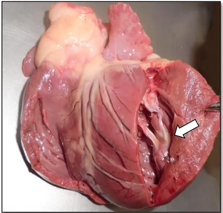
There are four heart chambers: right atrium, right ventricle, left atrium (LA) and left ventricle (LV). The right atrium receives the blood from the anterior and posterior vena cavae returning from the systemic circulation. Blood flows from the right atrium, across the tricuspid valve (right atrioventricular valve), and into the right ventricle. The outflow vessel of the right ventricle is the pulmonary artery, which is separated from the ventricle by the semilunar pulmonary valve. Blood returns to the heart from the lungs via four pulmonary veins that enter the left atrium. Blood flows from the LA, across the mitral valve (left atrioventricular valve), and into the LV, it is then ejected into the aorta across the aortic valve (Klabunde, 2012). A healthy dog’s heart is shown in figure 1.
6
Figure 1: Healthy dog’s heart from a Boxer with both ventricles sectioned, left-side view. Original
image, kindly provided by Doctor Graça Pires, professor of Anatomy in the Faculty of Veterinary Medicine, Lisbon’s University.
Legend: The white arrow indicates the point of insertion of the chorda tendinea in the papillary muscles, both of them components of the mitral valve apparatus.
For the purpose of maintaining a working circulatory system, the heart needs to ensure two fundamental mechanical functions. One is to fulfill the perfusion requirements of the metabolizing tissues by ejecting a sufficient amount of blood at the right pression into the aorta and pulmonary arteries. The other is to provide drainage of the capillary beds preserving an appropriate distribution of the circulating blood pool (Ljungvall & Häggström, 2016).
Approximately 10% of the dogs presented to primary care veterinary practices have heart disease, being Chronic Valvular Heart Disease (CVHD) the most common one (Atkins, et al., 2009). In CVHD, the left atrioventricular or mitral valve is more often affected and to a greater degree, although in approximately 30% of cases the right atrioventricular (tricuspid) valve is also involved (Borgarelli & Buchanan, 2012; Garncarz, Parzeniecka-Jaworska, Jank, & Łój, 2013). However, isolated degenerative disease of the tricuspid valve is uncommon. Thickening of the aortic and pulmonary valves is sometimes observed in older animals but rarely causes more than mild insufficiency (Ware, 2014).
DMVD of the left atrioventricular valve is characterized by the accumulation of glycosaminoglycans (myxomatous proliferation) and fibrosis of the valve leaflets and chordae tendineae (Häggström, 2010). The condition has been given numerous designations on the basis of its clinical and pathologic features. The terms chronic degenerative valvular disease, chronic valvular disease,
7
chronic valvular fibrosis, endocardiosis, myxomatous mitral valve disease, myxomatous valvular degeneration, myxomatous transformation, and mucoid degeneration all refer to the same disorder (Häggström, 2010; Abbott, 2016; Ljungvall & Häggström, 2016).
2.2 Mitral valve’s anatomy and histology
2.2.1 Anatomy
The mitral valve’s competence depends on the functional and structural performance of six basic components that form the mitral valve apparatus: 1) the posterior left atrial wall, 2) the mitral valve annulus, 3) the mitral valve leaflets, 4) the chordae tendineae, 5) the left ventricular papillary muscles, 6) and the associated left ventricular wall (see figure 2). Each of these elements act both independently and synergistically, working in a complex interplay with the objective of preserving the valve’s integrity (Perloff & Roberts, 1972; Fox, 2012).
Figure 2: Dorsolateral view of a normal mitral valve apparatus from two-year-old mongrel dog
(Reproduced with permission from Dr. Philip Fox)
Legend: Along the top frame are the valve leaflets attached to the myocardial tissue. Primary chordae
tendineae (small vertical arrows) and secondary chordae tendineae (broad arrow) attach the mitral leaflets to
the papillary muscle (P), Scale, mm (Fox, 2012).
There are two mitral valve leaflets, the anterior (septal) and the posterior (parietal), being the juncture where they are supported at their hinge points referred to as the mitral annulus. This discontinuous fibrous ring is a dynamic structure that suffers changes in shape and size during the cardiac cycle (Glasson, et al., 1996; Fox, 2012). In healthy animals, the mitral leaflets are thin, translucent structures, with a rough zone near their free margin where chordae tendineae attach, and a smooth zone towards the annular junction (Shimakura, Ishihara, Kawazoe, & Hashimoto, 1978; Fox, 2012; Abbott, 2016).
Intact mitral valve chordae tendineae mediate efficient and forceful ventricular contraction and optimize left ventricularsystolic performance. Each mitral valve leaflet is adjoined to the anterior and posterior papillary muscles or occasionally, directly to the ventricular wall by these fibrous chords.
8
Together, these structures work in a coordinated manner to prevent mitral valve prolapse and regurgitation (Oe, et al., 1993; Fox, 2012).
Chordae tendineae are categorized according to their insertion sites on the mitral valve’s leaflets, as
a result, there are two major sets of chordae. Primary (first-order or marginal) chordae arise from the papillary muscles and attach to the free margins of the leaflets, they are thin and most numerous. Secondary (second-order) chordae also ascend from the papillary muscles but insert near the intersection of the rough and smooth zone, on the ventricular aspect of the valve leaflets; they are larger and fewer in number. Both types of chordae, however, may originate from a common bifurcating stem (Rodriguez, et al., 2004; Fox, 2012).
Primary and secondary chordae serve different roles (Ibadia, Casali, Chassignolle, & Janier, 1997). The primary chordae maintain leaflet apposition and facilitate valve closure, whereas secondary
chordae are believed to be involved in maintaining normal LV size and geometry. When a primary chorda gets ruptured, it results in acute mitral regurgitation. Sectioning secondary chordae, in
contrast, does not produce mitral regurgitation, but can impact ventricular geometry and systolic function. (Nielsen, et al., 2001; Silbiger & Bazaz, 2009).
2.2.2 Histology
The mitral valve’s leaflets are heterogenous and laminated structures in which four distinct layers can be differentiated: atrialis, spongiosa, fibrosa, and ventricularis (layers from the atrial to the ventricular aspect) (see figure 3). These are most prominent at the leaflets mid-portion (Aupperle, et al., 2009).
Figure 3: Photomicrograph of the proximal third of the posterior mitral valve leaflet from a three-year-old German Shepherd dog. (Reproduced with permission from Dr. Philip Fox)
Legend: Layers of the mitral valve leaflets: atrialis (A), spongiosa (S), fibrosa (F) and ventricularis (V). CT stands for chorda tendinea. Haematoxylin and eosin stain. Bar = 200µm (Fox, 2012).
The atrialis consists in a thin layer of endothelial cells supported by scattered collagen fibers, elastic fibers, fibroblasts and smooth muscle cells. The spongiosa extends from the annulus to the free edge of the leaflet, and it is characterized by being a layer rich in proteoglycans and glycosaminoglycans, it also contains ground substance embedding a loose collection of collagen,
9
elastic fibers, fibroblasts, and Anichkov’s cells. The fibrosa is composed by a dense layer of compact collagen bundles with scattered fibroblasts that proximally has continuity with the annulus, while distally continues with the central core of chordae tendineae. The ventricularis, is a thin layer alike the atrialis but without smooth muscle cells (Aupperle, et al., 2009). Theendothelial covering of the ventricular aspect of the leaflets and the one covering the chordae tendineae are continuous, and the fibrous valvular layer is continuous with the fibrous cardiac skeleton (Fox, 2012). Amongst the roles of the fibrous skeleton are to internally reinforce the myocardium, to anchor the valve cusps, to avoid excessive dilation of valvular orifices, to serve as an insertion point for atrial and ventricular myocyte bundles, and to buffer conduction of electrical impulses between atria and ventricles (Evans & Lahunta, 2013).
2.3
Epidemiology
DMVD has the highest prevalence (percentage of a population with a particular disease at a given point in time) of all canine heart diseases, accounting for 75-80% of the cases of dogs with cardiac disease (Das & Tashjian, 1965; Egenvall, et al., 2006; Ljungvall & Häggström, 2016). Due to the fact that DMVD is a progressive disease, a clinically evident valvular dysfunction is preceded by subtle changes in the valve structure. As a result, the prevalence of DMVD reported by clinical studies is lower than that detected by postmortem examination (Abbott, 2016).
The disease is age related and the prevalence increases drastically in 4 to 5-year old dogs (Whitney, 1974). Concerning the sex, DMVD is approximately 1,5 times more common in males than in females, furthermore the disease seems to appear at a younger age and have a more rapid progression from mild to severe in males. Given the fact that the disease affects middle-aged to old dogs, the animal can succumb to other co-morbid conditions or old age before progressing into congestive heart failure (Thrusfield, Aitken, & Darke, 1985; Häggström, Hoglund, & Borgarelli, 2009; Borgarelli & Häggström; 2010).
DMVD can be encountered in all breeds, but the highest prevalence is found in small to medium-sized dog breeds, namely Cavalier King Charles Spaniels (CKCS), Dachshunds, Chihuahuas, Miniature Poodles, Miniature Pinchers, Beagles, Whippets and Cocker Spaniels. The coat color does not seem to have an influence in the frequency of the valve disease (Borgarelli & Buchanan, 2012). Even though most studies focus in smaller breeds, when larger breeds are taken into account German Shepherd dogs appear to be over-represented in the experimental groups. Additionally, larger dogs often experience faster disease progression allied with a greater myocardial dysfunction, and a more guarded prognosis (Madron, 1992; Borgarelli, et al., 2004). In small breed dogs, the progression is usually slow but can be, at times, unpredictable. CKCS are remarkably predisposed to developing DMVD at a younger age, nonetheless the time course of their disease progression to heart failure does not appear to be noticeably different from that of other small breed dogs (Keene, et al., 2019).
10
2.4
Etiology
First of all, it is important to keep in mind that a definitive etiology to the DMVD is, to this date, unknown. Despite that fact, several possible causes have been suggested throughout the years (Ljungvall & Häggström, 2016).
Since some dog breeds are more predisposed to an early onset of DMVD, it is believed that the disease has a strong genetic background, being heritable in at least some breeds, namely the CKCS and the Dachshund (Olsen, Fredholm, & Pedersen, 1999; Lewis, Swift, Woolliams, & Blott, 2011; Meursa, et al., 2018). DMVD has been suggested to be inherited as a polygenic threshold trait, which means that multiple genes influence the trait and a certain threshold has to be reached before the disease develops (Swenson, Häggström, Kvart, & Juneja, 1996; Olsen, et al., 1999). Moreover, an association between the presence and severity of DMVD in parents and offspring at a given age, has been demonstrated. In other words, when both parents have an early onset of the disease, the offspring will, on average, develop the disease at a younger age as well. On the contrary, when both parents have a late onset of DMVD, they will give birth to offspring that manifests the disease at an old age or never (Lewis, et al., 2011). With regard to the threshold, males have a seemingly lower one than females, as a consequence they are expected to manifest the disease at a younger age than females, within a family in which the offspring have approximately the same genotype (Ljungvall & Häggström, 2016). A genome-wide analyses of CKCS unveiled 2 loci associated with the development of DMVD, however, even though the chromosomal regions have been identified, no specific causative genes have been determined (Madsen, et al., 2011). These discoveries reinforce the hypothesis that a genetically determined process initiates the valve degeneration (Swenson, et al., 1996; Olsen, et al., 1999; Madsen, et al., 2011).
Another suggested cause for DMVD is related to phenotypic alterations in the valvular interstitial cell (VIC) population. A transformation seems to occur from an inactive fibroblast phenotype, which is responsible for normal valve structure by maintaining the extracellular valve matrix, to a more active myofibroblast type. This happens both in dogs with DMVD and in humans with the equivalent acquired valvular disease (Rabkin, et al., 2001; Black, French, Dukes-McEwan, & Corcoran, 2005). As it will be explained below in greater detail, the degenerative changes of DMVD are characterized by an overproduction and deposition of extracellular matrix with disruption of collagen content and organization in the mitral leaflets and chordae tendineae (Han, et al., 2008; Aupperle, et al., 2009). This remodeling, that happens within the mitral valve, is mediated by the referred activation of the normally quiescent VIC (Oyama & Chittur, 2006; Disatian, Ehrhart, Zimmerman, & Orton, 2008). Serotonin (5-hydroxytryptamine, 5-HT) has been linked to VIC activation in several species, including humans, rats, and dogs. Additionally, multiple studies have shown altered local 5HT signaling in dogs with DMVD (Oyama & Chittur, 2006; Connolly, et al., 2009; Disatian & Orton, 2009; Oyama & Levy, 2010; Scruggs, Disatian, & Orton, 2010; Orton, Lacerda, & MacLea, 2012).
11
Serotonin is a monoamine neurotransmitter known to control a wide range of biological functions (Jonnakuty & Gragnol, 2008). Most of 5-HT is synthesized in the intestine by the enterochromaffin cells, but there is also local synthesis of 5-HT in the heart (Ni & Watts, 2006; Lacerda, Maclea, Kisiday, & Orton, 2012). The 5-HT is then secreted into the blood stream, and nearly all of it is taken up by platelets via active transport and stored in its dense granules. Hence, serum concentration of serotonin results from an equilibrium between 5-HT synthesis, platelet uptake and storage, and metabolism (Launay, Schneider, Loric, Da Prada, & Kellermann, 2006). It has been shown that giving 5-HT injections to rats causes the development of valvular lesions (Gustafsson, et al., 2005). Moreover, valvulopathies in people have been associated with 5-HT-producing carcinoid tumors and the intake of serotonergic drugs (Lundin, Norheim, Landelius, Oberg, & Theodorsson-Norheim, 1988; Hendrikx, Van Dorpe, Flameng, & Daenen, 1996; Zanettini, et al., 2007; Gustafsson, Hauso, Drozdov, Kidd, & Modlin, 2008). There are many similarities between these 5-HT-induced valvular lesions and those in patients with DMVD, which further points towards serotonin having a role in the development of the disease (Disatian & Orton, 2009).
Furthermore, platelet 5-HT content is substantially higher in dogs with DMVD versus healthy non-CKCS dogs. When comparing healthy non-CKCS with healthy dogs of other breeds, it has been concluded that CKCS have a higher platelet 5-HT content, suggesting not only that platelet-derived 5HT is involved in the development of early stages of DMVD, but also that 5-HT may be genetically linked to the etiologic mechanisms of the disease in this breed. As for the 5-HT concentration in the mitral valve leaflets and left ventricle myocardium, it is also increased in affected dogs, denoting that altered tissue serotonin signaling is a specific feature of DMVD (Cremer, et al., 2014). However, serum 5-HT concentrations decrease with the increasing severity of the disease, which implies that even if serotonin primarily plays a role in the progression of early DMVD, it may be less involved when severe valvular lesions are already installed (Ljungvall, et al., 2013).
2.5
Natural History
2.5.1 Microscopic Alterations
As far as histopathological findings are concerned, a myxomatous degeneration starts to take place in the valve leaflets. This degeneration is characterized by giving origin to a disorganized and weakened connective tissue. There is a progressive expansion of the spongiosa layer that becomes thickened with increased extracellular matrix and proliferation of edematous ground substance. It is also verified that the normal arrangement of layered collagen in the fibrosa layer becomes disrupted and attenuated. In advanced stages, differentiating spongiosa and fibrosa layers can be challenging (see figure 4). Increased amounts of mucopolysaccharides, and glycosaminoglycans are frequently seen within affected valves (Buchanan, 1977; Han, et al., 2008; Hadian, Corcoran, & Bradshaw, 2010; Han, Black, Culshaw, French, & Corcoran, 2010). Regarding the heart chambers, changes in the composition and structure of the extracellular matrix and the myocytes are also present. In
12
advanced stages of DMVD the presence of myocardial fibrosis and intramyocardial arteriosclerosis have been reported, particularly in the papillary muscles (Falk, Jönsson, Olsen, & Pedersen, 2006).
Figure 4: Photomicrograph of the distal posterior mitral valve leaflet from a twelve-year-old Maltese
dog with severe Degenerative Mitral Valve Disease (DMVD) (Reproduced with permission from Dr. PhilipFox)
Legend: It can be observed the increased thickness of the spongiosa from glycosaminoglycan and proteoglycan deposition, and degeneration of the fibrosa. Left frame: Haematoxylin and eosin stain; bar = 1mm. Right frame: Masson trichrome stain; bar = 1 mm. This image demonstrates the total loss of the leaflet’s normal layered arrangement in DMVD. The white arrow points to a secondary chorda tendinea (Fox, 2012).
2.5.2 Macroscopic Alterations
DMVD is characterized by having a long pre-clinical asymptomatic period, in which the first morphologic changes are only visible on the necropsy (Borgarelli & Buchanan, 2012). Due to the evolving nature of the DMVD, with the advancing of time the lesions become more observable and, depending on the stage of the disease, the valve will present different macroscopic and microscopic appearances (Ljungvall & Häggström, 2016).
The first macroscopic modifications occur in the area of apposition of the leaflets, being generally more evident in the sites where chordae tendineae insert, nonetheless the chordae themselves can also be affected by the myxomatous degeneration. The normally thin and translucent free edges of the leaflets gradually become thickened and irregular with parts bulging towards the LA side (Han, et al., 2013; Corcoran, et al., 2004). Progressively the lesions spread to other zones of the leaflets and the bulging worsens. Succinctly, DMVD is macroscopically characterized by nodular distortion and thickening of the mitral leaflets and chordae tendineae, beginning with a few nodules at the free edges of the valve that later increase in number and coalesce to form plaque-like elevations (Whitney, 1974; Buchanan, 1977).
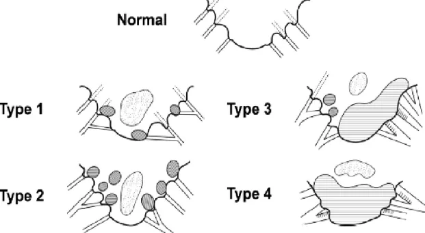
In 1974, Whitney tabulated the necropsy findings of several dogs with DMVD and elaborated a system to classify the different stages of degenerative mitral lesions, that demonstrates the evolving nature of the lesions. This system devises the lesions in four types (see figure 5). Type I lesions are those in which a few small edematous nodules can be found in the area of apposition of the leaflets,
13
at this point the chordae tendineae themselves are not affected and the valve maintains its competence. In Type II lesions the nodules are bigger but the chordae tendineae remain unaffected. As for Type III lesions, the nodules are larger and plaque-like elevations can be seen, moreover the
chordae tendineae are thickened and irregular proximally, being at this stage that evidence of
valvular incompetence starts to show. Finally, in Type IV lesions the valve is severely distorted and contracted, the chordae tendineae are affected in all surface and may even be ruptured, in addition there is evidence of valvular incompetence in the majority of the cases (Whitney, 1974).
In patients presenting severe cases of DMVD, the atrial wall may rupture to different extents causing hemopericardium and acute left heart failure. Endomyocardial splits, ruptured pectinate muscles in the atrial appendage and acquired atrial septal defects may be observed in such cases (Buchanan, 1972). In case of rupture of the atrial septum, although uncommon in dogs with DMVD, the acquired septal defect normally leads to right failure instead of acute left heart failure (Peddle & Buchanan, 2010).
Figure 5: Four grade classification of mitral valve lesions according to Whitney. (Reproduced with
permission from Dr. Michele Borgarelli)
Legend: The shaded areas represent nodular or diffuse thickening of the valve. The dotted areas represent zones of opacity caused by myxomatous degeneration (Borgarelli & Buchanan, 2012).
2.5.3 Physiopathology
All of the mentioned alterations in patients with DMVD generate a distortion of the valve architecture and chordae tendineae, which leads to a defective coaptation of the mitral leaflets during ventricular systole and a possible systolic atrial displacement of the valve leaflets. In healthy individuals, during early systole, the mitral leaflets are forced into apposition when the left ventricle pressure surpasses the left atrial pressure avoiding the retrograde ejection of blood. Additionally, the tethering effect of
14
the chordae tendineae averts the prolapse of the leaflets into the LA. In dogs with DMVD, due to the abnormal coaptation, not all of the left ventricular stroke volume is ejected through the aorta, a percentage flows backwards into the LA. Moreover, the elongation or rupture of the chordae
tendineae causes the leaflets to completely or partially prolapse, further aggravating the mitral
regurgitation (Beardow & Buchanan, 1993; Abbott, 2016). The importance of the regurgitation volume depends not only on the size of the regurgitant orifice, but also on the systolic pressure gradient between the left heart chambers (Buchanan, 1977; Ahmed, McGiffin, O'Rourke, & Dell'Italia, 2009).
The consequences of mitral regurgitation vary according to its severity. A mild regurgitation is not sufficient to induce changes in the heart size and function, given the fact that the small volume that goes backwards is effortlessly accepted by the LA and the volume going in the right direction is preserved. Meanwhile, with the progression of the valve’s lesions, the regurgitant volume becomes greater, triggering cardiac and non-cardiac compensatory mechanisms whose objective is to maintain the forward stroke volume (Baumgartner, 2017). In dogs with advanced stages of DMVD, more than 75% of the total LV stroke volume can be ejected back into the LA during systole (Kittleson & Brown, 2003).
Once the mitral valve starts leaking, the blood entering the LV during diastole comes not only from the lungs but also from the fraction that has been previously regurgitated into the LA, which causes an increase in the preload and a reduction of the after load (resistance to LV emptying) (O'Gara, et al., 2008). In order to keep an intra-atrial pressure lower than that of the ventricle, the LA starts to enlarge in the interest of accommodating the increasing regurgitant volume and ensuring the right direction of the blood flow. Since the extent of the LA’s enlargement is linked to the severity of the mitral regurgitation, it has been suggested that a major determinant factor to the degree of left-sided cardiac dilatation is the mitral regurgitation volume (Kittleson & Brown, 2003; Eriksson, Hansson, Häggström, Järvinen, & Lord, 2010). On the other hand, this capacity of enlargement relies on the compliance of the LA’s walls, which depends on the regurgitant volume increase rate, that itself is determined by the rate of progression of the DMVD. In practice, this means that a dog with slowly progressing DMVD can present an extremely enlarged atrium without pulmonary congestion or edema. Whereas in cases with acutely increased mitral regurgitation volume, the LA does not have time to properly adapt its size. An abrupt raising of the pressure inside the LA that reflects backwards in the hydrostatic pressure of the pulmonary capillaries and is responsible for the development of pulmonary congestion and edema, which is defined as an increase in the extravascular water content of the lungs. Rupture of principal chordae tendineae is an example of a situation that can lead to this succession of events. Hence, it is clear that he LA’s capacity to absorb the regurgitant volume is very important in protecting the pulmonary vascular bed from hypertension. Another compensatory mechanism that tries to delay the formation of a pulmonary edema is the development of a more effective lymphatic drainage of the pulmonary interstitium in chronic pulmonary congestion, since
15
overt pulmonary edema develops when the capacity of the pulmonary lymphatic system is exceeded (Staub, 1980; Murray, 2011; Borgarelli, et al., 2015 ; Ljungvall & Häggström, 2016).
As the degeneration of the valve advances, a progressively larger amount of blood moves ineffectually back and forth between the LV and the LA, diminishing the forward flow to the aorta. While the LA enlarges to accommodate the regurgitant volume, the LV has to compensate for the loss of forward stroke volume and deal with the high end-diastolic pressures and volumes. To do so, in response to the volume overload, the ventricle wall suffers an eccentric hypertrophy in which the ratio wall thickness and chamber size remains roughly unchanged. This hypertrophy maintains an adequate forward stroke volume and, at the same time, normalizes the pressure in the volume-overloaded LV (Carabello, 2002; Grossman & Paulus, 2013). The mechanism behind this is the Frank-Starling law, which dictates that when there is a greater end-diastolic volume the myocardium will be more stretched and, as a consequence, there will be an increased sarcomere length which enhances the sensitivity to calcium ions culminating in a much stronger contraction. In other words, the volume overloading causes an increase in the contractile strength and will increase the stroke volume (Komamura, et al., 1993). Hemodynamic wall stress has also been suggested to stimulate neurohormonal activation such as augmentation of the local production of angiotensin II that contributes to the myocardial hypertrophy. This production of angiotensin II is due to activation of the renin-angiotensin-aldosterone system by the increased secretion of renin in response to a decreased renal perfusion (Häggström, et al., 1997; Spinale, 2002).
All these cardiac and non-cardiac compensatory mechanisms try to provide the hemodynamic support needed to uphold the cardiac output in spite of the mitral regurgitation. Although beneficial at first, with the progression of the disease, they become contributing factors to the deterioration of the failing heart (Baumgartner, 2017). Concerning the cardiac remodeling, it is known that it will eventually impact the heart’s mechanical function. For instance, the change in the dimensions of the cardiac chambers leads to an alteration in the overall shape of the heart from elliptical to globular. Consequently, even if it allows a myocardial adaptation to the anomalous wall stress, the disruption of the normal geometry of the mitral annulus may contribute to further aggravate the mitral regurgitation (Grossman & Paulus, 2013). Additionally, chronic volume overloading reduces cardiac function and myocyte contractility. This can be explained by a subendocardial ischemia caused by an increase in oxygen demand due to increased intramyocardial tension, neurohormonal activation and the Frank-Starling mechanism. Another contributing factor to the subendocardial ischemia is the enlarged muscle mass outgrowing the vascular growth. During ischemia there is a production of oxygen free radicals as well as nitric oxide activation which further aggravates the myocardial injury (Jennings & Reimer, 1981; Prasad, et al., 1996). The deterioration of systolic myocardial function in these conditions is a state sometimes referred to as overload cardiomyopathy (Abbott, 2016). Nonetheless, the ventricular pump function is normally kept fairly well until late stages of DMVD, even in face of severe congestive signs (Ware, 2014). This explains why dogs with severe mitral
16
regurgitation have more commonly respiratory signs, caused by pulmonary congestion and edema, than signs caused by reduced forward cardiac output (Ljungvall & Häggström, 2016). As a consequence, death due to DMVD is most often mediated by congestive heart failure, even though sudden death can usually occur (Borgarelli, et al., 2008). Besides, overt heart failure tends to occur years after the start of the remodeling and myocardial disease, however, as demonstrated by a study, most cardiac enlargement takes place in the year preceding the onset of congestive heart failure (Lord, Hansson, Kvart, & Häggström, 2010).
2.5.4 Prognosis
Given the fact that the assessment of DMVD severity is not standardized, reports on the natural history and prognosis are rather variable (Keene, et al., 2019).
In a study including patients with several stages of DMVD, more than 70% of asymptomatic dogs with echocardiographic confirmation of the disease were alive at the end of 6.6 years of observation. The same study revealed that the survival time for those with moderate heart failure due to DMVD had a median survival time of 33 months and those with severe heart failure 9 months (Borgarelli, et al., 2008). Another study concluded that patients in pre-clinical stages had a survival time of 27.6 months and only 13% of dogs progressed to a more advanced stage during a 6.6 years period of time. Data coming also from this study demonstrated that over that period of observation the overall mortality was 27%, with cardiac deaths accounting for 11% (Borgarelli, et al., 2012). Concerning pre-clinical dogs, another study reported that 82% of them were still asymptomatic 12 months later (Moonarmart, et al., 2010). Many more studies can be found showing some minor variations between them (Borgarelli & Buchanan, 2012).
The differences amongst studies can be explained by inclusion of diverse criteria, different breeds, treatment and frequency of complications. Nonetheless, when taking into consideration all the studies, it can be concluded that patients with moderate heart failure due to DMVD receiving the appropriate medical treatment have fairly long clinical outcome, and the prognosis is relatively favorable for dogs with pre-clinical disease. Moreover, animals in asymptomatic stages tend to stay stable and not progress into heart failure when properly treated and monitored (Borgarelli & Buchanan, 2012). Further refinement of risk stratification schemes is needed to the development of a truly reliable method of evaluation, with good sensitivity and specificity (Keene, et al., 2019).
2.6 Clinical Signs and Physical Examination
There are usually no clinical signs in mild to moderate (ACVIM Stages A and B) cases of DMVD (Ljungvall & Häggström, 2016). In most dogs the disease is detected when a cardiac murmur is identified in a routine health care check-up or when managing a noncardiac illness (Abbott, 2016). This murmur is caused by mitral regurgitation, that is defined by is a systolic leakage of the mitral
17
valve. What causes the sound to which we call murmur is the blood flowing back from the left ventricle into the left atrium due to poor coaptation of the mitral leaflets (Chetboul, Bussadori, & Madron, 2016). Despite the fact that a systolic heart murmur is typically the most prominent clinical finding, a mid-systolic click may be heard in early stages of DMVD. It must be taken into account that the absence of an audible murmur cannot rule out mild regurgitation (Kvart & Häggström, 2002; Ljungvall, et al., 2009). Short physical stimulation, like a short run, can often augment the intensity of the murmur in early stages of the disease. In these patients the murmur may be intermittent and the point of its maximal intensity is on the left side of the thorax, over the mitral valve area (Ljungvall, Rishniw, Porciello, Ferasin, & Ohad, 2014; Reimann, et al., 2014). As the disease progresses, the intensity of the murmur augments as well as its duration, becoming gradually holosystolic and audible in both sides of the thorax (Ljungvall, et al., 2009). In patients with moderate or severe mitral regurgitation an exaggerated apical impulse on precordial palpation can be present, this impulse is usually referred to as precordial thrill (Abbott, 2016). The intensity of the heart murmur is of subjective evaluation but it can be classified as shown in table 1.
Table 1: Comparison of 4 level and 6 level murmur grading systems. Adapted from Rishniw, 2018.
Cough is typically one of the first clinical signs reported by the owner. Dogs with left mainstem bronchial compression, but without pulmonary congestion or edema, may have a dry and harsh cough with coughing spells at any time during the day, more pronounced during physical exercise or excitement. In contrast, when coughing is caused by pulmonary congestion or edema it may be moist and productive, associated with tachypnea and dyspnea. However, recent evidence seems to oppose to the traditional dogmatic approach that related cough to congestive heart failure (CHF) in dogs (Ljungvall & Häggström, 2016; Ferasin & Linney, 2019). When a dog shows signs of CHF the pulmonary sounds are usually more perceptible with crackles, snaps and popping sounds, best heard at the end of inspiration. In animals with fulminant pulmonary edema, the expectoration of pink froth is sometimes observed (Ohad, Rishniw, Ljungvall, Porciello, & Häggström, 2013; Abbott, 2016; Ljungvall & Häggström, 2016).
The first clinical signs of decompensated CHF (ACVIM Stage C) are generally mild but can worsen over a period of days or weeks. Bearing in mind that these signs are non-specific, the diagnostic challenge is to establish whether the DMVD is responsible for them. Dogs in decompensated CHF