Vol. 44, N. 2 : pp. 113 – 119, June, 2001
ISSN 1516-8913 Printed in Brazil
BRAZILIAN ARCHIVES OF
BIOLOGY AND TECHNOLOGY
A N I N T E R N A T I O N A L J O U R N A L
Microflora Dynamics in Earthworms Casts in an Artificial
Soil (Biosynthesol) Containing Lactic Acid Oligomers
Nathalie Alauzet
1, Sevastianos Roussos
2, Henri Garreau
1*and Michel Vert
11 CRBA CNRS-UPRESA 1465, Faculté de Pharmacie, 15 av. C. Flahault, 34060 Montpellier, France
2
Equipe de Mycologie-FMS, UR119-IRD, IFR-BAIM, Universités de Provence et de la Méditerranée, ESIL Case 925, 163, av. de Luminy, 13288 Marseille cedex 9, France
ABSTRACT
Studies were performed to appreciate the presence of micro-organisms able to degrade OLA, in earthworms casts or in the surroundings. Worms were grown in biosynthesol, an artificial soil. The counting of bacteria and fungi in earthworms casts and in biosynthesol without earthworms suggested that earthworms ate some of the
micro-organisms. The main filamentous fungi genera found were Aspergillus, Trichoderma, Fusarium and Penicillium.
Previous results in the literature have shown that some species from the Aspergillus and Fusarium genera were able to degrade OLA and other aliphatic esters. It could be suggested that these two genera and some bacteria were responsible for the pre-degradation of OLA, and that earthworms might eat them.
Key words: Earthworm, Eisenia andrei, biodegradation, lactic acid polymers, artificial soil, natural microflora
*
Author for correspondence
INTRODUCTION
Biodegradable plastics are considered as one of the solutions to reduce the nuisance of plastic wastes to the environment. Among degradable polymers, polylactic acids (PLA) are promising for industrial applications, and various companies such as Dow-Cargill are planning to produce and use as packagings stereocopolymers of the PLAX-type (acronym where X stands for the percentage in
L-lactyl units, according to Vert et al., 1981). The
abiotic hydrolysis of PLA is well documented (Li
et al., 1990a, Vert et al., 1994). In aqueous media,
many factors can contribute to modulate the degradation characteristics of PLAX polymers (Li
et al., 1990b). Ester hydrolysis is dependent on
autocatalysis by carboxylic chain ends and from diffusion-reaction phenomena involving absorbed
water and oligomeric molecules generated by degradation via their solubility in aqueous environment. When submitted to water and heat, high molecular weight (HMW) PLA degrades to low molecular weight PLA (oligomers) up to dimers and monomers of lactic acid. This explains why PLA can degrade in a humid and warm
medium like a compost (Buchanan et al., 1995,
Alauzet, 1999) or a vermicompost (Alauzet, 1999). HMW PLA has never been shown to be biodegraded by living organisms, although PLA 50 plates left 8 weeks in a soil were invaded up to the bulk by filamentous fungi after further incubation for 8 weeks under culture conditions
(Torres et al., 1996a). However some of the
by-products can be assimilated by micro-organisms
(Torres et al., 1996b, Karjomaa et al., 1998).
(Pseudomonas putida) and a fungus (Fusarium
moniliforme) that were able to bioassimilate lactic
acid oligomers (OLA) according to acronyms proposed by Vert and Guérin (1992). Furthermore,
some strains of the fungus Aspergillus niger have
been identified as able to degrade other aliphatic polyesters (Nishida and Tokiwa, 1993, Gonsalves
et al., 1992). Recent investigations (Alauzet, 1999)
have shown that earthworms placed into
biosynthesol (Bouché et al., 1998), an artificial
soil modified from Abdul Rida and Bouché (1997) and containing only OLA as carbon source, are able to bioassimilate OLA after a previous digestion by organisms. The micro-organisms found in the gut of earthworms seem to be the same as in the surrounding environment (Morgan, 1988), but their number fluctuate after passing through the earthworm gut (Parle, 1963). In this work, we wish to report the results of an investigation aimed at showing the presence of micro-organisms able to degrade OLA in earthworms casts; For this, a comparison quantitative of microflora present in earthworms casts and in biosynthesol without earthworm was made. Identification of filamentous fungi present in biosynthesol and in earthworms casts was also undertaken.
MATERIAL AND METHODS
Chemicals. OLA 50 (
M
p
=825,M
n
=500,I=1,68), and OLA 96 (
M
p
=730,M
n
=460,I=1,55) were synthesized by heating D,L-lactic acid and L-lactic acid (Sigma) in aqueous solution at 120°C for 18h under progressively reduced pressure up to 17 mmHg (Alauzet, 1999).
Culture media:
(1) Biosynthesol. The medium in which the
earthworms grew was modified from the biosynthesol used by Abdul Rida and Bouché (1997) according to Alauzet (1999)@. The
medium was composed of pure Levilite
(Prolabo) 45 g, 45 glass balls of 1.5 to 2 cm in diameter (Vetrotex, ref. E14,5g), glucose 0.9 g,
NH4NO3 0.1 g (nitrogen source), modified
Winogradsky solution 112 ml, OLA 50 or OLA 96 2 g(carbon source). The modified Winogradsky
solution was composed of MgSO4, 7 H2O 600
mg/l, MnSO4 20 mg/l, Fe2 (SO4) 80, (NH4)2SO4
200 mg/l, ZnSO4, 7 H2O 20 mg/l, CuSO4 20 mg/l,
Na2HPO4 4 g/l, KH2PO4 2.1 g/l, CaCO3 2 g/l.
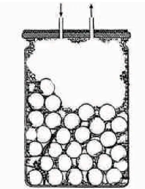
Biosynthesol was dispatched in one liter jars (Figure 1), agitated, then let non-sterile at 25°C. 10 earthworms per jar were added after 13 days of incubation when the pH of the medium was stabilized at 6.8/7.2.
(2) Plate Count Agar or PCA (Difco) with 0.1 g/l
of cycloheximide in Petri dishes was used for bacterial counting. For anaerobic bacteria, the Petri dishes were incubated in anaerobic jars, with Microcult A plaques (Merck). Cultures were incubated at 25°C for 2 and 7 days.
Figure 1 - Experimental device used for bioassimilation determination (biosynthesol).
(3) Potato Dextrose Agar or PDA (Difco), alone
was used in Petri dishes for genus identification, and with 0.05 g/l of Rose Bengal (Sigma) and 0.1 g/l of chloramphénicol for fungal counts.
Earthworms: An epigeic species, Eisenia andrei
Bouché 1972, was used. Adult worms, i.e. weighing more than 300 mg each, were collected from the Soil Zooecology Laboratory in Montpellier.
agitated for 3 min and the worms were removed from the water. Dilutions (1/10) were made from this suspension.
Earthworms casts: Earthworms casts were
regularly sampled in biosynthesol. Glass balls were first taken out, then the 10 earthworms were washed 3 times with sterile water and let 18 h in humidified Petri dishes. The casts present in Petri dishes were then introduced in 50 ml of physiological water, and homogenised using an ultraturrax. Dilutions (1/10) were made from this suspension. The weight of fresh casts was determined by weighing earthworms before and after defecation (after 18 h). Earthworms were then re-introduced into biosynthesol.
Biosynthesol: About 2 g of biosynthesol were
sampled, scraping from the bottom of the jar for optimal sampling. The 2 g were then split in 2 parts. About 1 g was weighed, dried at 105°C for 24 h to determine the dry weight, and the rest was also weighed, then mixed with 50 ml of physiological water and homogeinised with ultraturrax mixer. Dilutions (1/10) were made and microflora dynamics were evaluated.
Analysis: Numeration: For each dilution, 200 µl
were sampled and inoculated on Petri dishes containing PCA for bacterial counts (aerobic or anaerobic) and PDA for fungal counts. All Petri dishes were then incubated at 25°C. Microflora counting was done after 2 and 7 days of incubation.
Determination of fungi. From each Petri dish, the
main genus were isolated on a PDA medium without Rose Bengal. After 3-5 days, mycelia were sampled and observed with an optic microscope. Fungi genus were then determined through morphological criteria using identification keys such as the description of mycelia and of
asexual reproduction forms (Domsch et al., 1980).
RESULTS AND DISCUSSION
Natural microflora of earthworm epidermis: Before counting the microflora of earthworms casts, it was necessary to count the epidermal microflora, as the latter could contaminate the casts. The counting of epidermal microflora is shown in Table 1. Aerobic bacteria were predominant compared to the fungal population.
The results of epidermal microflora counting are taken into consideration in the calculation of earthworms casts microflora counting.
Table 1 - Counting of earthworms epidermal microflora (results are given in cultivable cells/single earthworm).
Aerobic Incubation time at 25°C
Microflora 2 days 6 days
bacteria 3.0 105 1.4 106
Fungi 6.0 103 8.0 103
Aerobic bacteria and fungi present in biosynthesol containing OLA 50 and OLA 96
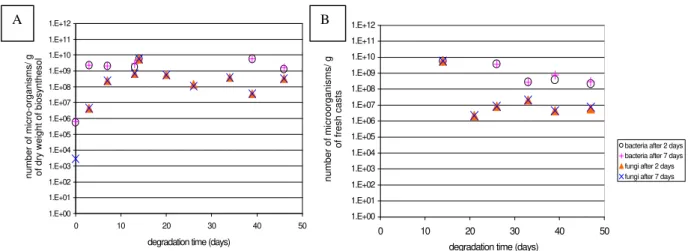
without earthworm: The counting of bacteria and
fungi that grow in biosynthesol in the presence of OLA 50 or 96 for 2 and 7 days of incubation at 25°C are shown in the Figure 2. The number of micro-organisms was more or less the same, whether after 2 or 7 days of Petri dishes incubation, indicating that those micro-organisms had a rapid growth. For bacteria, the growing phase was very short (less than 3 days). Their number then reached a stationary phase. For fungi, their growing phase lasted 7 days. Beyond, the population decreased regularly. Bacterial or fungal growth rates were comparable whether the sole carbon source was OLA 50 or OLA 96.
Aerobic bacteria and fungi present in earthworms casts in a biosynthesol containing
OLA 50 or OLA 96: In a biosynthesol without
earthworm, the number of bacteria and fungi was higher than in a real soil (Figure 3). A quantity of
109 to 1010 bacteria/g of biosynthesol was found,
instead of the expected 108-109 in a real soil, and
108 fungi/g of biosynthesol instead of the expected
106 (Prescott et al., 1996). After transit through the
earthworm gut, the number of bacteria and fungi also decreased and were close to the population found in normal soil. The number of fungi
decreased down to 106, and that of bacteria down
to 108. Therefore, earthworms seem to regulate the
1.E+00 1.E+01 1.E+02 1.E+03 1.E+04 1.E+05 1.E+06 1.E+07 1.E+08 1.E+09 1.E+10 1.E+11 1.E+12
0 10 20 30 40 50
degradation time (days)
number of microorganisms/ g
of fresh casts bacteria after 2 days
bacteria after 7 days fungi after 2 days fungi after 7 days
1.E+00 1.E+01 1.E+02 1.E+03 1.E+04 1.E+05 1.E+06 1.E+07 1.E+08 1.E+09 1.E+10 1.E+11 1.E+12
0 10 20 30 40 50
degradation time (days) number of micro-organisms/ g of dry weight of biosynthesol
A B
Figure 2 - Counting of micro-organisms versus time: A) in biosynthesol without earthworm, containing OLA 50 as sole carbon source B) in E. andrei casts, with OLA 50 as sole carbon source.
1.E+00 1.E+01 1.E+02 1.E+03 1.E+04 1.E+05 1.E+06 1.E+07 1.E+08 1.E+09 1.E+10 1.E+11 1.E+12
0 10 20 30 40 50
degradation time (days)
number of microorganisms/ g
of fresh casts bacteria after 2 days bacteria after 7 days fungi after 2 days fungi after 7 days
1.E+00 1.E+01 1.E+02 1.E+03 1.E+04 1.E+05 1.E+06 1.E+07 1.E+08 1.E+09 1.E+10 1.E+11 1.E+12
0 10 20 30 40 50
degradation time (days)
number of micro-organisms/ g of dry weight of biosynthesol
A B
Figure 3 - Counting of micro-organisms versus time: A) in biosynthesol without earthworm, containing OLA 96 as sole carbon source; B) in E. andrei casts, with OLA 96 as sole carbon source.
1.E+00 1.E+01 1.E+02 1.E+03 1.E+04 1.E+05 1.E+06 1.E+07 1.E+08 1.E+09 1.E+10 1.E+11
0 10 20 30 40 50 60 70 80
degradation time (days)
number of anaerobic bacteria/g of fresh casts
or g of dry biosynthesol
OLA 50 OLA 96 worms+OLA 50 worms+OLA 96
Table 2 - Percentage of the main fungal genus found in the biosynthesol or in earthworms casts. (a) in biosynthesol with OLA 50 as sole carbon source
(b) in biosynthesol with OLA 96 as sole carbon source (c) in E.andrei casts with OLA 50 as sole carbon source (d) in E.andrei casts with OLA 96 as sole carbon source
(a)
degradation time (days) 3 7 10 14 20 26 32 39 46 59
Aspergillus 100 100 100 87 57 7 31 13
Trichoderma 13 12
Penicillium 43 62 52 22 50
Fusarium 38 36 93 29 37
others 18
(b)
degradation time (days) 3 7 10 14 20 26 32 39 46 59
Aspergillus 100 100 100 66 50 16 14 5
Trichoderma 34 28 16 26 9 10
Penicillium 50 39 28 10 41 48
Fusarium 33 56 48 8 12
others 28 25
(c)
degradation time (days) 7 10 14 21 26 33 39 46 59 68
Aspergillus 100 65 5
Trichoderma 70 21 18 55 38.5 16
Penicillium 30 4 6.5 15 21
Fusarium 70 13 33 46.5 58
others 9 5.5
(d)
degradation time (days) 7 10 14 21 26 33 39 46 59 68
Aspergillus 33.3 21 3
Trichoderma 33.3 70 75 40 78 74 55
Penicillium 33.3 30 21 3 24
Fusarium 20 10 17 21
others 5 18 6 9
The results were comparable, whether the carbon source was OLA 50 or OLA 96.
Anaerobic bacteria in biosynthesol containing
OLA 50 and OLA 96 and in earthworms casts:
The presence of anaerobic micro-organisms, in biosynthesol as well as in earthworms casts, had been brought to the fore (figure 4). This shows that the artificial soil used (biosynthesol) contains
anaerobic microhabitats, and even earthworms can have anaerobic micro-organisms in their gut.
Evolution of fungi genus in biosynthesol containing OLA 50 and OLA 96 without
earthworms and in earthworm casts: Different
determined through microscope observation of mycelium and conidia. There were mainly 4 fungal genera present in biosynthesol (Table 2):
Aspergillus, Trichoderma, Penicillium and
Fusarium which were also found in earthworms
casts. This is not surprising as the genera
Aspergillus, Penicillium and Trichoderma are
frequently found in soils. Moreover, filamentous
fungi such as Fusarium and Penicillium show
tolerance towards lactic acid (Torres et al., 1999).
At the beginning of the incubation, there was only
Aspergillus, which might reflect a contamination
of the media. Later on, in worm-free biosynthesol,
other species appeared, namely Trichoderma on
the 20th day, Penicillium at the 26th day, and
Fusarium at the 34th day. The population of these
fungi was slightly increased when earthworms were added to the medium. The large deviation found for the distribution of the different genera was assigned to the heterogeneity of the solid medium. As a matter of fact, small areas of different colours could be seen in biosynthesol, proving that there were microhabitats. The sampling was rendered difficult, making data rather inaccurate. Some species disappeared, then appeared again, others seemed to fluctuate.
It is of interest to notice that the population of micro-organisms remained more or less the same from the taxonomic point of view in earthworm-free biosynthesol and in worm casts. These findings agree with those reported by Morgan (1988) who concluded that micro-organisms present in earthworms gut are the same as the ones in the surrounding soil, and that earthworms do not have their own commensal microflora.
The presence of Fusarium was particularly
interesting as, according to Torres et al. (1996c), at
least one fungus of this genus, F. moniliforme, is
able to bioassimilate OLA 50 and OLA 100. The
number of Fusarium remained stable regardless of
the presence of earthworms, their number being apparently greater when OLA 50 was the only carbon source. We have shown in a previous work that earthworms grown in biosynthesol with only OLA 50 or OLA 96 as a sole carbon source could gain weight (Alauzet,1999). Other data (Alauzet, 1999) suggested that earthworms were not responsible for the degradation. It seems that worm-associated micro-organisms pre-degrade OLA, then earthworms eat these micro-organisms
and can gain weight. The Fusarium strain found in
biosynthesol may be among the main responsible for degradation. However recent results suggested
that OLA 50 degrades during the first 27 days of earthworm incubation (Alauzet, 1999), when
Fusarium are not yet detected. One may thus
suppose that Aspergillus, which appeared very
soon and was the only fungus present at the beginning of the incubation, was one of the main filamentous fungi responsible for degradation together with bacteria.
In conclusion, the slight decrease of the number of fungi in the earthworms casts compared to the population found in biosynthesol shows that earthworms regulate the number of fungi mostly
by eating them. Among those fungi, Fusarium and
Aspergillus strains known as able to bioassimilate
PLA (Fusarium, Torres et al., 1996c) or other
aliphatic esters (Aspergillus, Nishida et Tokiwa,
1993, Gonsalves et al, 1992) were found. It is thus probable that OLA is first degraded by those fungi and also by bacteria, and then earthworms eat them. Further investigations are needed to determine the exact role of the micro-organisms and to explore whether the identified fungi and bacteria are really able to degrade OLA and to what extent. It is also needed to know whether earthworms can eat those particular fungi, in order to better understand the vermi-microbial interactions in degrading OLA.
ACKNOWLEDGEMENTS
We thank Pr. Marcel Bouché, from the Soil Zooecology Laboratory, INRA Montpellier, for
providing the E.andrei strains.
RESUMO
Já mostramos que a minhoca Eisenia andrei é
Os principais fungos filamentosos encontrados
foram Aspergillus, Trichoderma, Fusarium e
Penicillium. Resultados anteriores mostraram que
algumas espécies dos gêneros Aspergillus e
Fusarium são capazes de degradar o OLA e outros
ésteres alifáticos. Sugere-se que esses 2 gêneros e algumas bactérias são responsáveis pela pré-degradação do OLA, antes que as minhocas os comam.
REFERENCES
Alauzet, N. (1999), Etude de la dégradation de polyesters de type poly (acide lactique) en présence de vers de terre. PhD Thesis, Montpellier I University, France.
Abdul Rida, A.; M. and Bouché M. B. (1997), Earthworm toxicology: from acute to chronic tests.
Soil Biology and Biochemistry, 29, 699-703
Buchanan, C. M.; Dorsche, D. D.; Gardner, R. M.; Komarek, R. J. and White, A. W. (1995), Biodegradation of cellulose esters: composting of cellulose ester-diluent mixtures. Journal of
Macromolecular Science-Pure Applied Chemistry,
A32, 683-697
Cortez, J. and Bouché M. B. (1998), Field decomposition of leaf litters : earthworm-micro-organisms interactions. The plough-in effect. Soil Biology and Biochemistry, 30, 795-804
Domsch, K. H.; Gams, W. and Anderson, T. H. (1980),
Compendium of soil fungi, Academic Press, H. B.
Jovanovich Publ., London.
Gonsalves, K. E.; Chen, X. and Cameron, J. A. (1992), Degradation of non-alterning poly (ester-amides).
Macromolecules, 25, 3309-3312
Karjomaa, S.; Suortti, T.; Lempiainen, R.; Seli, J. F.; Itavaraa, M. and Steinbuechel, A. (1998), Microbial degradation of poly (lactic acid) oligomers. Biodegradable polymers and macromolecules.
Polymer Degradation and Stability, 59, 333-336. Li, S. M.; Garreau, H. and Vert, M. (1990a),
Structure-property relationships in the case of the degradation of massive poly (alpha-hydroxy acids) in aqueous media. Part 1: Poly (DL-lactic acid). Journal of Materials Science: Materials in Medicine, 1, 123-130
Li, S. M.; Garreau, H. and Vert, M. (1990b), Structure-property relationships in the case of the degradation of massive poly (alpha-hydroxy acids) in aqueous media. Part 3: Influence of the morphology of poly (L-lactic acid). Journal of Materials Science: Materials in Medicine, 1, 198-206
Morgan, M. H. (1988), The role of micro-organisms in the nutrition of Eisenia fetida. In: Earthworms in
waste and environmental management, C. A.
Edwards and E. F. Neuhauser (eds), 71-82
Nishida, H. and Tokiwa, Y. (1993), Distribution of poly (b-hydroxybutyrate) and poly (e-caprolactone) aerobic degradaing micro-organisms in different environments. Journal of Environmental Polymer Degradation, 1, 227-233
Parle, J. N. (1963), A microbiological study of earthworm casts. J. General. Microbiol., 31, 13-22 Prescott, L. M.; Harley, J. P. and Klein, D. A. (1996),
Microbiology. Wm. C. Brown Publishers, Dubuque , USA, 935p
Torres, A.; Li, S. M.; Roussos, S. and Vert, M. (1996a), Poly (lactic acid) degradation in soil or under controlled conditions. Journal of Applied Polymer Science , 62, 2295-2302.
Torres, A.; Li, S. M.; Roussos, S. and Vert, M. (1996b), Screening of micro-organisms for biodegradation of poly (lactic acid) and lactic acid-containing polymers.
Applied and Environmental Biology, 62, 2393-2397 Torres, A.; Li, S. M.; Roussos, S. and Vert, M. (1996c),
Degradation of L- and DL-lactic acid oligomers in the presence of Pseudomonas putida and Fusarium
moniliforme. Journal of Environmental Polymer
Degradation, 4, 213-223
Torres, A.; Li, S. M.; Roussos, S. and Vert, M. (1999), Microbial degradation of a poly (lactic acid) as a model of synthetic polymer degradation mechanisms in outdoor conditions. In: Biopolymers. Utilizing
nature's advanced materials, ACS Symposium Series
723, 218-226
Vert, M.; Li, S. M. and Garreau, H. (1981), Bioresorbable polyesters for bone surgery.
Makromolekulare Chemie, 5(suppl.), 30-41
Vert, M. and Guerin, P. (1992), Des biosystèmes aux matériaux polymères: une utopie?, Biofutur, Juin, 52-57
Vert, M.; Torres, A.; Li, S. M.; Roussos, S. and Garreau, H. (1994), The complexity of the degradation of poly(2-hydroxy-acid)-type aliphatic polyesters. In Biodegradable Plastics and Polymers, Y. Doi and K. Fukuda (Eds). Elsevier, Amsterdam, 11-23