Vol.57, n.1: pp. 138-144, January/February 2014
ISSN 1516-8913 Printed in Brazil BRAZILIAN ARCHIVES OF
BIOLOGY AND TECHNOLOGY
A N I N T E R N A T I O N A L J O U R N A L
Production
and
Characterization
of
Surface-active
Compounds from
Gordonia amicalis
Ani Beatriz Jackisch-Matsuura1*
,
Leonardo Silva Santos2, Marcos Nogueira Eberlin3, Andréia Fonseca de Faria6,Takeshi Matsuura4, Matthew James Grossman5and Lucia Regina Durrant 61Centro de Pesquisa Leônidas e Maria Deane/Fiocruz Amazônia, Manaus - AM - Brasil. 2Instituto de Química de
Recursos Naturales; Universidad de Talca; Talca - Chile.3Laboratório Thomson de Espectrometria de Massas; Instituto de Química; Universidade Estadual de Campinas; Campinas - SP - Brasil. 4Instituto de Ciências Biológicas; Universidade Federal do Amazonas; Manaus - AM - Brasil. 5BioSage; Lawrenceville, New Jersey –
USA. 6Faculdade de Ciências de Alimentos; Universidade Estadual de Campinas Campinas - SP - Brasil
ABSTRACT
Two methods were used to make crude preparations of surface-active compounds (SACs) produced by Gordonia amicalis grown on the medium containing 1% diesel oil. Using a 2:1 (v/v) solution of chloroform:methanol for extraction, Type I SACs were isolated and shown to produce oil in water (O/W) emulsions. Type II SACs were isolated by precipitation with ammonium sulfate and produced predominantly water in oil emulsions (W/O). The crude Type I and II preparations were able to produce a significant reduction in the surface tension of water; however, the crude Type II preparation had 10-25 fold higher emulsification activity than the Type I preparation.
Both SAC preparations were analyzed by the TLC and each produced two distinct bands with Rf 0.44 and 0.62 and
Rf 0.52 and 0.62, respectively. The partially purified SACs were characterized by the ESI(+)-MS, FT-IR and NMR.
In each one of these fractions, a mixture of 10 oligomers was found consisting of a series of compounds, with masses from 502 to 899, differing in molecular mass by a repeating unit of 44 Daltons. The mass spectra of these compounds did not appear to match other known biosurfactants and could represent a novel class of these compounds.
Key words: Bioemulsifier, bioremediation, biosurfactant, Gordonia amicalis, industrial chemicals, microbial surfactant production
*Author for correspondence: [email protected]
INTRODUCTION
Gordonia strains have received considerable
attention recently due to their ability to degrade a wide variety of xenobiotic and environmental pollutants such as alkanes, polyisoprenes and aromatic hydrocarbons as well as for their ability
to desulfurize dibenzothiophene and
benzothiophene. The production of surface-active compounds (SACs) by Gordonia sp. has been associated with their ability to degrade the
hydrophobic compounds (Arenskötter et al. 2004). However, very little information is currently available about the SACs produced by the
Gordonia members. In biomedicine,
moisturizing properties and skin compatibility (Brown 1991; Stanghellini and Miller 1997; Banat et al. 2000; Singh et al. 2007).
This work aimed to study the production and isolation of two different preparations of SAC’s using a Gordonia amicalis strain isolated from
diesel-contaminated soil and the partial
characterization of the structures of the
compounds.
MATERIALS AND METHODS
16s Sequence Analysis of the Biosurfactant Producing Isolate
G. amicalis strain DRM 190-07 was isolated from the soil contaminated with diesel oil collected near the REPLAN petroleum refinery in Campinas, São Paulo, Brazil. This strain was originally identified by morphological and biochemical analysis as
Planococcus citreus strain CCT 4018 (Jacobucci et al. 2001; Jacobucci et al. 2009). The isolate has been subsequently reclassified as G. amicalis based on 16S rDNA sequence analysis. Total DNA from an isolated colony was purified with the Qiagen genomic DNA extraction kit (Qiagen, CA, USA) according to the manufacturer’s instructions. Primers for amplification of 16S rDNA were p 27F
(5′-AGA GTT TGA TCM TGG CTC AG-3′ (′M =
A or C)) and p 1401R (5’-GCG TGT GTA CAA GAC CC-3’), homologous to the conservative ends of bacterial 16S rDNA. Amplicons obtained from the 16S rDNA were purified (GFX PCR DNA and Gel Band Purification kit GE Health Care, USA) and sequenced with a MegaBACE automated sequencing system (1000 GE Health Care, USA). The sequence (1293 bp) was compared to the Ribosomal Database Project (RDP) data base and a phylogenetic tree was produced using a range of
Gordonia 16S sequences obtained from the
GeneBank database using the RDP online tools “Seqmatch” and “Tree Builder” (Cole et al. 2007; Cole et al. 2009). Comparison of the sequence against the GeneBank database revealed that it was 100% identical to that of a G. amicalis isolated previously and identified as a dibenzothiophene-desulphurizing actinomycete (11, 16S Genebank
sequence ID AF101418.1, RDP identifier
S000428902). Figure 1 show a phylogenetic tree based on alignment of the 16S sequence of the surfactant producing isolate and other Gordonia
sequences obtained from the RDP
(http://rdp.cme.msu.edu/) database.
Figure 1 - Phylogenetic tree of the 16S sequence
Gordonia amicalis surfactant producing isolate (indicated as CPQBA 190-07 DRM) and 16S sequences from a variety of other Gordonia species. Numbers on the tree are bootstrap values. Distance is indicated by the scale bar.
Biosurfactant Production and Isolation
The bacterium was grown on GYP medium (2% glucose, 0.5% yeast extract; 1% peptone; 2% agar) at 30oC for 72 h, after which the cells were harvested and cell suspensions with OD610nm = 2.0
were prepared and used to inoculate 7 L of liquid medium (1mL/50mL medium) in a BioFlo III fermenter (New Brunswick Scientific) with 14 L
capacity. The medium contained (%) 0.05 MgSO4,
0.3 NaNO3, 0.1 KH2PO4, 0.1 yeast extract and
0.03 peptone, and supplemented with 1% diesel oil. The inoculated medium was incubated at 30oC with 250 rpm agitation and 0.3 vvm aeration for 72 h (Rapp and Backhaus 1992). Culture broth was made cell free by centrifugation at 16,192 xg for 15 minutes, followed by filtration through a Whatman 1 filter. Crude extracts of Type I SACs
were obtained by extraction with
chloroform/methanol (2:1, v/v) (Rocha et al. 1992) while Type II SACs were obtained by precipitation with ammonium sulfate (Navon-Venezia et al. 1995). Crude extracts of both Type I and II SACs were lyophilized and maintained at 4oC until used.
Surface Tension Measurement
Hamburg, Germany) at 20oC by the Du Nouy method using a platinum-iridium ring.
Emulsification Test
Emulsification was evaluated using SAC
preparation solutions as described for surface tension measurements in 1.0 cm diameter test tubes containing 3.5 mL of the Type I, or Type II solutions and 2.0 mL of toluene as emulsification substrate. Optical density was measured at 610 nm with a spectrophotometer before and after vigorous mixing for one minute on a vortex mixer. The ∆OD was reported as oil-in-water (O/W) emulsification activity. After 24 h, the height of the emulsion layer that formed above the aqueous phase was measured and reported as water-in-oil (W/O) emulsification activity and expressed in cm.
Water-in-oil emulsification activities were
classified based on the emulsion height formed as high (≥ 1.8 cm), moderate (1 to 1.7 cm) and low (<1 cm). Oil-in-water emulsions were classified as high OD610nm≥1.2, moderate OD610nm 0.7 to 1.1 and
low OD610nm 0.1 to 0.6.
Comparison of G. amicalis’s Sacs with Commercial Surfactants
Surface tension measurements and emulsification activities of the SACs isolated from G. amicalis’s growth media were compared to those obtained
with the synthetic surfactants such as
Triton X-100®
(t-octylphenoxypoly-ethoxyethanol), Niaproof®(Sodium
7-ethyl-2-methyl-4-undecyl sulfate) and Span 20®(Sorbitan monolaurate), and the biosurfactant Surfactin (all
obtained from Sigma) and used at the
concentration of 106 µg/mL, 415 µg/mL and 516 µg/mL, and 200 µg/mL, respectively.
Antimicrobial activity
Type I and II extracts were assayed for antimicrobial activity by the diffusion method in agar at concentrations of 10, 100, 1000 and 10000 mg/L. Ten µL of each SAC solution were added to a 6.0 mm paper filter disk and tested against
Staphylococcus aureus, Bacillus cereus,
Pseudomonas aeruginosa, Escherichia coli,
Listeria monocytogenes, Mycobacterium
smegmatis, Candida albicans and Aspergillus flavus.
Thin-Layer Chromatography
Thin-layer chromatography (TLC) was carried out on silica gel 60 F 254 plates (Merck). Crude
extracts (2.5 mg) of the SACs were dissolved in 100 µL of methanol. Ten microliter aliquots of the
methanol solutions were developed with
chloroform/MeOH/NH4OH (65:25:4). After
air-drying, the SACs were identified by spraying the plates with Rhodamine 6G (0.005%) and visualizing under UV light and by UV visualization alone. After TLC treatment, the partially purified SAC’s were obtained by scraping the bands off the plates and re-suspended in the TLC eluting phase solution. The solubilized SAC’s were transferred to a fresh tube to remove silica gel and evaporated under reduced pressure to remove the solvent. The partially purified SACs were used for subsequent analysis.
Electrospray Ionization Mass Spectrometry (ESI(+)-MS), FT-IR AND NMR
The fractions of the crude extracts separated by thin layer chromatography were identified and characterized by ESI(+)-MS, FT-IR and NMR. ESI(+)-MS analysis used a high-resolution Q-TOF mass spectrometer (Micromass, UK). Fourier Transform Infrared spectroscopy (FT-IR) was performed in a Nicolet Impact 410 using NaCl, or KBr cell films. The absorbance bands were expressed in cm-1. Nuclear Magnetic Resonance analyses were performed in a Varian Gemini-2000 (300 MHz, 7.9 Tesla) equipment and chemical shifts were expressed in (δ) ppm values using tetramethylsilane (TMS) as the internal standard for 1H NMR.
RESULTS AND DISCUSSION
The surface tension of the G. amicalis cell-free culture broth was reduced to 37 mN/m after 72 h of cultivation and formed O/W emulsions. For comparison, water was used, which had a surface tension of 72 mN/m at 25 ºC. Crude Type I SACs (isolated by chloroform-methanol extraction) were obtained with a yield of 0.53 g/L. As is shown in Table 1, 5.0 mg/mL of the crude Type I preparation reduced the surface tension of water to 37 mN/m and had high oil in water (O/W) emulsion activity (OD610nm = 2). Crude extracts of
was no significant production of an O/W emulsion. The surface tension of water was reduced to 55.6 mN/m by the Type II preparation at 0.2 mg/L (Table 1).
Table 1 - Emulsification activity and surface tension of solutions of commercially available surfactants and
Gordonia amicalis’s SACs tested in different
concentrations.
SACs
Emulsion O/W (OD610nm)
Emulsion W/O (cm)
ST (mN/m)
Crude Extract Type I (5000 µg/mL)
>2.0 - 37
Crude Extract Type I (1000 µg/mL)
1.3 42.9
Crude Extract Type II (200 µg/mL)
- 2.3 55.6
Surfactin (200 µg/mL) - 2.1 32.6 Triton X-100
(106 µg/mL)
- 2.2 31
Span 20 (516 µg/mL) - 1.0 29.3 Niaproof (415 µg/mL) - 2.2 53.8
Triton X-100, Span 20, Niaproof and Surfactin showed similar W/O emulsification abilities as the Type II extract, totally emulsifying all the oil substrate, with the exception of Span 20, which
emulsified only 50% of the substrate at a concentration of 516 µg/mL. However, it lowered the surface tension of water to 29.3 mN/m. None of the commercial surfactants produced significant O/W emulsification activity at the concentrations used here.
Two distinct spots were detected by the TLC from the Type I and II crude extracts. Extract Type I produced spots with Rf 0.44 and 0.62; Type II
produced spots with Rf 0.52 and 0.62. The SACs
recovered from the Type I band with Rf 0.62 were
tested for their ability to reduce the surface tension of water and a decrease to 35 mN/m was observed at a concentration of 1.5 mg/mL. In contrast, the Type I SACs from the TLC band with Rf 0.44
were less effective than those from band Rf 0.62 at
reducing the surface tension of water. Both the Rf
0.44 and 0.62 SAC’s produced O/W emulsions; however those from Rf 0.44 were more effective.
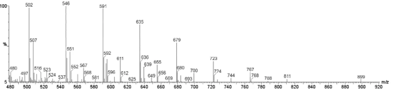
The partially purified SAC from the Type I Rf 0.62
band was further characterized by ESI(+)-MS and FT-IR and 1H NMR spectroscopy. In the ESI(+)-MS of the Type I Rf 0.62 band, a homologue series
of 10 ions separated by 44 m/z units dominated from m/z 502 to m/z 899 (Fig. 2).
Figure 2 - ESI(+)-MS spectrum for the sample of Rf 0.62 from the extract Type I produced by
Gordonia amicalis.
The results suggested that these were oligomers containing a repeating unit of 44 Da. The 13C isotopologue ions with its m/z value 1 unit higher (m/z 591 and 592 for instance) showed that these species were singly charged protonated molecules, that is [M + H]+ions. To our knowledge a biosurfactant with a similar ESI(+)-MS subunit pattern for which -(CH2CH2O)n- or an isomeric
unit such as -(CH2CH(OH))n- seems likely and has
not previously been reported (Nitschke et al. 2004; Nitschke et al. 2005; Jacques et al. 2007; de Araújo et al. 2011).
FT-IR analysis of the Type I SACs from the Rf
Figure 3 - FT-IR spectrum for the sample of Rf 0.62
from the extract Type I produced by G. amicalis.
1
H NMR (300 MHz, CDCl3) analysis of the Type I
SACs from the Rf 0.62 band showed the presence
of unsaturated carbon bonds =CH (δ 5.39), saturated carbon bonds –CH (δ 0.4 –2,5) and amide bonds (δ 8.60).A structure consistent with the observed data and a 44 Da repeated was, therefore, indeed [–(CH2-CHOH)-]n. Alternatively,
the 44 Da repeating unit could result from
acetylation (CH3-CO-, 43 Da) of a carbon double
bound, which would be expected to also result in the addition of a hydrogen atom to one of the other carbon atom of the double band, resulting in an increase in mass of 44 Da.
Two possible structures for the [M + H]+ ion of
m/z 502, based on the combined analytical data discussed above, were:
1) CH3–(CH2)14–(CH=CH–CH2–CH2)3–CH=CH–
CH2-CHOH–CH2–CONH2
2) CH3–(CH2)7–CH=CH–(CH2)2– CH=CH–(CH2
-CHOH)6–CONH2
ESI(+)-MS analysis of the TLC Type II band with Rf0.62 produced a similar spectrum as that from
the Type I band with Rf0.62. Figure 4 shows the
spectrum of the Type II SAC's from the TLC band with Rf 0.52. Again, a homologous mixture of 10 oligomers with a m/z separation of 44 units was observed. However, the SACs from the Rf 0.52
band showed relevant changes in relative abundances with, for instance, more abundant ions of m/z 546 and 590. 1H NMR analysis of this sample (Rf 0.52) showed similar characteristics as
that obtained in the spectra of band with Rf 0.62,
suggesting the same basic structure.
Figure 4 - ESI(+)-MS spectrum for the sample of Rf 0.52 from the extract Type II produced by
Gordonia amicalis.
The crude extracts of the Type I and Type II SACs did not present antimicrobial activity at any of the
concentrations assayed. The absence of
antimicrobial activity is desirable for in-situ
bioremediation to avoid the disruption of indigenous microbial communities that typically
provide most of the biodegradation activity in contaminated sites.
In the genus Gordonia, the most extensively studied species for the production of SACs is
the production of at least two types of SACs byG. amicalis; however, structural characterization of these compounds was not performed. The present study showed that, depending on the isolation technique used, the G. amicalis used in this study yielded two types of SACs (Type I and II) with different activities, one which reduced the surface tension of water and produced O/W emulsions (Type I) and one that primarily produced W/O emulsions (Type II). The analysis by ESI(+)-MS, FT-IR and 1H NMR indicated that the structure of the Type I and II SAC’s were very similar. Both the preparations contained a homologous series of oligomers, differing by a structural repeating unit of 44 Da. The data indicated that the compounds contained C, N and O, and carbonyl, hydroxyl and amide bonds. The spectroscopic and spectrometric data of these compounds did not to match with other known biosurfactants and could represent a novel class of these interesting compounds. To elucidate further the structure of the identified SACs, the results showed that modified sugars and amino acids were possibly attached via the amide group. Lipids containing amide linkages to polar head groups are common in the cell membranes and lipopolysaccharides of Gram-negative bacteria in the form of ornithine-containing lipids and lipid A. They are present in the biosurfactant emulsion produced by Acinetobacter calcoaceticus RAG-1(Gautam et al. 2006). Ornithine lipids with amide linkages have also been found in Mycobacterium
(Lanéelle et al. 1990).
CONCLUSIONS
The specific activity of the crude SAC preparations was similar to the commercial surfactants tested in this study in terms of emulsification activity. It was likely that the superficial activity could be significantly increased with further purification. The ability to produce two different types of SAC activities by G. amicalis could be advantageous in applications in
the chemical, cosmetic, food, biomedical
applications and bioremediation industries.
ACKNOWLEDGMENTS
This work was supported by FAPESP (00/05092-0 and 98/11906-9).
REFERENCES
Arenskötter M, Broker D, Steinbuchel A. Biology of the metabolically diverse genus Gordonia. Appl
Environ Microbiol. 2004; 70: 3195-3204.
Banat IM, Makkar RS, Cameotra SS. Potential commercial applications of microbial surfactants.
Appl Microbiol Biotechnol. 2000; 53: 495-508.
Brown MJ. Biosurfactants for cosmetic applications. Int J Cosmet Sci. 1991; 3: 61-64.
Cole JR, Chai B, Farris RJ, Wang Q, Kulam-Syed-Mohideen AS, McGarrell D, et al. The ribosomal database project (RDP-II): introducing myRDP space and quality controlled public data. Nucleic Acids Res.
2007; 35: D169-D172.
Cole JR, Wang Q, Cardenas E, Fish J, Chai B, Farris RJ, et al. The Ribosomal Database Project: improved alignments and new tools for rRNA analysis. Nucleic
Acids Res. 2009; 37: D141-D145.
de Araújo MEMB, Campos PRB, Noso TM, Alberici RM, Cunha IBS, Simas RC, et al. Response surface modeling of the production of structured lipids from soybean oil using Rhizomucor miehei lipase. Food
Chem. 2011; 127: 28-33.
Dogan I, Pagilla KR, Webster DA, Stark BC. Expression of Vitreoscilla hemoglobin in Gordonia
amarae enhances biosurfactant production. J Ind
Microbiol Biotechnol. 2006; 33: 693–700.
Franzetti A, Bestetti G, Caredda P, La Colla P, Tamburini E. Surface-active compounds and their role in the access to hydrocarbons in Gordonia
strains. FEMS Microbiol Ecol. 2008; 63: 238-248. Gautam KK, Tyagi VK. Microbial surfactants: A
review. J Olea Sci. 2006; 55: 155-166.
Iwahori K, Tokutomi T, Miyata N, Fujita M. Formation of stable foam by the cells and culture supernatant of
Gordonia (Nocardia) amarae. J Biosci Bioeng. 2001; 92: 77-79.
Jacobucci DFC, Oriani MRG, Durrant LR. Reducing COD level on oily effluent by utilizing biosurfactant-producing bacteria. Braz Arch Biol Technol. 2009; 52:1037-1042.
Jacobucci DFC, Vasconcelos CK, Matsuura ABJ, Falconi FA, Durrant LR. Degradation of diesel oil by biosurfactant-producing bacterial strains. AEHS
Contaminated Soil, Sediment and Water. 2001; 8:
31-34.
Jacques RJS, Santos EC, Haddad R, Catharino RR, Eberlin MN, Bento FM, Camargo FAD. Mass spectrometry analysis of surface tension reducing substances produced by a path-degrading
Pseudomonas citronellolis strain. Braz J Microbiol.
2007; 39: 352-353.
Navon-Venezia S, Zosim Z, Gottlieb A, Legmann R, Carmeli S, Ron EZ, Rosenberg E. Alasan, a new bioemulsifier from Acinetobacter radioresistens.
Appl Environ Microbiol. 1995; 61: 3240-3244.
Nitschke M, Costa SGVAO, Haddad R, Gonçalves LAG, Eberlin MN, Contiero J. Oil Wastes as Unconventional Substrates for Rhamnolipid Biosurfactant Production by Pseudomonas aeruginosa LBI. Biotechnol Prog. 2005; 21: 1562-1566.
Nitschke M, Haddad R, Costa GN, Gilioli R, Meurer EC, Gatti MSV, Eberlin MN, Höehr NF, Pastore GM. Structural Characterization and Biological Properties of a Lipopeptide Surfactant Produced by Bacillus subtilis on Cassava Wastewater Medium. Food Sci Biotechnol. 2004; 13: 591-596.
Pagilla KR, Sood A, Kim H. Gordonia (Nocardia)
amarae foaming due to biosurfactant production.
Water Sci Technol. 2002; 46: 519-24.
Rapp P, Backhaus S. Formation of extracellular lipases by filamentous fungi, yeast and bacteria. Enzyme
Microb Technol. 1992; 14: 938-943.
Rocha C, San-Blas F, San-Blas GE, Vierma L. Biosurfactant production by two isolates of
Pseudomonas aeruginosa. World J Microbiol
Biotechnol. 1992; 8: 125-128.
Singh A, Van Hamme JD, Ward OP. Surfactants in microbiology and biotechnology: Part 2 - Application aspects. Biotechnol Adv. 2007; 25: 99-121.
Stanghellini ME, Miller RM. Biosurfactants - Their identity and potential efficacy in the biological control of zoosporic plant pathogens. Plant Dis. 1997; 81: 4-12.