Kinetic study on microstructural changes during convective air drying of grapes
8
0
0
Texto
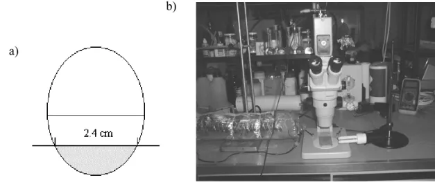
(2) INTRODUCTION Drying is probably the oldest method of preserving fruits and initially was used as a way of storing foods for the low season. With the development of new preservation methods and easiness to obtain fresh fruits in the market, drying is currently regarded as another method for diversifying products for consumer's convenience. Although it has lost its importance facing the new technologies, sun drying in particular is widely used in developing countries. One of the major sun drying products are raisins (1). The most common grape (Vitis vinifera) varieties turned into raisins are Thompson Seedless, Black Corinth and Muscat of Alexandria (2). Raisins are usually included in breakfast cereals, dairy, bakery and confectionery products, and more recently nutritional bars. Air drying leads to major changes in the fruit structure and texture, such as fruit 'softening' or loss of fruit firmness (3). Texture of dried fruits is a very important organoleptic characteristic. Physical properties of foods and texture in particular may be related to their microstructure (4). The initial work of Reeve (5) and Crafts (6) reported the effect of drying on fruit and vegetable microstructure, and considered shrinkage of tissues as a major physical change. Long neglected in the formulation of drying models, shrinkage is recognized now as an important consequence of fruit drying that has to be accounted for, since it modifies the shape and dimension of products (7), which in turn affects the mass transport phenomena. Cellular shrinkage causes modifications in the global structure of fruits, and is directly related to the loss of water during drying. According to Hills and Remigereau (8), who studied the air-drying of parenchyma apple tissue, drying results in loss of water from the vacuolar compartment, with minor changes in the water content of either the cytoplasm or the cell wall compartments. Water loss during drying causes loss of turgor pressure, which depends on the osmotic pressure and induces plasmolysis (9), affecting cellular wall integrity. In cellular tissues it is the turgor pressure that keeps them firm, thus it is related to fruit texture. Physical cellular changes may be quantified by image analysis (3). Nowadays, several microscopic equipments with high magnification and resolution power, can be applied to study food microstructure (e.g. Scanning Electron Microscopy is widely used). Microscopy is a powerful tool for studying food microstructure, especially if complemented with techniques of image analysis (4). Stereo-microscopy has limited magnification, but due to its large focal distance it allows the observation of large fruit pieces during drying while being a non-invasive method. Preparative techniques are fast and do not originate many alterations of the sample. The information obtained from the microscope is then suitable for quantification, by using software for image analysis (10). The main goal of this research was to introduce a microstructural approach in drying of fruits. Observing what happens at the microscopic level during drying will result not only on qualitative information, but also on quantitative data suitable for modeling. Therefore, a more specific objective was to quantify physical microstructural changes during air drying of grapes quarters, and in particular to quantify cellular shrinkage.. MATERIALS AND METHODS Samples. Fresh grapes (Vitis vinifera) from the Ruby variety, were bought from a local market and stored in a refrigerator for two weeks. Water content was determined using a vacuum oven, at 70°C until a stable weight value was reached. Original grapes had a mean water content of 80.63% ± 0.14 (w/w) on wet basis. Grape quarters of approximately 2.4 cm diameter were cut with a razor blade, as schematically presented in Figure 1a). Cellular walls were stained in a solution of methylene-blue 0.034%, during 15 seconds prior to drying.. 2.
(3) Drying experiments. Experiments were carried out using a drying stage mounted under the microscope (Figure 1b). Two electrical resistances and a fan were mounted inside a stainless steel tube 10 cm diameter and thermally insulated with glass fibre, creating a forced hot airflow. Temperature was monitored with a digital thermometer (Delta Ohm HD8802, Padova, Italy) and a thermocouple (type K). Air relative humidity and velocity were measured with a hygrometer (Rotronic AM3, New York, USA) and an anemometer (Airflow LCA 6000,Buckinghamshire, England), respectively. A grape quarter was held on the top of four pins stuck into a support, and placed under the microscope objective. Drying experiments were carried out at 20°C (49.2% mean air relative humidity, RH), 30°C (36.1% RH), 40°C (12.5% RH), 50°C (10.4% RH) and 60°C (2.2% RH), for approximately 2 hours and with a constant air velocity of 1.6 ± 0.1 m/s. Two replicate runs were carried out for each condition. b) a). Figure 1. a) Schematic draw to cut the sample. Shadowed part was used for drying. b) Stereo-microscope with video camera and air-drying tube. Microstructure and Image Analysis. A group of cells was continuously focused along drying time, with a stereo-microscope (Olympus SZ11, Tokyo, Japan) using a magnification of 25 X. A digital color video-camera (Sony Exwave Had, Tokyo, Japan) was attached to the microscope and then connected to a TV monitor and a personal computer. Image acquisition was done with an interface (Logitech Snappy, Rancho Cordova, USA). Images were captured at given time intervals with Snappy software. This technique allows monitoring the same group of cells during the entire drying process. For each experiment, a group of 20 cells was selected for further amplification and analysis with Paint Shop Pro 4.12 and UTHSCA Image Tool 2.0 (University of Texas Health Science Center, Texas, USA) software. Each cell was delimited and several geometrical parameters were determined using the specified software: area, perimeter, major and minor axis length, Feret diameter, elongation, roundness and compactness. The Feret diameter (FD) corresponds to the square root of the area divided by π. Elongation is calculated as the ratio of the major axis to the minor axis length. Roundness is equal to 4π times the area divided by the square of the perimeter and compactness equals FD divided by the major axis length. Elongation, roundness and compactness are indicators of the sphericity of the cell. Cells with shape closer to a perfect circle present values of elongation, roundness and compactness closer to one. At selected times during each experiment, each geometric cellular parameter was evaluated for all the selected 20 cells and a mean value was calculated.. 3.
(4) Data Analysis. In order to eliminate initial variability between different fruits and even different cell groups, every cellular parameter was normalized (i.e. divided by its initial value, at zero time). Experimental data for each cellular parameter related to dimension, were mathematically modeled. The effect of drying time, at constant temperature, was described by a first order kinetics: p (1) = exp ( − k T t) p o where p is the cellular parameter value, po its initial value at time zero, t the time and kT the rate of change constant at a given temperature (T). Arrhenius law was used to model the temperature effect on the rate of change constant: Ea 1 1 ) ( − k = k ref exp− R T Tref . (2). where kref is the rate of change constant at a reference temperature, Tref, Ea the activation energy, R the Universal gas constant and T the absolute temperature. A one-step non-linear regression was simultaneously performed to all data (11, 12) using the statistical software STATA version 3.0 (Texas, USA). For every cellular parameter related to dimension, a rate of change constant at reference temperature (40°C), an activation energy and the corresponding 95% confidence intervals were obtained. For cellular parameters related with shape a two-way analysis of variance was performed for unbalanced data at a 95% confidence level.. RESULTS AND DISCUSSION As expected, a gradual overall shrinkage of the grape cells was observed with drying time (Figure 2). Disruption of cellular walls with consequent cellular collapse, was clear in some experiments. At the beginning of drying the surface became more brilliant with the gradual increase of liquid water.. Figure 2. Images of grape cells shrinkage at 40°C, as a function of time. Results obtained from image analysis presented two distinct behaviors. Cellular parameters directly related to dimensions (area, perimeter, major and minor axis length and Feret diameter) clearly. 4.
(5) decreased along drying time. The remaining parameters (elongation, roundness and compactness), related to the cells shape, presented normalized values randomly distributed around one, thus no significant changes were noticed. Experiments were concluded before drying equilibrium had been reached, since at longer drying times visualization of cellular walls became more difficult. Cellular walls collapsed due to the material characteristics and the drying process. This phenomenon also reported by Reeve (5) is accelerated at higher temperatures. Cellular area was found to be the most interesting parameter to be studied, because it is directly related to shrinkage. Cellular area as a function of time showed an exponential decrease and was well described by a first order model (Eq. 1), with higher temperatures leading to an acceleration of the rate of area decrease (Figure 3). Thus, higher temperatures increase cellular shrinkage of grapes. As an example, a change in temperature from 20 to 60°C increased the rate constant for area change by 350%. 1,1. 20°C 30°C 40°C 50°C 60°C. a) area / initial area. 1,0. 0,9 0,08. b). 0,06. 0,8. 0,04 0,02 0. 0,7. 0,6. 0,7. 0,8. 0,9. 1. 1,1. -0,02 -0,04. pre dic t e d v a lue s. 0,6 0. 50. 100. 150 time (min). 200. 250. 300. Figure 3. a) Experimental cellular area as a function of time and temperature. Continuous lines represent model predicted values. b) Model residuals as a function of predicted values. Other cellular parameters directly related to dimension have a similar behavior: their magnitude also decreases with drying time and higher temperatures accelerate their rate of change. This effect of temperature on rate constant was well described by the Arrhenius model (Eq. 2). Experimental values for cellular area, model predicted values and corresponding residuals, obtained from one-step non-linear regression, can be observed in Figure 3. Kinetic cellular parameters are compiled in Table 1, together with corresponding standard errors, for each parameter related to cellular dimension. The magnitude of the values obtained is 10-3 min-1 and 3000 J/mol for the rate of change at 40°C and activation energy, respectively. Table 1 Rate of change at 40°C and activation energy, for each cellular parameter related to dimensions (kinetic parameters were obtained by a one-step non-linear regression of all data). Cellular parameters Area Perimeter Major axis Minor axis FD. k40°C x 10-3 (min-1). Ea (kJ/mol). 1.46 ± 0.05 0.89 ± 0.05 0.88 ± 0.05 0.95 ± 0.05 0.91 ± 0.04. 3.1 ± 0.3 3.1 ± 0.4 2.9 ± 0.3 3.6 ± 0.7 3.3 ± 0.3. 5.
(6) Cellular parameters directly related to shape did not show any tendency with drying time. Bolin and Huxsoll (3) reported that the roundness index (inverse of roundness) for cells of apple rings increased with drying, however for grape cells this was not observed. Compactness and roundness remained practically unchanged (Figure 4).. Roundness / Initial Roundness. 1,2. 20°C 30°C 40°C 50°C 60°C. 1,1. 1,0. 0,9. 0,8 0. 50. 100. 150 200 tim e (m in). 250. 300. Figure 4. Experimental cellular roundness as a function of time and temperature. Cellular elongation embraced a wider range of values when compared to roundness or compactness. This may be due to a higher sensitivity of the elongation mathematical expression, to cell changes during drying. Elongation seemed to present a smooth tendency to increase along drying time. Wang and Brennan (7) had observed the elongation of cellular walls by microscopy during potato drying experiments. However, for these experiments with grapes the table of single factor ANOVA showed no significant differences. Therefore, experimental results for grape cells did not follow the behavior previously described for some cells of agricultural materials. The results of this research were obtained for cells of grape quarter tissues, therefore conclusions have to be carefully extrapolated for the case of drying intact agricultural products.. CONCLUSIONS Stereo-microscopy has proved to be a useful non-invasive tool in studying drying at microscopic level. This technique allows monitoring the same cells from a quarter of fruit tissue during the entire drying process. Cellular shape and dimensions were quantified during drying, thanks to powerful techniques of image analysis. It was observed that the cells dimensions suffered modifications during drying, but their shape remained unchanged. A kinetic approach was successfully applied to model these microstructural changes. Area, perimeter, major and minor axis length and Feret diameter presented an exponential decrease with drying time and data were well described by a first-order model. Within the studied range, temperature increased the rate of cellular shrinkage in grape quarters and this effect followed an Arrhenius type behavior. Under the drying conditions used, it was not possible to observe any consistent trend of cellular elongation, roundness or compactness with time and no generalization on the effect of temperature on these parameters could thus be made. The study of drying at microscopic level will certainly contribute to better understand its mechanisms.. 6.
(7) ACKNOWLEDGEMENTS Part of this research was supported by the RED ALFA: "Food Quality in Food Engineering” (ref:5-0130-9) and the CYTED XI.13: "Relaciones Estructura-Propriedad en la Deshidratacion y Almacenaje de Alimentos Dehidratados" projects. The author Inês N. Ramos would like to acknowledge PRAXIS XXI PhD grant no. 18543/98 to Fundação para a Ciência e a Tecnologia, Portugal. The authors would like also to acknowledge the contribution of Dr. Laura Cadoche during the initial set-up of the experiments.. NOTATION Ea FD k p R RH T t. activation energy (J/mol) Feret diameter rate of change constant (min-1) cellular parameter (area, perimeter, major and minor axis length, Feret diameter, elongation, roundness and compactness) universal gas constant (J/mol K) relative humidity (%) absolute temperature (K) time (min). Subscript o initial value ref at reference temperature T at temperature T. REFERENCES AND NOTES (1) Ratti, C.; Mujumdar, A. S. Drying of Fruits. In Processing Fruits: Science and Technology; Somogyi, L. P., Ramaswamy, H. S., Hui, Y. H., Eds.; Technomic Publishing: Lancaster, 1996; Vol. 1, pp 185220. (2) Patil, V. K.; Chakrawar, V. R.; Narwadkar, P. R.; Shinde, G. S. Grape. In Handbook of Food Science and Technology; Salunkhe, D. K., Kadam, S. S., Eds.; Marcel Dekker: New York, 1995; pp 7-38. (3) Bolin, H. R.; Huxsoll, C. C. Scanning Electron Microscope / Image Analyzer Determination of Dimensional Postharvest Changes in Fruit Cells. J. Food Sci. 1987, 6, 52, 1649-1650. (4) Aguilera, J. M.; Stanley, D. W. Microstructural Principles of Food Processing and Engineering; Aspen Publishers: Gaithersburg, 1999; 2nd ed. (5) Reeve, R. M. A. Microscopic Study of the Physical Changes in Carrots and Potatoes During Dehydration. Food Res. 1943, 8, 128-136. (6) Crafts, A. S. Cellular Changes in Certain Fruits and Vegetables During Blanching and Dehydration. Food Res. 1944, 9, 442-452. (7) Wang, N.; Brennan, J. G. Changes in Structure, Density and Porosity of Potato During Dehydration. J. Food Eng. 1995, 24, 61-76. (8) Hills, B. P.; Remigereau, B. NMR Studies of Changes in Subcellular Water Compartmentation in Parenchyma Apple Tissue During Drying and Freezing. Int. J. Food Sci. Technol. 1997, 32, 51-61.. 7.
(8) (9) Jewell, G. G. Fruits and Vegetables. In Food Microscopy; Vaughan, J. G., Ed.; Academic Press: London, 1979; pp 1-34. (10) Aguilera, J. M.; Lillford, P. J. Microstructural and Imaging Analysis as Related to Food Engineering. In Food Engineering 2000; Fito, P., Ortega-Rodríguez, E., Barbosa-Cánovas, G., Eds.; Chapman & Hall: London, 1997; pp 23-38. (11) Lund, D. B. Considerations in Modelling Food Processes. Food Technol. 1983, 37, 92-94. (12) Arabshahi, A.; Lund, D. B. Considerations in Calculating Kinetics Parameters from Experimental Data. J. Food Process Eng. 1985, 7, 239-251.. 8.
(9)
Imagem


Documentos relacionados