doi: 10.3389/fpls.2019.00694
Edited by: José Manuel Martínez-Rivas, Instituto de la Grasa (IG), Spain Reviewed by: Eddo Rugini, Università degli Studi della Tuscia, Italy Enrique Martinez Force, Spanish National Research Council (CSIC), Spain *Correspondence: Maria Doroteia Campos [email protected]
Specialty section: This article was submitted to Crop and Product Physiology, a section of the journal Frontiers in Plant Science Received: 06 March 2019 Accepted: 08 May 2019 Published: 29 May 2019 Citation: Campos MD, Zellama MS, Varanda C, Materatski P, Peixe A, Chaouachi M and Félix MR (2019) Establishment of a Sensitive qPCR Methodology for Detection of the Olive-Infecting Viruses in Portuguese and Tunisian Orchards. Front. Plant Sci. 10:694. doi: 10.3389/fpls.2019.00694
Establishment of a Sensitive qPCR
Methodology for Detection of the
Olive-Infecting Viruses in Portuguese
and Tunisian Orchards
Maria Doroteia Campos1* , Mohamed Salem Zellama2, Carla Varanda1,
Patrick Materatski1, Augusto Peixe3, Maher Chaouachi2and Maria do Rosário Félix3
1ICAAM – Instituto de Ciências Agrárias e Ambientais Mediterrânicas, Instituto de Investigação e Formação Avançada,
Universidade de Évora, Évora, Portugal,2Laboratoire de Recherche “Bioressources: Biologie Intégrative & Valorisation,”
Institut Supérieur de Biotechnologie de Monastir, Université de Monastir, Monastir, Tunisia,3Departamento de Fitotecnia,
ICAAM – Instituto de Ciências Agrárias e Ambientais Mediterrânicas, Escola de Ciências e Tecnologia, Universidade de Évora, Évora, Portugal
Sensitive detection of viruses in olive orchards is actually of main importance since these pathogenic agents cannot be treated, their dissemination is quite easy, and they can have eventual negative effects on olive oil quality. The work presented here describes the development and application of a new SYBR R
Green-based real-time quantitative PCR (qPCR) analysis for specific and reliable quantification of highly spread olive tree viruses: Olive latent virus 1 (OLV-1), Tobacco necrosis virus D (TNV-D), Olive mild mosaic virus (OMMV), and Olive leaf yellowing-associated virus (OLYaV). qPCR methodology revealed high specificity and sensitivity, estimated in the range of 0.8–8 copies of the virus genome, for the studied viruses. For validation of the method, total RNA and double strand RNA (dsRNA) from naturally infected trees were used. In a first trial, dsRNAs from trees of cv. “Galega vulgar” from a Portuguese orchard, were subjected to qPCR and from the 30 samples tested, 26 were TNV-D and/or OMMV-positive and 25 were OLV-1 positive. In a second trial, total RNA from trees of different cultivars from Tunisian orchards, were here tested by qPCR and all viruses were detected. From the 33 samples studied, the most prevalent virus detected in Tunisia orchards was OLV-1 (3OLV-1 samples diagnosed), followed by OLYaV (20 samples diagnosed), and finally the combination in last TNV-D and/or OMMV (12 samples diagnosed). In both trials, qPCR demonstrated to be effective and sensitive, even when using total RNA as template. qPCR through the use of a SYBR R
Green methodology enabled, for the first time, a reliable, sensitive, and reproducible estimation of virus accumulation in infected olive trees, in which viruses are usually in low titres, that will allow gaining new insights in virus biology essential for disease control and give an important contribution for establishment of sanitary certification of olive propagative material.
INTRODUCTION
Olive trees are susceptible to several pathogens that may affect the yield and quality of their products with important economic impact, such as diseases causes by fungi (i.e., Colletotrichum spp. and Spilocaea oleagina) (Salman, 2017; Materatski et al., 2018), and more recently the emergence of Xylella fastidiosa, first noticed in 2013, and responsible for a severe outbreak of Olive quick decline syndrome (Saponari et al., 2013). Regarding virus infection, plants infected usually present morphological and physiological alterations, which always incurs in inferior performance such as the decreased host biomass and crop yield loss, with chloroplast known to be highly involved in those processes (Zhao et al., 2016). Olive trees are affected by several viruses, with 15 olive viruses identified until now (Félix et al., 2012). Viral symptoms on olive trees include bumpy fruits, leaf yellowing, vein banding, and vein clearing, (Martelli, 1999;
Albanese et al., 2012). These symptoms are non-specific and difficult to recognize, and often occur in virus-infected olive trees without apparent symptoms, which makes viral diagnosis in the field impossible to perform (Martelli et al., 2002;Alabdullah et al., 2010;Martelli, 2011;Zellama et al., 2018). A decrease in oil yield and maturity index in virus infected olives was also reported, as well as elevated total phenols in olive oil content from infected olives when compared to healthy fruits (Godena et al., 2012). Nevertheless, the Olive leaf yellowing associated virus (OLYaV) was not found to have a negative interference in oil yield and quality (Fontana et al., 2019).
Virus surveys carried in Mediterranean countries and others, concerning eight olive-infecting viruses (SLRSV, Strawberry latent ringspot virus; ArMV, Arabis mosaic virus; CLRV, Cherry leafroll virus; OLRSV, Olive latent ringspot virus; CMV, Cucumber mosaic virus; OLYaV, Olive leaf yellowing-associated virus; OLV-1,Olive latent virus 1; OLV-2, Olive latent virus 2), have shown high levels of infection, such as 75% in Tunisia, 51% in Syria, 33% in Italy, 31% in Lebanon, 25% in Croatia, and 31% of necroviruses infection in Portugal (Al Abdullah et al., 2005;Fadel et al., 2005; Faggioli et al., 2005; Varanda et al., 2010; Luigi et al., 2011; Zellama et al., 2018). However, the high presence of double stranded RNA (dsRNA) in olive orchards suggests even higher levels of infection (Félix et al., 2002; Saponari et al., 2002) that may be identified with new and more sensitive diagnostic techniques.
Phytosanitary certification programs depend on robust diagnostic procedures for detection of the olive viruses, since their spread will mainly depend on preventive measures such as the use of pathogen-tested propagative material (Albanese et al., 2012). Following the EU regulation on the marketing of fruit plants intended for fruit production, namely the Commission Directive 93/48/EEC (EEC Directive, 1993), the Conformitas Agraria Communitatis (CAC) category, applicable to olive plant material, demands that plants must be free of all viruses (Annex to the 93/48/EEC). The Italian certification of propagative material (DM 20/11/2006), followed by most of the Mediterranean countries, including the countries from the North of Africa, imposes the absence of several viruses such as ArMV, CLRV, SLRSV, CMV, OLV-1, OLV-2, OLYaV, andTobacco necrosis
virus senso lato, in which Tobacco necrosis virus D (TNV-D) and Olive mild mosaic virus (OMMV) are included (Albanese et al., 2012). The adoption of such certification programs requires the use of extremely sensitive diagnostic techniques for viral detection, which are the basis for valid programs. The low viral titres in olive tissues are the major constraint of the techniques, and do not always allow the successful, accurate and reproducible detection (Zellama et al., 2018).
Traditionally, unreliability techniques based on serology tests (ELISA) have been used for the detection of viruses (Martelli, 1999;Bertolini et al., 2001). More recently, molecular biology-based methods that include the highly laborious dsRNA analysis have been performed, once dsRNA reduces the constraint caused by the common low viral concentration in trees (Varanda et al., 2010, 2014;Zellama et al., 2018). RT-PCR has demonstrated to be the most rapid, sensitive, and reliable technique (Varanda et al., 2010; Luigi et al., 2011; Albanese et al., 2012; Zellama et al., 2018), using different templates as viral targets such as TNA, total RNA or dsRNA. The advancements of the molecular virology and biotechnology have witnessed major breakthroughs in the recent years resulting in highly sensitive and effective technologies/methods (Yadav and Khurana, 2016). In plant virology, real-time quantitative PCR (qPCR) is increasingly being used to improve sensitivity and accuracy while maintaining reliability (Poojari et al., 2016; Malandraki et al., 2017). The use of qPCR instrumentation presents several advantages, since it requires considerably short hands-on time, and detection of amplified products is automated, simple, and reproducible (Espy et al., 2006).
In the work described here, three new SYBR R
Green qPCR assays were developed for reliable and sensitive detection of OLV-1, TNV-D and/or OMMV, and OLYaV. The work involved the use of specific primers for each target virus, using different templates (total RNA and dsRNA). The main performance criteria were tested such as the sensitivity of the technique and the specificity of the primers. qPCR was validated through application to plant material from different olive orchards, from Portugal and Tunisia.
MATERIALS AND METHODS
Virus Isolates and Plant Material
Virus reference isolates of OLV-1, TNV-D, and OMMV were obtained according to Varanda et al. (2014). For OLYaV, since this virus is not mechanically transmitted (van Regenmortel et al., 2000), a sequenced cDNA obtained from an infected olive tree was used as reference (Zellama et al., 2018).
For validation and comparative analysis of virus infection, two different experiments were conducted. In the first one, samples were collected from 30 symptomatic or asymptomatic olive trees from an orchard under traditional management. The cultivar studied was the native cultivar “Galega vulgar,” and the orchard was located in Alentejo region (south Portugal).
The second experiment was performed in samples collected from a total of 33 symptomatic or asymptomatic olive trees. These samples were randomly selected from the 280 collected
by Zellama et al. (2018), but with the care to select 3 trees per each orchard. The sampled trees belonged to 11 orchards, under different types of management: three traditional, six semi-intensive, and two intensive. The orchards were located in several regions covering whole Tunisia. Sampled trees belonged to the Tunisian native cultivars “Chemlali,” “Chétoui,” “Meski,” and to the introduced cultivars “Picholine,” “Koroneiki,” and “Arbequina.” All samples consisted of four cuttings of ca. 20 cm in length collected from 2-year stems from each quadrant of the canopy of each tree, that were kept in plastic bags at 4◦
C, until further use.
Nucleic Acids Extraction and Reverse
Transcription
In the first experiment, double stranded RNAs (dsRNAs) were extracted from 300 mg of cortical scrapings according to
Morris and Dodds (1979). dsRNAs were visualized by 0.8% agarose gel electrophoresis prior to denaturation by heating at 100◦
C for 5 min, followed by 15 min on ice. cDNA was produced by RevertAid H Minus Reverse Transcriptase (Thermo Scientific), according to manufacturer’s instructions, with random hexamers (Promega).
In the second experiment, total RNA was extracted from 25 mg of cortical scrapings from cuttings that were mixed together and homogenized using liquid nitrogen. Total RNA was extracted using the RNeasy Plant Mini Kit (Qiagen), following the manufacturer’s instructions. The quantification of RNA and the evaluation of its purity were determined in a NanoDrop-2000C spectrophotometer (Thermo Scientific). RNA integrity was evaluated by denaturing gel electrophoresis. Total RNA (500 ng) was reverse transcribed with the Maxima First Strand cDNA Synthesis Kit (Thermo Scientific), according to manufacturer’s instructions.
Conventional PCR Assays
For conventional PCR assays, primers used for OLV-1 and OLYaV were the same as in Zellama et al. (2018) (Table 1). For the detection of OMMV and TNV-D, a single set of primers that allow the amplification of both TNV-D and OMMV was used (Table 1; Cardoso et al., 2004). 2 µl of cDNA were used in PCR carried out in 1x DreamTaq Buffer (Thermo Scientific), 0.2 mM dNTPs, 0.5µM of each primer and 2.5 U of DreamTaq
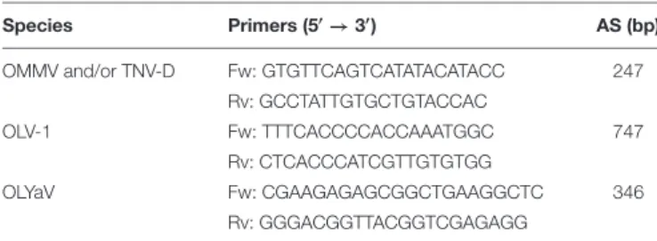
TABLE 1 | Primers used on conventional PCR assays.
Species Primers (50→
30
) AS (bp)
OMMV and/or TNV-D Fw: GTGTTCAGTCATATACATACC 247
Rv: GCCTATTGTGCTGTACCAC
OLV-1 Fw: TTTCACCCCACCAAATGGC 747
Rv: CTCACCCATCGTTGTGTGG
OLYaV Fw: CGAAGAGAGCGGCTGAAGGCTC 346
Rv: GGGACGGTTACGGTCGAGAGG
Description of forward (Fw) and reverse (Rv) primers used for specific detection of Olive mild mosaic virus (OMMV) and/or Tobacco necrosis virus D (TNV-D) (Cardoso et al., 2004), Olive latent virus 1 (OLV-1) and Olive leaf yellowing-associated virus (OLYaV) in the conventional PCR assays (Zellama et al., 2018). AS, amplicon size.
DNA Polymerase (Thermo Scientific) in a total volume of 50µl. Amplifications were carried out in a Thermal Cycler (Bio-Rad) at 95◦
C for 1 min, 35 cycles at 95◦
C for 30 s, 54◦
C (for OLV1 and both TNV-D and OMMV) or 58◦
C (for OLYaV) for 1 min, 72◦ C for 1 min, and a final extension step of 72◦
C for 10 min. Amplified products were analyzed by electrophoresis in 1% agarose gel.
SYBR
RGreen qPCR Assays
These assays were performed in both experiments. For TNV-D and/or OMMV it was used the already referred primer set (Cardoso et al., 2004). For OLV-1 a gene-specific primer set was designed in a conserved region of the capsid protein (CP), after alignment of all full-genome and CP sequences collected from NCBI GenBank database (accession n◦
KF804054). For OLYaV the same procedure was followed, but the primers were designed in the conserved gene coding the heat shock protein 90 (accession n◦
AJ844555). The Primer Express 3.0 Software (Applied Biosystems) was used for the design of the primers, using the default parameters of the software, and their specificity was tested in silico using basic local alignment search tool (BLAST) at the National Center for Biotechnology Information (NCBI)1. All qPCR primers are listed in Table 2.
qPCRs were carried out on a 7500 Real Time PCR System (Applied Biosystems) with SYBR Green q-PCR Master Mix (Nzytech). 5µL of first-strand cDNA (previously diluted 1:10) and 560 nM of each specific primer were used, performing in a total 18 µl reaction volume. The quantification cycle (Cq) values were acquired for each sample with the following cycling conditions: 10 min at 95◦
C for initial denaturation, an amplification program of 40 cycles at 95◦
C for 15 s and 60◦
C for 1 min. Three technical replicates were considered for each sample. Virus reference isolates and no template controls were included in all plates. The fluorescence threshold was manually set above the background level. The specificity of qPCR reactions was evaluated by melting curve analysis. The identity of each amplicon was confirmed by Sanger sequencing. Specific amplification of target viral cDNA was confirmed by cloning PCR amplicons into pGem R
-T Easy vector (Promega) and used to transformEscherichia coli JM109 (Promega) competent cells, by standard methodologies. Each amplicon, sequenced through Sanger procedure, was identified by comparison with corresponding virus sequences available from GenBank.
The specificity of each SYBR R
Green assay against the cDNA of the other virus reference isolates was also tested by qPCR.
For each specific viral fragment, to determine the amplification efficiencies of the primers, standard curves were generated from 10-fold dilution series of plasmid DNA (each viral amplicon were cloned into a plasmid vector, as described above), used to draw a nine-point calibration curve to validate each assay in the dynamic range chosen (0.8 to 8E7 target copies). Amplification efficiencies were calculated through the equation E = (10(−1/slope) − 1) × 100, as well as slope and linearity (coefficient of determination, R2). Detection sensitivities of the assays were determined from the Cq of the lowest plasmid dilution that fell within the
TABLE 2 | Primers used on qPCR assays.
Species Accession ID Primers (50→30) AS (bp) References
OMMV and/or TNV-D AY616760 Fw: GTGTTCAGTCATATACATACC 247 Cardoso et al. (2004)
Rv: GCCTATTGTGCTGTACCAC
OLV-1 KF804054 Fw: GGGGTATGATGGTGCTATGG 162 This work
Rv: ACTCCGCAATATCCGTTCTG
OLYaV AJ844555 Fw: GCTTATCTACTACGCCGATCTTGTC 71 This work
Rv-AAGAGTGGATCCATCTAGATCGAAA
Description of forward (Fw) and reverse (Rv) primers used for specific detection of Olive mild mosaic virus (OMMV) and/or Tobacco necrosis virus D (TNV-D), Olive latent virus 1 (OLV-1) and Olive leaf yellowing-associated virus (OLYaV), in the qPCR assays. AS, amplicon size.
linear standard curve. The method performed for absolute DNA quantification was based on the determination of the absolute number of target copies (TCN) previously described byCampos et al. (2018).
RESULTS
Linearity, Sensitivity, and Specificity of
qPCR Assays
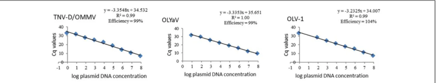
Standard curves of all studied olive viruses were constructed based on Cq values obtained from a 10-fold dilution series of the target plasmid DNA obtained from each reference isolate, in the dynamic range that was chosen. Linear relationships between Cq values and log plasmid DNA were obtained for all viruses, with regression coefficients (R2) above 0.99 (Figure 1). As expected, similar standard curves were obtained when OMMV and TNV-D were used as targets, once a single set of primers for amplification of both viruses was used. The efficiencies (E) for all virus-specific primer pairs ranged from 99% to 104% (Figure 1).
The detection sensitivity of each assay was determined from the Cq of the lowest plasmid dilution that fell with standard curve, that enable to detect 8 target copies of OLYaV and 0.8 target copies of TNV-D/OMMV and OLV-1 (Table 3). The Cq values that correspond to the detected target copies are indicated in Table 3.
The specificity of the assays was confirmed in silico, performing homology search similarities against NCBI databases, and experimentally, with each reference virus isolates targeted
only by the respective SYBR R
Green assay (not shown). Also, the specificity of the primers and their combinations were verified via cloning and sequencing of amplified products.
Validation of Real-Time qPCR Assays
Using Field Samples
To evaluate the practical robustness and accuracy of the qPCR assays, two experiments were conducted to test the presence of the viruses using field samples. Additionally, conventional PCR assays were also performed. It was also studied if total RNA template as viral target was suitable for virus detection through qPCR.
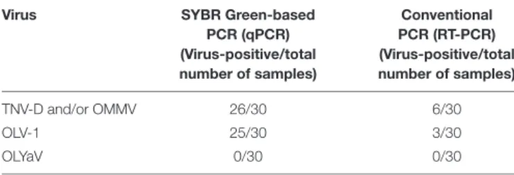
In the first experiment, concerning the use of dsRNA from 30 field-collected samples from a Portuguese olive orchard, OLYaV was not detected with neither qPCR nor conventional PCR. For the other studied viruses, qPCR revealed 26 positive samples for TNV-D and/or OMMV and 25 positive samples for OLV-1
TABLE 3 | Detection limits of each of the three qPCR assays based on determination of copy numbers.
Virus Cq value Copy number
TNV-D/OMMV 33.27 0.8
OLV-1 33.36 0.8
OLYaV 32.07 8
The detection range was determined from standard curves generated from the dilution series of respective recombinant plasmid DNA. The mean Cq values of positive samples were represented with respective copy numbers. TNV-D, Tobacco necrosis virus D; OMMV, Olive mild mosaic virus; OLV-1, Olive latent virus 1; OLYaV, Olive leaf yellowing-associated virus.
FIGURE 1 | Standard curves of olive viruses constructed based on Cq values obtained from a ten-fold dilution series of each target plasmid DNA in the dynamic range of 0.8 to 8E7 target copies. TNV-D, Tobacco necrosis virus; OMMV, Olive mild mosaic virus; OLV-1, Olive latent virus 1; OLYaV, Olive leaf yellowing-associated virus; R2, regression coefficients.
(Table 4 and Supplementary Table 1). Using conventional PCR, only six samples were positive for TNV-D and/or OMMV and three samples were positive to OLV-1.
To demonstrate the suitability of the qPCR methodology in different olive material and corroborate qPCR sensitivity, a second experiment was performed in 33 field-collected samples from 11 olive orchards, in several regions of Tunisia. For the qPCR experiment it was used as template total RNA from cortical scrapings from cuttings, instead of the highly laborious protocol for extraction of dsRNA used for conventional PCR. The results revealed that through qPCR and using total RNA as template, it was possible to detect all the studied viruses (Table 5 and Supplementary Table 1). qPCR revealed that the most prevalent virus detected in Tunisia orchards was OLV-1 (31 samples diagnosed), followed by OLYaV (20 samples diagnosed), and finally the combination in last TNV-D and/or OMMV (12 samples diagnosed). Results on conventional PCR revealed that 11 samples were OLV-1 positive, 10 samples were TNV-D and/or OMMV-positive and 10 samples were OLYaV positive, as determined byZellama et al. (2018).
Both experiments performed here demonstrate qPCR tests as suitable for detection of virus infecting olive.
DISCUSSION
Since there is no known source of genetic resistance to olive viruses, a key component of viral disease management is the propagation of virus-free plant materials. The main approach
TABLE 4 | Comparison of SYBR R
Green-based real-time quantitative reverse transcription PCR assays with conventional PCR, for virus detection in field-collected samples from a Portuguese olive orchard, with cv. “Galega vulgar.”
Virus SYBR Green-based
PCR (qPCR) (Virus-positive/total number of samples) Conventional PCR (RT-PCR) (Virus-positive/total number of samples) TNV-D and/or OMMV 26/30 6/30 OLV-1 25/30 3/30 OLYaV 0/30 0/30
TNV-D, Tobacco necrosis virus D; OMMV, Olive mild mosaic vírus; OLV-1, Olive latent virus 1; OLYaV, Olive leaf yellowing-associated virus.
TABLE 5 | Comparison of SYBR R
Green-based real-time quantitative reverse transcription PCR assays with conventional PCR, for virus detection in field-collected samples from 11 olive orchards, in several regions of Tunisia.
Virus SYBR Green-based
PCR (qPCR) (Virus-positive/total number of samples) Conventional PCR (RT-PCR) (Virus-positive/total number of samples) TNV-D and/or OMMV 12/33 10/33 OLV-1 31/33 11/33 OLYaV 20/33 10/33
Conventional PCR results included in Zellama et al. (2018). TNV-D, Tobacco necrosis virus D; OMMV, Olive mild mosaic vírus; OLV-1, Olive latent virus 1; OLYaV, Olive leaf yellowing-associated virus.
used to obtain, propagate and commercialize plants free from harmful pathogens is through phytosanitary selection and certification programs. The obtention of pathogen-free material from infected trees is possible to achieve through sanitation treatments such as heat therapy, meristem tip culture and micrografting, although the use of these methodologies for virus elimination in olive is still limited (Albanese et al., 2012). Thus, sensitive, reliable and cost-effective assays for the detection of olive viruses are critical and of major importance to guarantee a sustainable production of the orchards. The use of the highly sensitive qPCR arises as an extremely useful tool for studying various agents of infectious. In plant pathology, this technology is increasingly being used for studying various causal agents of plant diseases, including viruses (Poojari et al., 2016;
Malandraki et al., 2017;Abdullah et al., 2018;Baaijens et al., 2018;
Campos et al., 2019).
The work performed here, describes for the first time the use of a SYBR R
Green-based qPCR methodology for specific and reliable quantitative detection of highly spread olive viruses. Moreover, despite the low viral titres of the olive viruses (Martelli, 1999), qPCR turns possible the use of total RNA for the detection of viruses, instead of the highly laborious dsRNA extraction essential for RT-PCR (Varanda et al., 2010, 2014; Félix et al., 2012; Zellama et al., 2018). The primers used in the experiments successfully differentiated the target pathogens, and the results obtained show that qPCR is not only a faster method, but most importantly, a sensitive method for the detection and quantification of olive viruses directly from woody plant material. It was used a primer set that allow the simultaneous detection of TNV-D and OMMV (designed in the CP gene, see Cardoso et al., 2004), and newly designed primers targeting OLV-1 and OLYaV, that revealed E, R2 and slope consistent with the acceptance criteria (Broeders et al., 2014), confirming the accuracy and linear response of the assays over a wide range of dilutions, and suggesting absence of PCR inhibitors. These performance criteria further demonstrate that the developed assays, additionally to detection, constitute a reliable quantitative tool suitable for population or competition studies among the studied viruses in mixed infections in a variety of olive hosts. Conventional PCR conditions are different from qPCR conditions, with different optimized protocols and primers (only TNV-D and/or OMMV primer set was common to both methodologies), and consequently with not comparable results. Nevertheless, the idea of the higher sensitivity qPCR methodology, even with the low viral titres in olive tissues, is here reinforced. In the Portuguese cv. “Galega vulgar” the viruses OLV-1, TNV-D and/or OMMV were identified while OLYaV was not detected. OLV-1, TNV-D and/or OMMV were already identified in Portugal by RT-PCR methodology, although with lower rates (Cardoso et al., 2004, 2009; Varanda et al., 2010). In Tunisian orchards, independently of the cultivar, type of management or region, OLYaV, OLV-1, TNV-D and/or OMMV were detected, confirming previous RT-PCR results (Zellama et al., 2018), although qPCR registered a higher level of infection for the viruses under study. These results reinforce the idea of the sensitivity of the qPCR methodology, regardless of cultivar or region.
qPCR combined with the SYBR R
Green methodology enabled, for the first time, a high level of reliability, sensitivity and reproducibility regarding the estimation of virus accumulation in infected olive trees, being an efficient tool for the early diagnosis of the diseases, and for the improvement of the viral diagnosis. The results achieved will allow gaining new insights in virus biology, essential for disease control. Furthermore, this robust diagnostic procedure will give an important contribute for the establishment of phytosanitary certification programs of olive and high-quality productions.
DATA AVAILABILITY
The datasets generated for this study are available on request to the corresponding author.
AUTHOR CONTRIBUTIONS
MDC and MRF: conceptualization. MDC: formal analysis. MDC and AP: funding acquisition. MDC, MZ, and CV: methodology.
MDC, MZ, CV, and PM: resources. MRF and MC: supervision. MDC: writing, original draft preparation. MDC, MZ, CV, PM, AP, MC, and MRF: writing, review and editing.
FUNDING
This work was financially supported by FEDER and National Funds through the ALENTEJO 2020 (Regional Operational Program of the Alentejo), Operação ALT20-03-0145-FEDER-000014 – “Valorização das Variedades de Oliveira Portuguesas (OLEAVALOR)”, and by National Funds through FCT – Foundation for Science and Technology under the Project UID/AGR/00115/2019.
SUPPLEMENTARY MATERIAL
The Supplementary Material for this article can be found online at: https://www.frontiersin.org/articles/10.3389/fpls.2019.00694/ full#supplementary-material
REFERENCES
Abdullah, A. S., Turo, C., Moffat, C. S., Lopez-Ruiz, F. J., Gibberd, M. R., Hamblin, J., et al. (2018). Real-time PCR for diagnosing and quantifying co-infection by two globally distributed fungal pathogens of wheat.Front. Plant Sci. 9:1086. doi: 10.3389/fpls.2018.01086
Al Abdullah, A., El Beaino, T., Saponari, M., Hallak, H., and Digiaro, M. (2005). Preliminary evaluation of the status of olive-infecting viruses in Syria.EPPO Bull. 35, 249–252. doi: 10.1111/j.1365-2338.2005.00818.x
Alabdullah, A., Minafra, A., Elbeaino, T., Saponari, M., Savino, V., and Martelli, G. P. (2010). Complete nucleotide sequence and genome organization of olive latent virus 3, a new putative member of the family Tymoviridae.Virus Res. 152, 10–18. doi: 10.1016/j.virusres.2010.05.010
Albanese, G., Saponari, M., and Faggioli, F. (2012). “Phytosanitary certification,” in Olive Germoplasm-The Olive Cultivation, Table Olive and Olive Oil Industry in Italy, ed. I. Muzzalupo (Rijeka: InTech), 107–132.
Baaijens, R., Urbez-Torres, J. R. R., Liu, M., Ayres, M., Sosnowski, M. R., and Savocchia, S. (2018). Molecular methods to detect and quantify botryosphaeriaceae inocula associated with grapevine dieback in Australia. Plant Dis. 102, 1489–1499. doi: 10.1094/PDIS-11-17-1854-RE
Bertolini, E., Olmos, A., Martínez, M. C., Gorris, M. T., and Cambra, M. (2001). Single-step multiplex RT-PCR for simultaneous and colourimetric detection of six RNA viruses in olive trees.J. Virol. Methods 96, 33–41. doi: 10.1016/S0166-0934(01)00313-5
Broeders, S., Huber, I., Grohmann, L., Berben, G., Taverniers, I., Mazzara, M., et al. (2014). Guidelines for validation of qualitative real-time PCR methods.Trends Food Sci. Technol. 37, 115–126. doi: 10.1016/j.tifs.2014.03.008
Campos, M. D., Patanita, M., Campos, C., Materatski, P., Varanda, C. M. R., Brito, I., et al. (2019). Detection and quantification of Fusarium spp. (F. oxysporum, F. verticillioides, F. graminearum) and magnaporthiopsis maydis in maize using real-time PCR targeting the ITS region.Agronomy 9:45. doi: 10.3390/ agronomy9010045
Campos, M. D., Valadas, V., Campos, C., Morello, L., Cardoso, G., Braglia, L., et al. (2018). A TaqMan real-time PCR method based on alternative oxidase genes for detection of plant species in animal feed samples.PLoS One 13:e0190668. doi: 10.1371/journal.pone.0190668
Cardoso, J. M. S., Félix, M. R., Clara, M. I., and Oliveira, S. (2009). Complete genome sequence of a tobacco necrosis virus D isolate from olive trees.Arch. Virol. 154, 1169–1172. doi: 10.1007/s00705-009-0414-9
Cardoso, J. M. S., Félix, M. R., Oliveira, S., and Clara, M. I. E. (2004). A tobacco necrosis virus D isolate from Olea europaea L.: viral characterization and coat
protein sequence analysis.Arch. Virol. 149, 1129–1138. doi: 10.1007/s00705-003-0258-7
Espy, M. J., Uhl, J. R., Sloan, L. M., Buckwalter, S. P., Jones, M. F., Vetter, E. A., et al. (2006). Real-time PCR in clinical microbiology: applications for routine routine laboratory testing.Clin. Microbiol. Rev. 19, 165–256. doi: 10.1128/CMR.00022-06
Fadel, C., Digiaro, M., Choueiri, E., El Beaino, T., Saponari, M., Savino, V., et al. (2005). On the presence and distribution of olive viruses in Lebanon.OEPP EPPO Bull. 35, 33–36. doi: 10.1111/j.1365-2338.2005.00776.x
Faggioli, F., Ferretti, L., Albanese, G., Sciarroni, R., Pasquini, G., Lumia, V., et al. (2005). Distribution of olive tree viruses in italy as revealed by one-step RT-PCR.J. Plant Pathol. 87, 49–55.
Félix, M., Clara, M., Leitão, F., and Fernandes Serrano, J. (2002). Virus incidence in four olea europaea cultivars evaluated by mechanical inoculation and immunological assays.Acta Hortic. 586, 721–724. doi: 10.17660/ActaHortic. 2002.586.154
Félix, R., Varanda, C. M. R., and Clara, M. I. E. (2012). Biology and molecular characterization of necroviruses affecting olea europaea L.: a review. Eur. J. Plant Pathol. 133, 247–259. doi: 10.1007/s10658-011-9907-y
Fontana, A., Piscopo, A., Bruno, A., De Tiberini, A., Muzzalupo, I., and Albanese, G. (2019). Impact of olive leaf yellowing associated virus on olive (Olea europaea L.) oil.Eur. J. Lipid Sci. Technol. 121:1800472. doi: 10.1002/ejlt. 201800472
Godena, S., Bendini, A., Giambanelli, E., Cerretani, L., Ermi´c, D., and Ermi´c, E. (2012). Cherry leafroll virus: impact on olive fruit and virgin olive oil quality. Eur. J. Lipid Sci. Technol. 114, 535–541. doi: 10.1002/ejlt.201100277
Luigi, M., Godena, S., Dermi´c, E., Barba, M., and Faggioli, F. (2011). Detection of viruses in olive trees in Croatian Istria.Phytopathol. Mediterr. 50, 150–153. Malandraki, I., Beris, D., Isaioglou, I., Olmos, A., Varveri, C., and Vassilakos, N.
(2017). Simultaneous detection of three pome fruit tree viruses by one-step multiplex quantitative RT-PCR.PLoS One 12:e0180877. doi: 10.1371/journal. pone.0180877
Martelli, G. P. (1999). Infectious diseases and certification of olive: an overview. Bull. OEPP EPPO 29, 127–133. doi: 10.1111/j.1365-2338.1999.tb00806.x Martelli, G. P. (2011). “Infectious diseases of olive,” inOlive Diseases and Disorders,
eds L. Schena, G. E. Agosteo, and S. O. Cacciola (Tri-vandrum: Transworld Research Network), 71–78.
Martelli, G. P., Salerno, M., Savino, V., and Prota, U. (2002). An appraisal of diseases and pathogens of olive.Acta Hortic. 586, 701–708. doi: 10.17660/ ActaHortic.2002.586.150
Materatski, P., Varanda, C., Carvalho, T., Campos, M. D., and Rei, F. (2018). Diversity of colletotrichum species associated with olive anthracnose and new perspectives on controlling the disease in Portugal.Agronomy 8:301. doi: 10. 3390/agronomy8120301
Morris, T. J., and Dodds, J. A. (1979). Isolation and analysis of double-stranded RNA from virus-infected plant and fungal tissue.Phytopathology 69, 854–858. doi: 10.1094/Phyto-69-854
Poojari, S., Alabi, O. J., Okubara, P. A., and Naidu, R. A. (2016). SYBR R
Green-based real-time quantitative reverse-transcription PCR for detection and discrimination of grapevine viruses.J. Virol. Methods 235, 112–118. doi: 10. 1016/j.jviromet.2016.05.013
Salman, M. (2017). Biological control of spilocaea oleagina, the causal agent of olive leaf spot disease, using antagonistic bacteria.J. Plant Pathol. 99, 741–744. Saponari, M., Alkowni, R., Grieco, F., Pantaleo, V., Savino, V., Martelli, G. P., et al.
(2002). Detection of olive-infecting viruses in the mediterranean basin.Acta Hortic. 586, 787–790. doi: 10.17660/ActaHortic.2002.586.170
Saponari, M., Boscia, D., Nigro, F., and Martelli, G. P. (2013). Fungal species associated with a severe decline of olive in identification of dna sequences related to xylella fastidiosa in oleander, almond and olive trees exhibiting leaf scorch symptoms in apulia (southern italy).J. Plant Pathol. 95, 659–668. van Regenmortel, M. H. V., Fauquet, C. M., Bishop, D. H. L., Carstens, E. B.,
Estes, M. K., Lemon, S. M., et al. (2000).Virus Taxonomy. Seventh Report of the International Committee on Taxonomy of Viruses. San Diego, CA: Academic Press.
Varanda, C., Cardoso, J. M. S., do Rosário Félix, M., Oliveira, S., and Clara, M. I. (2010). Multiplex RT-PCR for detection and identification of three necroviruses
that infect olive trees.Eur. J. Plant Pathol. 127, 161–164. doi: 10.1007/s10658-010-9593-1
Varanda, C. M. R., Nolasco, G., Clara, M. I., and Félix, M. R. (2014). Genetic diversity of the coat protein of olive latent virus 1 isolates.Arch. Virol. 159, 1351–1357. doi: 10.1007/s00705-013-1953-7
Yadav, N., and Khurana, S. M. P. (2016). “Plant Virus Detection and Diagnosis: Progress and Challenges,” inFrontier Discoveries and Innovations in Interdisciplinary Microbiology, ed. P. Shukla (New Delhi: Springer).
Zellama, M. S., Varanda, C. M. R., Materatski, P., Nabi, N., Hafsa, A. B., Saamali, B. M., et al. (2018).An Integrated Approach for Understanding the High Infection Rates of Olive Viruses in Tunisia. Amsterdam: Springer Netherlands, doi: 10. 1007/s10658-018-01620-y
Zhao, J., Zhang, X., Hong, Y., Liu, Y., and Liu, Y. (2016). Chloroplast in plant-virus interaction.Front. Microbiol. 7:1565. doi: 10.3389/fmicb.2016.01565
Conflict of Interest Statement: The authors declare that the research was conducted in the absence of any commercial or financial relationships that could be construed as a potential conflict of interest.
Copyright © 2019 Campos, Zellama, Varanda, Materatski, Peixe, Chaouachi and Félix. This is an open-access article distributed under the terms of the Creative Commons Attribution License (CC BY). The use, distribution or reproduction in other forums is permitted, provided the original author(s) and the copyright owner(s) are credited and that the original publication in this journal is cited, in accordance with accepted academic practice. No use, distribution or reproduction is permitted which does not comply with these terms.