Linkage of cytosolic peroxiredoxin 2 to erythrocyte membrane imposed by hydrogen
peroxide-induced oxidative stress
Susana Rocha
a,b, Elísio Costa
a,b,c, Susana Coimbra
a,b,d, Henrique Nascimento
a,b, Cristina Catarino
a,b,
Petronila Rocha-Pereira
b,e, Alexandre Quintanilha
b,f, Luís Belo
a,b, Alice Santos-Silva
a,b,⁎
a
Faculdade de Farmácia, Universidade do Porto, Porto, Portugal
b
Instituto de Biologia Molecular e Celular (IBMC), Universidade do Porto, Porto, Portugal
c
Instituto de Ciências da Saúde, Universidade Católica Portuguesa, Porto, Portugal
d
Centro de Investigação das Tecnologias da Saúde (CITS), Instituto Politécnico da Saúde Norte, Gandra-Paredes, Portugal
eCentro de Investigação em Ciências da Saúde (CICS), Universidade da Beira Interior, Covilhã, Portugal f
Instituto de Ciências Biomédicas Abel Salazar (ICBAS), Universidade do Porto, Porto, Portugal
Peroxiredoxin 2 Erythrocyte membrane Oxidative stress Hemoglobin autoxidation
Human erythrocyte peroxiredoxin 2 (Prx2) is a typical 2-cys cytosolic peroxiredoxin with thiol-dependent hydrogen peroxide scavenger activity. In a previous work, we reported Prx2 erythrocyte membrane linkage in some Hereditary Spherocytosis patients and that it seemed to be related to oxidative stress. The aim of the present work was to determine if Prx2 linkage to erythrocyte membrane could be induced by oxidative stress mediated by H2O2and to further understand how and why this process occurs. We performed in vitro assays
in which catalase or both Hb autoxidation and catalase were inhibited, under H2O2-induced oxidative stress
conditions. Erythrocyte membrane linked Prx2 was detected by immunoblotting and quantified by densitometry. As oxidative stress markers, we determined membrane bound hemoglobin and lipid peroxidation, and we found that their values increased with H2O2concentration. Prx2 linkage to the
membrane also rose with increasing H2O2concentration, and was only observed when the oxidized form of
the enzyme was present in the cytosol. Oxidized Hb and Prx2 membrane linkages appear to be independent processes, although, both result from oxidative stress and may be useful as oxidative stress and/or erythrocyte damage/senescence markers.
Introduction
The erythrocyte is a very unique cell, not only because of its biconcave shape, but also, because it is a cell without nuclei or other organelles. Its main function is the transport/exchange of gases through the human body. Inherent to its function in transporting oxygen, a high content in hemoglobin (Hb) (95% of total protein in erythrocyte) and, therefore in iron, turns the red blood cell (RBC) into a preferential place for reactive oxygen species (ROS) formation. Accumulation of ROS within the cell can lead to lipid peroxidation, protein carbonylation and Hb denaturation, the reason why there is a variety of antioxidant systems to counteract the effects of ROS, namely, high quantities of enzymes, such as superoxide dismutase (SOD), catalase and peroxidases, specifically glutathione peroxidase (GSHPx) and peroxiredoxin 2 (Prx2). The latter is one of the most abundant cytoplasmatic proteins of the erythrocytes[1–3], although the way it operates in connection with other RBC antioxidant systems
is still poorly clarified; however, some recent studies indicate that Prx2 plays a preponderant role in maintaining low endogenous levels of hydrogen peroxide produced by Hb autoxidation[4,5].
Peroxiredoxins are a ubiquitous family of peroxidases with antioxidant proprieties. There are 6 types of mammalian peroxiredo-xins (I to VI), present in various tissues, and classified according to the presence of a conserved amino acid sequence and catalysis mechanism
[6,7]. Human erythrocyte Prx2 is a typical 2-cys peroxiredoxin, with a thiol-dependent hydrogen peroxide scavenger activity regulated by the thioredoxin (Trx)/Trx reductase/NADPH reducing system[7,8]. This protein has a molecular mass of 21.8 kDa[3,6]and exists in the cytoplasm as dimers and decamers in dynamic equilibrium[6,9,10]. Although cellular location of Prx2 is considered to be cytosolic, it can also be linked to the erythrocyte membrane[1,11–16]where it could be involved in mechanisms of defense against lipid peroxidation[11,17].
We recently reported[15]the linkage of Prx2 to the membrane of circulating RBCs in some Hereditary Spherocytosis (HS) patients; moreover, this linkage was correlated with higher amounts of membrane bound hemoglobin (MBH), a known marker of oxidative stress. In fact, several studies have reported a rise in MBH, both in physiologic (physical exercise[18], pregnancy[19,20]) and pathologic (myocardial infarction[21], stroke[22], psoriasis[23]) oxidative stress
⁎ Corresponding author. Serviço de Bioquímica, Faculdade de Farmácia da Uni-versidade do Porto, Rua Aníbal Cunha, 164, 4050-047, Porto, Portugal. Fax: +351 222 003 977.
E-mail address:assilva@ff.up.pt(A. Santos-Silva).
Abstract
related conditions. Senescent RBCs, that present high values of MBH
[24], are smaller, denser and with a higher membrane rigidity. These changes and the decreased enzyme activity in older RBCs, favor their splenic removal; in fact, the hypoxic environment and the vascular network that older RBCs have to face through spleen circulation, by testing the RBC metabolic activity (to overwhelming oxidative stress) and cell deformability, may condition their sequestration and removal. The aim of the present work was to determine if Prx2 linkage to the RBC membrane could be induced by oxidative stress conditions, and to understand how this process occurs. We performed in vitro assays in which catalase or both Hb autoxidation and catalase were inhibited, under hydrogen peroxide (H2O2)-induced oxidative stress conditions.
Materials and methods
Preparation of erythrocyte suspension
Blood samples from healthy volunteers (n = 3) were collected using heparin as anticoagulant.
Plasma and buffy-coat were discarded and red blood cells were washed in phosphate buffered saline (PBS) pH 7.4. The number of cells/ml in the obtained erythrocyte suspensions was determined using a hemocytometer, and, afterwards adjusted for 5 × 10⁸cell/ml with PBS, pH 7.4.
In vitro assays
Erythrocyte suspensions (5 × 10⁸cell/ml; 10 ml PBS, pH 7.4), under the following different assay conditions, were incubated during 30 min at 37 °C, under gentle agitation:
RBCs under H2O2-induced oxidative stress
Erythrocytes were incubated with hydrogen peroxide 25 μM,
50μM and 75 μM (the concentration of H2O2 stock solution was measured according to Low et al.[5]). A control tube containing only RBCs was used.
RBCs under H2O2-induced oxidative stress and catalase inhibition Erythrocytes were incubated with 100 mM sodium azide (NaN3) and H2O225μM, 50 μM and 75 μM. A control tube containing only RBCs and 100 mM NaN3was used.
RBCs under H2O2-induced oxidative stress and inhibition of catalase and Hb autoxidation
Before incubation, the erythrocytes were saturated with carbon monoxide (CO) and carboxyhemoglobin formation was determined by spectrophotometry[25]. Afterwards, RBCs were incubated with NaN3100 mM and H2O225μM, 50 μM and 75 μM. A control tube containing only RBCs saturated with CO and 100 mM NaN3was used. After incubation (for all in vitro assays), we removed an aliquot of each RBC suspension, in order to evaluate the oxidative status of cytosolic Prx2. Those erythrocyte aliquots were adjusted for 5 × 10⁶ cell/ml with PBS, pH 7.4 (2 ml); to these RBC suspensions N-ethyl
maleimide (NEM) was added (final concentration 100 mM) and,
afterwards, an incubation of 15 min at 37 °C, under gentle agitation, was performed.
The remaining suspension was processed to obtain erythrocyte membranes.
Preparation of erythrocyte membrane suspensions and hemolysates
To ensure minimal RBC membrane preparation variability, the protocol was performed by the same individual during the course of this study.
To prepare the erythrocyte membranes for electrophoretic analy-sis, RBCs were washed in saline solution, after the 30 minute
incubation period. The RBCs were submitted to hypotonic lyses, according to Dodge et al.[26]. The obtained membranes were washed in Dodge buffer, adding in thefirst two washes phenylmethylsulpho-nylfluoride, as a protease inhibitor, at a final concentration of 0.1 mM. The hemolysates were obtained after the 15 minute incubation period with NEM by hypotonic lyses with Dodge buffer of the pelleted erythrocytes, previously washed with saline solution.
The protein concentration of the erythrocyte membrane suspen-sions and of the hemolysates were determined by the Bradford's method[27].
To prepare erythrocyte membranes for electrophoresis, the membrane suspensions were treated with an equal volume of a solubilization buffer (0.125M Tris HCl pH 6.8, 4% sodium dodecil sulphate (SDS), 20% glycerol, 10% 2-mercaptoethanol) and heat denatured, when working in reducing conditions.
In order to determine the redox form in which Prx2 was present in the RBC cytosol, oxidizing conditions were utilized for the preparation of the hemolysate suspensions (solubilization buffer without a reducing agent)[5].
Immunoblot analysis for Prx2 in RBC membranes and hemolysates
Erythrocyte membrane suspensions and hemolysates were sub-mitted to SDS-PAGE in a 12% acrylamide gel (30μg protein/lane), using the discontinuous Laemmli system[28]. An internal control of human recombinant Prx2 protein (250 ng) (LabFrontier) was applied in all electrophoresis. Proteins were electrophoretically transferred from SDS gels to a nitrocellulose sheet with a porosity of 0.2 μm[29]. Additional reactive sites on the nitrocellulose were blocked by incubation in low fat dry milk (5% w/v) and 0.1% Triton-X 100 in PBS pH 7.0, for 1 h at room temperature and under gentle rotation. Prx2 immunoblot was then carried out by incubating the nitrocellulose with monoclonal antibodies anti-human Prx2 (dilution 1:3000), produced in mouse (LabFrontier), for 5 h; washing of the nitrocellulose was followed by incubation with anti-mouse IgG peroxidase linked (Sigma-Aldrich) for 1 h (dilution 1:3000). An enhanced chemilumi-nescence method (SuperSignal West Pico Chemiluminescent Sub-strate, Pierce) and a X-rayfilm (Kodak) were used to develop the immunoblot. The immunoblots were scanned (Darkroom CN UV/wl, BioCaptMW version 99, Vilber Lourmat) and the amount of Prx2 linked to erythrocyte membrane was determined by densitometry (Bio1D++ version99, Vilber Lourmat, France) by comparison with the internal control. The amount of Prx2 of each sample was determined as a ratio between the sample value and the internal control.
Membrane bound hemoglobin
The MBH was measured by spectrophotometry, after protein dissociation with Triton-X 100 (5% w/v in Dodge buffer); the absorbance was measured at 415 nm and this value was corrected by the background absorbance at 700 nm; this value and protein concentration were used to estimate MBH percentage[21].
Erythrocyte membrane lipoperoxidation
Erythrocyte membrane lipid peroxidation was determined using the thiobarbituric acid (TBA) test according to Mihara and Uchiyama
[30]. The ratios of malonaldehyde/protein concentration (mg) (MDA/prot) for each RBC membrane suspension were calculated to obtain uniform data.
Spectral analysis of hemolysates
To analyze the Hb spectral outline, the absorption spectrum (400– 700 nm) was determined, by spectroscopy (plate reader PowerWave XS, Bio Tek), in 1:50 dilutions of the hemolysates.
Results
To test our hypothesis that the linkage of Prx2 to the RBC membrane could be induced by oxidative stress, and to better understand how it occurs, we performed in vitro assays, exposing erythrocyte suspensions to different oxidative conditions.
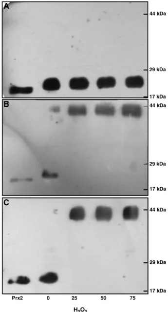
InFig. 1, we present immunoblots for Prx2 of RBC membranes prepared from the erythrocyte suspensions incubated without and with increasing H2O2concentrations (Fig. 1A), with the addition of a catalase inhibitor (Fig. 1B) and in conditions that both catalase and Hb autoxidation were inhibited (Fig. 1C). Using the same incubation conditions and locking Prx2 redox forms present within the cell by means of incubation with an oxidizing agent (NEM), the typical immunoblots of Prx2 in the hemolysates are presented in Fig. 2. When no H2O2was added to the RBC suspension, we found that Prx2 was not linked to the RBC membrane for all tested conditions (Fig. 1) and that Prx2 was present in the cytosol as monomers, the active reduced form of Prx2 (Fig. 2); for growing peroxide concentrations, Prx2 was detected in the RBC membrane in an increasing manner when catalase was inhibited (Figs. 1B and C) and, when catalase was active, Prx2 was not detected in the RBC membrane (Fig. 1A). By analysis of the immunoblots of the hemolysates, we observed that the monomeric form of Prx2 was present, as referred, in all suspensions without H2O2(Fig. 2) and in those where catalase was not inhibited (Fig. 2A); in the presence of H2O2 and NaN3, the dimeric oxidized form of Prx2 was detected in the cytosol (Fig. 2B and C) and increasing amounts of Prx2 were linked to the RBC membrane (Fig. 1B and C).
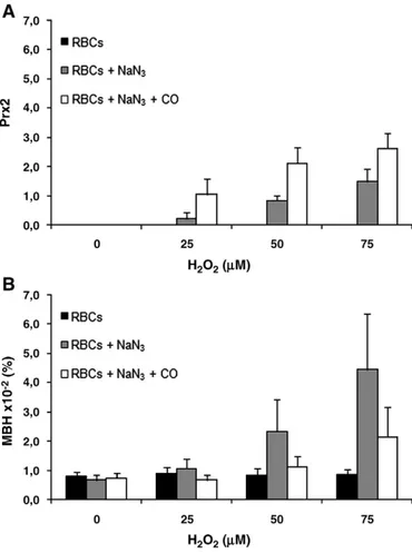
By using an internal control of human Prx2 in the immunoblots, we evaluated the amount of Prx2 linked to the RBC membrane in the different assay conditions (Fig. 3A). We also evaluated the MBH in the several studied conditions (Fig. 3B). We found that without H2O2, no Prx2 was detected in the RBC membrane and the value of MBH was similar for all assay conditions. When H2O2was added in increasing concentrations to RBCs, in the suspensions with active catalase, no Prx2 was linked to the membrane and the MBH amounts were similar for all concentrations of H2O2(Fig. 3A and B); in the suspensions incubated with NaN3 and H2O2, both Prx2 and MBH levels increased with H2O2concentration. When catalase and Hb autoxidation were inhibited, Prx2 membrane linkage increased also with H2O2concentration and, the amount of Prx2 linked to the
membrane was higher than that observed when only catalase was inhibited. The opposite was observed for MBH, though its values presented also an increase with H2O2concentration.
Regarding erythrocyte membrane lipoperoxidation, the levels of MDA/prot increased with H2O2concentration for all assay conditions and in the RBC suspensions without active catalase this increment was even higher, especially when associated with inhibition of Hb autoxidation by CO (Table 1).
The Hb spectral analysis of the hemolysates, showed a rise in the absorption peak characteristic of methemoglobin (metHb) (630 nm) with increasing H2O2 concentration for the RBC suspensions incubated with NaN3, and with both CO and NaN3; this peak was
Fig. 1. Immunoblots for peroxiredoxin 2 (Prx2) of erythrocyte membranes prepared from the RBC suspensions under the different assay conditions. All suspensions (5 × 10⁸
cell/ml in PBS, pH 7.4) were incubated for 30 min at 37 °C under gentle agitation, without and with increasing concentrations of H2O2(25, 50 and 75μM) (A); plus NaN3
(100 mM) to inhibit catalase (B) and, in order to inhibit both catalase and Hb autoxidation, RBC suspensions previously saturated with CO were incubated with NaN3
(100 mM) (C). SDS-PAGE of erythrocyte hemolysates performed in a 12% acrylamide gel, under reducing conditions. An internal control of human recombinant Prx2 was always used. Molecular weight (MW) markers are presented on the right.
Fig. 2. Peroxiredoxin 2 (Prx2) immunoblots of hemolysates obtained after subjecting erythrocytes to the different assay conditions. All RBC suspensions (5 × 10⁸
cell/ml in PBS, pH 7.4) were incubated for 30 min at 37 °C under gentle agitation, without and with increasing concentrations of H2O2(25, 50 and 75μM) (A); plus NaN3(100 mM) to
inhibit catalase (B) and, in order to inhibit both catalase and Hb autoxidation, RBC suspensions saturated with CO were incubated with NaN3(100 mM) (C). Afterwards,
aliquots of each suspension (5 × 10⁶
cell/ml) were incubated (15 min at 37 °C under gentle agitation) after addition of NEM (100 mM), in order to lock the Prx2 redox forms present in the cytosol of the erythrocytes. SDS-PAGE of erythrocyte hemolysates performed in a 12% acrylamide gel, under oxidizing conditions. An internal control of human recombinant Prx2 was always used. Molecular weight (MW) markers are presented on the right.
not observed in the hemolysates from the suspensions with active catalase or from the suspensions incubated without H2O2 (data not shown).
Discussion
In this work, we confirmed the hypothesis we recently proposed
[15] that the linkage of Prx2 to the erythrocyte membrane in circulating RBCs of Hereditary Spherocytosis patients could result from the development of oxidative stress within the cell, since we were able to cause the linkage of Prx2 to the membrane in vitro through H2O2-mediated oxidative stress. Whether this linkage is a simple result of oxidative stress damage to Prx2 or that it can serve a beneficial propose in vivo is still unknown.
In erythrocytes, Hb is particularly prone to go through an autoxidation process by which superoxide anion, H2O2and oxidized Hb (metHb) are produced. In a normal mature erythrocyte the antioxidant systems are able of detoxifying the cell of ROS and oxidized hemoglobin is usually regenerated to the reduced functional form by metHb reductases, one dependent on NADH and the other on NADPH, as co-factors [31,32]. However, under oxidative stress conditions, metHb formation increases and this Hb oxidized form binds to the cytoplasmatic domain of the erythrocyte membrane band
3 protein [33–36]. The rise of ROS within the cell also induces membrane lipid peroxidation[37]. Both oxidative changes may trigger RBC membrane damage, such as decreased membrane transporter activity, and changes in membrane permeability[38,39].
Although Prx2 is usually described as being localized in RBC cytosol, performing its role in the antioxidant systems, our data show that the linkage of Prx2 to the erythrocyte membrane may be triggered under H2O2-induced oxidative stress conditions (Fig. 1). Moreover, the binding of Prx2 to the membrane only occurred when its dimeric oxidized form was present in the cytoplasm (Figs. 1and
2). The active form of Prx2 is monomeric and the enzyme acquires the dimeric (inactive) form when it is oxidized during the reaction with H2O2[7]. Low et al.[5]reported that in the presence of excess H2O2, Prx2 becomes inactivated since it can only be regenerated to
monomers when H2O2 levels decrease, thus leading to an
accumulation of the dimeric form. In our study, when catalase
was active (incubations without NaN3), cytoplasmatic Prx2
remained in the monomeric active form (Fig. 2) and did not bind to the RBC membrane (Figs. 1and3A), confirming that catalase is indeed responsible for scavenging high/exogenous quantities of H2O2 [4,5,40]. This is strengthened by the results observed in the other oxidative stress markers. Actually, MBH (Fig. 3B), lipoperox-idation (Table 1) and Hb spectra in the hemolysates (data not shown) were unaffected by H2O2 (for the tested concentrations), maintaining basal values throughout increasing oxidative stress conditions, suggesting that catalase and the other antioxidant defenses effectively protected the erythrocyte suspensions from exogenous H2O2oxidative damage. Thesefindings are in accordance with the literature[4,41].
In a previous work[15], we hypothesized that Prx2 binding to the membrane could be achieved through its linkage to oxidized Hb, as it was reported elsewhere that Prx2 is able to bind to Hb[42]. In the present work, we analyzed and compared the results obtained for the erythrocyte suspensions where catalase or both catalase and Hb autoxidation were inhibited. MBH showed to be a good marker of Hb oxidation [18–24,43,44], as its rise was correlated with H2O2 concentration (Fig. 3B) and with the appearance of the absorption peak characteristic of metHb (data not shown). However, it appears that oxidized Hb and Prx2 membrane linkages are two separate processes given that, when we were able to cause diminished Hb linkage to the membrane (CO saturated suspensions, where Hb autoxidation and, consequently, metHb formation where impaired), higher amounts of Prx2 were found in the membrane, as compared to those where Hb autoxidation was not inhibited. Thisfinding probably reflects the higher susceptibility of Prx2 to H2O2in situations where Hb is made unavailable to react with H2O2, rendering its effects upon Prx2 much more pronounced. Moreover, if Prx2 needed to be linked to MBH in order to bind to the RBC membrane, we would not have found more membrane linked Prx2 when less MBH was present, as was the case.
Fig. 3. Erythrocyte membrane linked peroxiredoxin 2 (Prx2) (A) and membrane bound hemoglobin (MBH) (B) amounts versus hydrogen peroxide concentration for the different assay conditions applied to erythrocyte suspensions. Prx2 levels were determined by densitometric analysis of the erythrocyte membrane immunoblots (ratio between each Prx2 sample and the used internal control of human recombinant Prx2); MBH was determined by a spectroscopic method. Mean value and standard error mean (n = 3) are presented. (
▪
RBCs— erythrocyte suspensions incubated without and with H2O2; RBCs + NaN3— erythrocyte suspensions incubated without and withH2O2 and inhibition of catalase;□ RBCs + NaN3+ CO— erythrocyte suspensions
incubated without and with H2O2and inhibition of both catalase and Hb autoxidation).
Table 1
Lipid peroxidation of erythrocyte membranes (n = 3) for the different incubation conditions applied to erythrocyte suspensions, according to hydrogen peroxide concentration. MDA/prot (×10− 2) RBCs 2.3 ± 0.5 2.2 ± 1.0 3.3 ± 1.7 5.0 ± 2.1 RBCs + NaN3 3.4 ± 0.3 5.3 ± 0.7 5.6 ± 0.8 6.5 ± 1.7 RBCs + NaN3+ CO 3.0 ± 0.7 4.2 ± 1.0 7.4 ± 0.7 10.6 ± 2.3 H2O2(μM) 0 25 50 75
MDA— malonaldehyde; prot — protein concentration of erythrocyte membrane suspensions (mg); Incubation conditions: RBCs— 5×10⁸
cell/ml; RBCs + NaN3— 5×10⁸
cell/ml + 100 mM NaN3; RBCs+ NaN3+ CO— 5×10⁸cell/ml + 100 mM NaN3+ CO.
Cha et al.[11]suggested that Prx2 membrane linkage could be a mechanism used by the erythrocyte for its own protection against the deleterious effects of MBH; furthermore, it has been described that peroxiredoxins (Prx2 included) are capable of scavenging peroxynitrite and organic peroxides, such as lipid hydroperoxides
[7,45–47]. As we used reducing conditions, we were not able to determine in which form (monomer/dimer) Prx2 was linked to the membrane; actually, further studies are needed to evaluate the active/inactive state of Prx2 when it links to the membrane. In this regard, our lipoperoxidation data show that erythrocyte membrane lipid peroxidation rises with oxidative stress (Table 1), as has been described elsewhere [48]. However, we cannot forget that, in the studied conditions, Prx2 was inactive in the cytoplasm and, in the membrane the depletion in NADPH would render a possible active form also inactive. Therefore, MDA/prot values were increased and surprisingly more so in those membranes with more Prx2, as we increase H2O2concentration. The difference between values of MDA/ prot in suspensions subjected to the same H2O2concentration, when referring to both erythrocyte suspensions with inactive catalase, may possibly be explained by the fact that the presence of Prx2 could contribute to enhance membrane structure destabilization (since Prx2 is not a typical RBC membrane protein), rendering RBC membranes more susceptible to lipid hydroperoxide attack. This could explain why we found increased evidence of lipoperoxidation in the membranes with highest amount of linked Prx2 (Table 1and
Fig. 3A).
In conclusion, our data showed that the development of oxidative stress within the erythrocyte triggers oxidation of Hb, which links to the membrane, and is also responsible by the inactivation of Prx2 in the cytosol, that is accompanied by its linkage to the membrane. Both process seem to be independent and could be useful markers of oxidative stress and/or of RBC damage/senescence. Moreover, in accordance with the literature, catalase seems crucial in scavenging high levels of H2O2, but there are other antioxidant systems, such as Prx2, that seem to be more important in scavenging low H2O2levels. Further studies in oxidative stress related pathologies could be valuable to better clarify the role of the linkage of Prx2 to the erythrocyte membrane, since it can occur in circulating RBCs, namely, in Hereditary Spherocytosis[15]. Nonetheless, we demonstrate for the first time that the linkage of cytosolic Prx2 to the erythrocyte membrane can be induced by oxidative stress.
Acknowledgments
This study was supported by a PhD grant (SFRH/BD/22442/2005) attributed to S. Rocha by Fundação para a Ciência e Tecnologia (FCT) and Fundo Social Europeu (FSE).
References
[1] R.B. Moore, M.V. Mankad, S.K. Shriver, et al., Reconstitution of Ca2+
-dependent K+
transport in erythrocyte membrane vesicles requires a cytoplasmic protein, J. Biol. Chem. 266 (1991) 18964–18968.
[2] T. Rabilloud, R. Berthier, M. Vinçon, et al., Early events in erythroid differentiation: accumulation of the acidic peroxidoxin (PRP/TSA/NKEF-B), Biochem. J. 312 (1995) 699–705.
[3] E. Schröder, J.A. Littlechild, A.A. Lebedev, et al., Crystal structure of decameric 2-cys peroxiredoxinfrom human erythrocytes at 1.7 A° resolution, Structure 8 (2000) 605–615.
[4] R.M. Johnson, G. Goyette Jr., Y. Ravindranath, Y.S. Ho, Hemoglobin autoxidation and regulation of endogenous H2O2levels in erythrocytes, Free Radic. Biol. Med.
39 (2005) 1407–1417.
[5] F.M. Low, M.B. Hampton, A.V. Peskin, C.C. Winterbourn, Peroxiredoxin 2 function as a non-catalytic scavenger of low level hydrogen peroxide in the erythrocyte, Blood 109 (6) (2007) 2611–2617.
[6] J.R. Harris, E. Schröder, M.N. Isupov, et al., Comparison of the decameric structure of peroxiredoxin-II by transmission electron microscopy and X-ray crystal-lography, Biochim. Biophys. Acta 1547 (2001) 221–234.
[7] Z.A. Wood, E. Schröder, J.R. Harris, L.B. Poole, Structure, mechanism and regulation of peroxiredoxins, Trends Biochem. Sci. 28 (2003) 32–40.
[8] M.K. Cha, I.H. Kim, Thioredoxin-linked peroxidade from human red blood cell: evidence for the existence of thioredoxin and thioredoxin reductase in human red blood cell, Biochem. Biophys. Res. Commun. 217 (3) (1995) 900–907. [9] P. Kristensen, D.E. Rasmussen, B.I. Kristensen, Properties of thiol-specific
anti-oxidant protein or calpromotin in solution, Biochem. Biophys. Res. Commun. 262 (1999) 127–131.
[10] E. Schröder, A.C. Willis, C.P. Ponting, Porcine natural-killer enhancing factor-B: oligomerization and identification as a calpain substrate in vitro, Biochim. Biophys. Acta 1384 (1998) 279–291.
[11] M.K. Cha, C.H. Yun, I.H. Kim, Interaction of human thiol-specific antioxidant protein 1 with erythrocyte plasma membrane, Biochemistry, 39 (2000) 6944–6950. [12] R.B. Moore, S.K. Shriver, L.D. Jenkins, et al., Calpromotin, a cytoplasmic protein, is
associated with the formation of dense cells in sickle cell anemia, Am. J. Hematol. 56 (1997) 100–106.
[13] S.C. Murphy, B.U. Samuel, T. Harrison, et al., Erythrocyte detergent-resistant membrane proteins: their characterization and selective uptake during malarial infection, Blood 103 (5) (2004) 1920–1928.
[14] G.A. Plishker, D. Chevalier, L. Seinsoth, R.B. Moore, Calcium activated potassium transport and high molecular weight forms of calpromotin, J. Biol. Chem. 267 (1992) 21839–21843.
[15] S. Rocha, R.M.P. Vitorino, F.M. Lemos-Amado, et al., Presence of cytosolic peroxiredoxin 2 in the erythrocyte membrane of Hereditary Spherocytosis patients, Blood Cells Mol. Diseases 41 (2008) 5–9.
[16] T.Y. Low, T.K. Seow, M.C. Chung, Separation of human erythrocyte membrane associated proteins with one-dimensional and two-dimensional gel electro-phoresis followed by identification with matrix-assisted laser desorption/ ionization-time offlight mass spectrometry, Proteomics 2 (2002) 1229–1239. [17] Y.S. Lim, M.K. Cha, C.H. Yun, et al., Purification and characterization of
thiol-specific antioxidant protein from human red blood cell: a new type of antioxidant protein,, Biochem. Biophys. Res. Commun. 199 (1994) 199–206.
[18] A. Santos-Silva, I. Rebelo, E.M.B. Castro, et al., Leukocyte activation, erythrocyte damage, lipid profile and oxidative stress imposed by high competition physical exercise in adolescents, Clin. Chim. Acta 306 (1–2) (2001) 119–126.
[19] L. Belo, I. Rebelo, E.M.B. Castro, et al., Band 3 as a marker of erythrocyte changes in pregnancy, Eur. J. Haematol. 69 (3) (2002) 145–151.
[20] C.Catarino, I. Rebelo, L. Belo, et al., Erythrocyte changes in preeclampsia: relationship between maternal and cord blood erythrocyte damage, J. Perinat. Med. 37 (1) (2009) 19–27.
[21] A. Santos-Silva, E.M.B. Castro, N.A. Teixeira, et al., Altered erythrocyte membrane band 3 profile as a marker in patients at risk for cardiovascular disease, Atherosclerosis 116 (2) (1995) 199–209.
[22] A. Santos-Silva, I. Rebelo, E. Castro, et al., Erythrocyte damage and leukocyte activation in ischemic stroke, Clin. Chim. Acta 320 (2002) 29–35.
[23] P. Rocha-Pereira, A. Santos-Silva, I. Rebelo, et al., Erythrocyte damage in mild and severe psoriasis, Br. J. Dermatol. 150 (2) (2004) 232–244.
[24] A. Santos-Silva, E.M.B. Castro, N.A. Teixeira, et al., Erythrocyte membrane band 3 profile imposed by cellular aging, by activated neutrophils and by neutrophilic elastase, Clin. Chim. Acta 275 (1998) 185–196.
[25] W.G. Zijlstra, A. Buursma, W.P. Meeuwsen-van der Roest, Absorption spectra of human fetal and adult oxyhemoglobin, de-oxyhemoglobin, carboxyhemoglobin, and methemoglobin, Clin. Chem. 37 (1991) 1633–1638.
[26] J.T. Dodge, C. Mitchell, D.J. Hanahan, The preparation and chemical characteristics of hemoglobin-free ghosts of human erythrocytes, Arch. Biochem. Biophys. 100 (1963) 119–130.
[27] M.M. Bradford, A rapid and sensitive method for the quantification of microgram quantities of protein utilizing the principle of the protein–dye binding, Anal. Biochem. 72 (1976) 248–254.
[28] U.K. Laemmli, Cleavage of structural proteins during the assembly of the head of the bacteriophage T, Nature 227 (1970) 680–685.
[29] H. Towbin, T. Stahelin, J. Gordon, Electrophoretic transfer of proteins from polycrylamide gels to nitrocellulose sheets: procedure and some applications, Proc. Natl. Acad. Sci. U. S. A. 76 (9) (1979) 4350–4354.
[30] M. Mihara, M. Uchiyama, Determination of malonaldehyde precursor in tissues by thiobarbituric acid test, Anal. Biochem. 86 (1978) 271–278.
[31] T. Katsube, N. Sakamoto, Y. Kobayashi, et al., Exonic point mutations in NADH-cytochrome B5 reductase genes of homozygotes for hereditary methemoglobi-nemia, types I and III: putative mechanisms of tissue-dependent enzyme deficiency, Am. J. Hum. Genet. 48 (1991) 799–808.
[32] F. Xu, K.S. Quandt, D.E. Hultquist, Characterization of NADPH-dependent methemoglobin reductase as a heme-binding protein present in erythrocytes and liver, Proc. Natl. Acad. Sci. U. S. A. 89 (1992) 2130–2134.
[33] A.A. Demehin, O.O. Abugo, R. Jayakumar, et al., Binding of hemoglobin to red cell membranes with eosin-5-maleimide-labeled band 3: analysis of centrifugation andfluorescence data, Biochemistry 41 (2002) 8630–8637.
[34] J.M. Salhany, K.A. Cordes, R.L. Sloan, Characterization of the pH dependence of hemoglobin binding to band 3. Evidence for a pH-dependent conformational change within the hemoglobin-band 3 complex, Biochim. Biophys. Acta 1371 (1998) 107–113.
[35] N. Shaklai, J. Yguerabide, H.M. Ranney, Classification and localization of hemoglobin binding sites on the red blood cell membrane, Biochemistry 16 (1977) 5593–5597.
[36] J.A. Walder, R. Chatterjee, T.L. Steck, et al., The interaction of hemoglobin with the cytoplasmic domain of band 3 of the human erythrocyte membrane, J. Biol. Chem. 259 (1984) 10238–10246.
[37] C. Rice-Evans, E. Baysal, Iron-mediated oxidative stress in erythrocytes, Biochem. J. 244 (1987) 191–196.
[38] J.M. Rifkind, O.O. Abugo, A. Levy, et al., Formation of free radicals under hypoxia, in: P.W. Hochachka, P.L. Lutz, T. Sick, M. Rosenthal, G. van den Thillart (Eds.), Surviving Hypoxia: Mechanism of Control and Adaptation, CRC Press, Boca Raton FL, 1993, pp. 509–525.
[39] J.M. Rifkind, L. Zhang, A. Levy, P.T. Manoharan, The hypoxic stress on erythrocytes associated with superoxide formation, Free Radic. Res. Commun. 12–13 (Pt. 2) (1991) 645–652.
[40] E. Nagababu, F.J. Chrest, J.M. Rifkind, Hydrogen-peroxide-induced heme degrada-tion in red blood cells: the protective roles of catalase and glutathione peroxidase, Biochim. Biophys. Acta 1620 (2003) 211–217.
[41] H.S. Jacob, S.H. Ingbar, J.H. Jandl, Oxidative hemolysis and erythrocyte metabolism in hereditary acatalasia, J. Clin. Invest. 44 (1965) 1187–1199.
[42] K.M. Stuhlmeier, J.J. Kao, P. Wallbrant, et al., Antioxidant protein 2 prevents methemoglobin formation in erythrocyte hemolysates, Eur. J. Biochemistry 270 (2003) 334–341.
[43] E. Costa, S. Rocha, P. Rocha-Pereira, et al., Band 3 profile as a marker of erythro-cyte changes in chronic kidney disease patients, Open Clin. Chem. J. 1 (2008) 57–63.
[44] S. Coimbra, E. Castro, P. Rocha-Pereira, et al., The effect of green tea in oxidative stress, Clin. Nutr. 25 (2006) 790–796.
[45] R. Bryk, P. Griffin, C. Nathan, Peroxynitrite reductase activity of bacterial peroxiredoxins, Nature 407 (2000) 211–215.
[46] P. Cordray, K. Doyle, K. Edes, et al., Oxidation of 2-Cys-peroxiredoxins by arachidonic acid peroxide metabolites of lipoxygenases and cyclooxygenase-2, J. Biol. Chem. 282 (2007) 32623–32629.
[47] A.V. Peskin, F.M. Low, L.N. Paton, et al., The high reactivity of peroxiredoxin 2 with H(2)O(2) is not reflected in its reaction with other oxidants and thiol reagents, J. Biol. Chem. 282 (2007) 11885–11892.
[48] J.M. Rifkind, R.S. Ajmani, J. Heim, Impaired hemorheology in the aged associated with oxidative stress, Adv. Exp. Med. Biol. 428 (1997) 7–13.