University of Lisbon
Institute of Pharmacology and Neuroscience, Faculty of Medicine Unit of Neuroscience, Institute of Molecular Medicine
Setting GABA levels: GABA transporters
modulation by adenosine receptors
Ana Sofia Cristóvão Ferreira
Tese orientada pela Professora Doutora Ana Maria Sebastião PhD in Biomedical Sciences
Speciality in Neuroscience
Todas as afirmações efectuadas no presente documento são da exclusiva responsabilidade do seu autor, não cabendo qualquer responsabilidade à Faculdade de Medicina de Lisboa pelos conteúdos nele apresentados.
A impressão desta dissertação foi aprovada pelo
conselho científico da Faculdade de Medicina de Lisboa
em reunião de 15 de Maio de 2012.
The experimental work described in this thesis was performed at the Institute of Pharmacology and Neuroscience, Faculty of Medicine and Unit of Neuroscience, Institute of Molecular Medicine, under de supervision of Professor Ana Maria Sebastião.
O trabalho experimental descrito nesta tese foi realizado no Instituto de Farmacologia e Neurociências, Faculdade de Medicina e Unidade de Neurociências, Instituto de Medicina Molecular, sob orientação da Professora Doutora Ana Maria Sebastião.
The scientific content of the present thesis has been included in the publication of the following original articles:
Cristóvão-Ferreira S, Navarro G, Brugarolas M, Pérez-Capote K, Vaz SH, Fattorini G, Conti F, Lluis C, Ribeiro JA, McCormick PJ, Casadó V, Franco R, Sebastião AM (2011) Modulation of GABA transport by adenosine A1R-A2AR heteromers, which are coupled to both Gs- and G(i/o)-proteins. J Neurosci. 31, 15629-15639.
Cristóvão-Ferreira S, Vaz SH, Ribeiro JA, Sebastião AM (2009) Adenosine A2A receptors enhance GABA transport into nerve terminals by restraining PKC inhibition of GAT-1. J Neurochem. 109, 336-347.
Only the experiments performed by the author of this thesis were included in the corresponding results chapter. Experiments performed by other co-authors (the minority of the papers content) are referred as such and discussed.
Other publications closely related to the content of this thesis: Sebastião AM, Cristóvão-Ferreira S, Ribeiro JA (2012) Downstream pathways of adenosine in Adenosine: a key Link between Metabolism and CNS Activity. Edited by Masino SA and Boison D.
Vaz SH, Jørgensen TN, Cristóvão-Ferreira S, Duflot S, Ribeiro JA, Gether U, Sebastião AM (2011) Brain-derived neurotrophic factor (BDNF) enhances GABA transport by modulating the trafficking of GABA transporter-1 (GAT-1) from the plasma membrane of rat cortical astrocytes. J Biol Chem. 286, 40464-40476.
Vaz SH, Cristóvão-Ferreira S, Ribeiro JA, Sebastião AM (2008) Brain-derived neurotrophic factor inhibits GABA uptake by the rat hippocampal nerve terminals. Brain Res. 1219, 19-25.
“Tudo o que sabemos é uma impressão nossa, e tudo o que somos é uma impressão alheia”
In “O Livro do Desassossego”, Bernardo Soares
1 Introduction ... 1
1.1 GABA ... 1
1.1.1 GABA Receptors ... 2
1.1.2 GABA metabolism ... 6
1.1.3 GABA transporters ... 8
1.2 The tripartite synapse and glial cells... 24
1.2.1 Astrocytes ... 24
1.3 Adenosine ... 35
1.3.1 Adenosine synthesis... 37
1.3.2 Nucleoside transporters ... 40
1.3.3 Adenosine degradation... 43
1.3.4 Adenosine levels regulation at brain ... 47
1.3.5 Adenine nucleotides, adenosine and signaling ... 48
1.3.6 Adenosine receptors and signaling pathways ... 50
1.3.7 A1-A2A adenosine receptors interaction... 56
1.4 Heteromers of G protein coupled receptors ... 57
2 Aims... 65
3 Techniques ... 67
3.1 Isolated presynaptic terminals... 67
3.4 BRET – bioluminescence resonance energy transfer ... 70 3.5 GTP-γ-[35S]-assay... 73 4 Methods... 77 4.1 Reagents... 77 4.2 Experimental protocols... 79 4.2.1 Synaptosomes isolation ... 79
4.2.2 Cell lines and primary astrocytic cultures... 80
4.2.3 [3H]GABA uptake assays... 80
4.2.4 Biotinylation assays... 82 4.2.5 Western Blot ... 84 4.2.6 Immunocytochemistry... 85 4.2.7 BRET ... 85 4.2.8 [35S] GTP-γ- S assay... 86 4.3 Statistical analysis ... 87 5 Results ... 89
5.1 Adenosine modulation of GAT-1-mediated GABA uptake by synaptosomes ... 89
5.1.1 Rationale ... 89
5.1.2 Adenosine A2A receptors tonically enhance GAT1 -mediated GABA transport... 89
5.1.3 Adenosine A1 and A2B receptors do not affect GAT-1 mediated GABA transport... 95
the A2A receptors agonist... 97
5.1.5 PKC constitutively inhibits GABA transport, and prevents A2A receptors -mediated facilitation of GABA
transport 99 5.1.6 PKA and PKC interaction ... 103 5.1.7 Discussion... 107 5.2 Modulation of astrocytic GABA Transport by Adenosine A1–A2A Receptor Heteromers... 113
5.2.1 Rationale ... 113 5.2.2 Endogenous adenosine tonically modulates GABA uptake 114
5.2.3 Adenosine A1 receptors activation decreased and
adenosine A2A receptors activation enhanced GABA uptake 117
5.2.4 Adenosine A1 -A2A receptor heteromers in
astrocytes... 123 5.2.5 Adenosine A1 or A2A receptors activation, but not its
blockade, leads to internalization of the A1-A2A receptor
heteromers ... 128 5.2.6 The adenosine A1-A2A receptor heteromer is coupled
to Gi/0 and Gs proteins... 131
5.2.7 The A1-A2A receptor heteromer signals through
AC/PKA pathway ... 138 5.2.8 Discussion... 141
7 Future perspectives ... 153
8 Acknowledgments ... 157
9 References ... 161
Figure 1.1 – Schematic representation of the evidence of GABA as a
neurotransmitter in mammalian cerebral cortex... 2
Figure 1.2 – Schematic representation of a GABAA receptor. ... 3
Figure 1.3 – Schematic representation of GABAC receptor. ... 4
Figure 1.4 – Schematic representation of GABAB receptor ... 6
Figure 1.5 – Schematic representation of glutamate/GABA-glutamine cycle in GABAergic synapse. ... 8
Figure 1.6 – Schematic representation of a GABAergic synapse... 10
Figure 1.7 – Schematic representation of GABA transport ... 11
Figure 1.8 – Schematic representation of GAT-1... 13
Figure 1.9 – Chemical structure of adenosine. ... 35
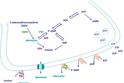
Figure 1.10 – Schematic representation of adenosine metabolism. 47 Figure 1.11 – Distribution of adenosine A1 and A2A receptors ... 51
Figure 1.12 – Associated pathways to adenosine receptors ... 55
Figure 3.1 – Electronic microscopic visualization of a synaptosome 68 Figure 3.2 – BRET- bioluminence resonance energy transfer ... 72
Figure 3.3 – G protein activation ... 74
Figure 5.1 – Adenosine, through adenosine A2A receptors, enhances GABA transport into nerve endings by increasing the surface density of GAT-1 and maximum transport rate. ... 94
Figure 5.3 – Adenosine A1 and A2B receptors do not affect GAT-1 mediated GABA transport into nerve endings. ... 96 Figure 5.4 – Influence of the AC/PKA transduction pathway ... 98 Figure 5.5 – Influence of the PLC/PKC transduction pathway upon GABA transport ... 102 Figure 5.6 – Influence of PKC activity upon the A2AR mediated facilitation of GABA transport... 103 Figure 5.7 – PKC and PKA interaction ... 106 Figure 5.8 – Schematic representation of the influence of A2AR upon GAT-1 mediated GABA transport into nerve endings... 111 Figure 5.9 – Adenosine receptor activation modulates [3H]GABA uptake in astrocytes... 117 Figure 5.10 – Adenosine A1 receptors activation decreased Vmax of GAT-1 and GAT-3 while adenosine A2A receptors activation led to an enhancement of Vmax of both GAT-1 and GAT-3 ... 118 Figure 5.11 – Inhibition of [3H]GABA uptake is promoted by
adenosine A1 receptors. ... 121 Figure 5.12 – Adenosine A2A receptors activation facilitates
[3H]GABA uptake ... 122 Figure 5.13 – Adenosine A1-A2A receptors heteromers in
A1-A2A receptors heteromers... 131 Figure 5.15 – [35S]GTP-γ-S assays suggest the involvement of both Gs and Gi/0 in the adenosine A1-A2A receptors heteromer... 135 Figure 5.16 – Blockade of Gi/0 proteins by PTx prevents both effects mediated by adenosine A1 and A2A receptors ... 136 Figure 5.17 – Blockade of Gs proteins by ChTx prevents both effects mediated by adenosine A1 and A2A receptors ... 137 Figure 5.18 – Adenosine A1-A2A receptors heteromer signaling. . 140 Figure 5.19 – Schematic representation of adenosine A1-A2A
receptors heteromer function ... 148 Figure 6.1 – Schematic representation of adenosine effect upon GABA transport at tripartite synapse ... 152
AC – adenylate cyclase Ach – acetylcholine
ADA – adenosine deaminase ADK – adenosine Kinase
ADP – adenosinse diphosphate
AMPA – α-amino-3-hydroxy-5-methyl-4-isoxazolepropionic acid AMP – adenosine 5’-monophosphate
ANOVA – analysis of variance AOAA – aminooxyacetic acid ATP – adenosine 5’-triphosphate
BAPTA – 1,2-bis(o-aminophenoxy)ethane-N,N,N',N'-tetraacetic acid BDNF – brain derived neurotrophic factor
bFGF – basic-fibroblast growth factor BGT-1 – Betaine/GABA transporter 1
BRET – bioluminescence resonance energy transfer
BRET50 – BRET constant (corresponds to the acceptor/donor ratio at
which 50% of BRETmax is reached)
BRETmax – BRET constant (maximal BRET signal)
BSA – bovine serum albumin CADO – 2-chloro-adenosine
cDNA – complementar DNA
CGS 21680 –
4-[2-[[6-amino-9-(N-ethyl-β-D-ribofuranuronamidosyl)-9H-purin-yl]amino]ethyl] benzenepropanoic acid hydrochloride
CHO cells – chinese hamster ovary cells ChTx – Cholera Toxin
CNS – central nervous system
CNT1 – concentrative nucleoside transporter 1 CNT6 – concentrative nucleoside transporter 6 CNTs – concentrative nucleoside transporters CPA – N6-cyclopentyladenosine
DAG – diacylglycerol
DDT1MF-2 cells – smooth muscle cell line DMEM – Dulbecco’s Modified Eagles Medium DMSO – dimethylsulfoxide
DPCPX – 8-cyclopentyl-1,3-dipropylxanthine DTT – DL-Dithiothreitol
EDTA – Ethylenediamine tetraacetic acid
eEPSCs – evoked excitatory post-synaptic currents eIPSCs – evoked inhibitory post-synaptic currents
ENT2 – equilibrative nucleoside transporter 2 ENT4 – equilibrative nucleoside transporter 4 ENTs – equilibrative nucleoside transporters ERK – extracellular signal-regulated kinase FBS – foetal bovine serum
FITC – Fluorescein Isothiocyanate
FRET – fluorescent resonance energy transfer GABA – gamma–aminobutyric acid
GABA-T – GABA transaminase GAD – glutamate decarboxylase GAT-1 – GABA transporter 1 GAT-2 – GABA transporter 2 GAT-3 – GABA transporter 3 GATs –GABA transporters
GDP – guanosine 5’- diphosphate
GF109203X – 2-[1-(3-dimethylaminopropyl)indol-3-yl]-3-(indol-3-yl) maleimide
GFAP – glial fibrillary acidic protein GLAST–glutamate aspartate transporter GLT-1 – glial glutamate transporter 1
GS –glutamine synthetase GTP – guanosine 5’- triphosphate
H-89 – N-[2-(p-Bromocinnamylamino)ethyl]- 5- isoquinolinesulfonamide dihydrochoride
HEK 293 cells – Human Embryonic Kidney 293 cells
HEPES – 4-(2-hydroxyethyl)-1-piperazineethanesulfonic acid [3H]GABA – 4-amino-n-[2,3-3H] butyric acid
[35S]GTP – guanosine 5’-(g-[35S]-thio)triphosphate IC50 – pharmacological constant (half maximal inhibitory
concentration) IMP–inosine monophosphate IP3–inositol trisphosphate JNK – jun-N-terminal kinase KHR – Krebs-Henseleit-Ringer KM – Michaelis-Menten constant KO –knockout L-DOPA – levodopa LPS – lipopolysaccharide
MAP – mitogen-activated protein
mRNA–messenger RNA
MRS 1706 - N-(4-acetylphenyl)-2-[4-(2,3,6,7-tetrahydro-2,6-dioxo-1,3-dipropyl-1H-purinyl)phenoxy]acetamide)
NECA – 5'-N-ethylcarboxamidoadenosine
NFκB – nuclear factor kappa-light-chain-enhancer of activated B cells NGF – nerve growth factor
NGS – normal goat serum
NMDA –N-Methyl-D-aspartic acid NOS – NO synthase
NP40 –nonyl phenoxypolyethoxylethanol 5’-NT – 5´-nucleotidase
p38 – protein 38
PAG – phosphate-activated glutaminase PDD – phorbol-12,13-didecanoate
PIP2 –Phosphatidylinositol-4,5-bisphosphate
PKA – protein kinase A (protein kinase cAMP dependent) PKC – protein kinase C
PLA2 –phospholipase A2
PTx – Pertussis toxin PLC – phospholipase C
RIPA – radioimmunoprecipitation assay R-PIA – R-phenylisopropyladenosine
R-SNAP – R - Soluble NSF Attachment Protein R-SNARE – R-SNAP receptor
RLuc – Renilla luciferase
Rp-cAMPs – Rp-Adenosine 3′,5′-cyclic monophosphorothioate triethylammonium salt hydrate
SAH – S-adenosylhomocystein
SAHH – S-adenosylhomocystein hydrolase SAPK – stress-activated protein kinase
SCH 58261 – 2-(2-furanyl)-7-(2-phenylethyl)-7H-pyrazolo[4,3-e][1,2,4]triazolo[1,5-c]pyrimidin-5-amine
SDS – Sodium dodecyl sulfate
SDS-PAGE– sodium dodecyl sulfate polyacrylamide gel electrophoresis
SEM – Standard error of the mean
sEPSCs – spontaneous excitatory post-synaptic currents sIPSCs – spontaneous inhibitory post-synaptic currents SKF 89976A – hydrochloride
(1-(4,4-diphenyl-3-butenyl)-3-piperidinecarboxylic acid hydrochloride SLMV – synaptic-like microvesicles
TCA – tricarboxylic acids TEA – triethylamine
TEMED – Tetramethylethylenediamine TGF ββββ – transforming growth factor β TNF αααα – tumor necrosis factor-α
TRIS – tris(hydroxymethyl)-aminomethane TrKB – tropomyosin related kinase B
TRITC – Tetramethylrhodamine isothiocyanate
U73122 – (1-[6-[[(17β)-3-methoxyestra-1,3,5(10)-trien-17-yl]amino]hexyl]-1H-pyrrole-2,5-dione)
VAMP3 – Vesicle-associated membrane protein 3 vGAT–vesicular GABA transporter
vGLUT – vesicular glutamate transporter Vmax – maximal velocity
VRACs–volume regulated anion channels YFP – yellow fluorescent protein
Gamma-aminobutyric acid is the main inhibitory neurotransmitter in central nervous system. To assure a controlled GABAergic transmission GABA must be quickly removed from synapse, which occurs through specific transporters expressed both in pre-synaptic terminal (GAT-1) and surrounding astrocytes (GAT-1 and GAT-3). Adenosine, which is a well-known neuromodulator, promotes GABA release in the hippocampus (Cunha and Ribeiro, 2000). The main goal of this thesis was to identify a possible role of adenosine upon GABA uptake into pre-synaptic terminals and into astrocytes.
In pre-synaptic terminals, the removal of endogenous adenosine by ADA inhibited GABA uptake, an effect that was mimicked by the blockade of A2A receptors. Thus, endogenous adenosine, through the
activation of A2A receptors, promotes GABA uptake into pre-synaptic
terminals. Experiments with activators and inhibitors of transduction pathways revealed that AC/cAMP/PKA is the transduction pathway associated with this effect. In fact, the activation of PKA restrains an inhibitory tonic influence mediated by PKC, resulting in an increase of GABA uptake. The increase of transport rate occurs through the enhancement of the expression of GAT-1 at the surface membrane.
In astrocytes, adenosine has a biphasic effect upon GABA uptake. This biphasic effect is mediated by the adenosine A1-A2A receptors
heteromers, whose presence in astrocytes was identified in this work. The adenosine A1-A2A receptor heteromer is coupled to both
protomer, which through Gi/0 protein inhibit AC, decreasing PKA
activity and consequently, GABA uptake mediated by both GAT-1 and GAT-3. When adenosine levels rise, adenosine activates preferentially the A2A protomer, which through Gs activity enhances
PKA activity, promoting GABA uptake into astrocytes. The adenosine A1-A2A receptors heteromer can be viewed as a unique entity since it
reaches and leaves the membrane as an entire complex and all the components have to be available for the heteromer be functional. Moreover, this work clearly showed that the heteromer is associated with both Gs and Gi/0, beinga tetramer of A1-A1-A2A-A2A, rather than
an A1-A2A complex coupled to Gq as previously thought.
Globally, at tripartite synapse level, adenosine controls the final destination of GABA after its release into the synapse. At low levels, adenosine will inhibit uptake into astrocytes but promote the uptake of GABA into presynaptic terminals, therefore facilitating the inhibitory phasic and tonic transmission. At higher levels of adenosine, GABA uptake into astrocytes will be favoured, which may decrease GABAergic tonic transmission, therefore facilitating excitability. In conclusion, this work highlighted a yet unknown mechanism through which adenosine may contribute to the switch between inhibition and excitation, through a concerted cross-talk between astrocytes and inhibitory neurons.
O ácido gama-aminobutírico (GABA) é o principal neurotransmissor inibitório do Sistema Nervoso Central (SNC). Uma vez na sinapse o GABA é rapidamente recaptado através de transportadores específicos expressos pelos neurónios mas também pelas células da glia, que envolvem a sinapse.
A rápida recaptação de GABA pelos transportadores permite um controlo adequado dos níveis de GABA na sinapse, o que é fundamental para a limitação temporal e espacial da transmissão inibitória. Este controlo é assegurado por transportadores específicos expressos no terminal pré-sináptico e nos astrócitos que envolvem a sinapse. Actualmente são conhecidos quatro transportadores de GABA: três de alta afinidade (1, 2, GAT-3) e um de baixa afinidade (BGT-1). O GAT-1 é o principal transportador de GABA, sendo expresso pelos neurónios e também pelas células da glia. Por sua vez, O GAT-3 é o principal transportador das células gliais. Em conjunto, estes dois transportadores são os responsáveis pela recaptação de GABA nas sinapses do sistema nervoso central.
A adenosina é um conhecido neuromodulador, que desempenha, ao nível do sistema nervoso central, um vasto leque de acções, que resulta numa inibição tónica do SNC. De entre as funções desempenhadas, a adenosina modula a concentração de neurotransmissores da sinapse, regulando quer a sua libertação vesicular quer a sua recaptação por transportadores.
diferentes de receptores: dois de alta afinidade (A1 e A2A) e dois de
baixa afinidade (A2B e A3). Os receptores A1 e A3 são receptores
inibitórios, estando classicamente acoplados a proteínas Gi/0. Por seu
lado, os receptores do subtipo A2 são receptores excitatórios,
estando geralmente acoplados a proteínas Gs.
Tendo como ponto de partida o efeito modulador da adenosina através da activação dos receptores A2A, sobre libertação de GABA
no hipocampo (Cunha e Ribeiro, 2000), este trabalho teve como principal objectivo a identificação de um possível efeito modulador da adenosina sobre a recaptação de GABA para os terminais pré-sinápticos e também para os astrócitos, através dos transportadores GAT-1 e GAT-3.
Os resultados obtidos neste trabalham indicam claramente que a adenosina através da activação dos receptores A2A promove a
recaptação de [3H]GABA para os terminais pré-sinápticos mediada pelo transportador GAT-1. Com o objectivo de determinar o efeito da adenosina endógena, os terminais pré-sinápticos (sinaptossomas) foram incubados com adenosina desaminase (ADA, 1U/ml), tendo sido observada uma diminuição da recaptação de [3H]GABA. A ADA degrada a adenosina em inosina, um metabolito inactivo para os receptores de adenosina, permitindo assim inferir o efeito da adenosina endógena. O efeito inibitório causado pela remoção da adenosina endógena foi mimetizado pelo bloqueio dos receptores A2A com o antagonista selectivo SCH 58261 (50 nM). O envolvimento
receptores A2A pelo CGS 21680 promoveu a recaptação de
[3H]GABA, mediada pelo transportador GAT-1.
O envolvimento de outros subtipos de receptores de adenosina, especificamente os subtipos A1 e A2B, foi avaliado através do uso de
agonistas e antagonistas selectivos. Tanto a activação como o bloqueio dos receptores A1 ou A2B não causaram qualquer efeito
sobre a recaptação de [3H]GABA mediada por GAT-1 em sinaptossomas.
Ensaios de biotinilação, realizados após incubação dos sinaptossomas com o agonista selectivo dos receptores A2A,
mostraram que o efeito excitatório da adenosina sobre a recaptação de [3H]GABA ocorre através do aumento do número de transportadores GAT-1 na membrana celular. Este efeito foi confirmado por curvas de saturação do transportador GAT-1, que mostraram um aumento da velocidade máxima do transportador após incubação com o agonista selectivo dos receptores A2A, o que é
indicativo de um aumento do número de transportadores GAT-1 na membrana celular.
A via de transdução de sinal associada ao efeito dos receptores A2A
foi identificada através de ensaios de recaptação realizados na presença de bloqueadores e activadores de cinases intracelulares. O bloqueio da proteína cinase do tipo A (PKA) pelo H-89 (1µM) preveniu totalmente o efeito do CGS 21680, enquanto a activação da adenilato ciclase (AC) pela forskolin (10µM) imitou a acção do
recaptação de [3H]GABA mediada por GAT-1, através da activação da via da PKA.
Foi ainda testado o envolvimento da via de sinalização mediada pela proteína cinase do tipo C (PKC). O bloqueio da PKC pelo GF109203X (1µM) promoveu a recaptação de [3H]GABA mediada por GAT-1. Em concordância, a activação da PKC por ésteres de forbol (12,13- didecanoato de forbol – PDD 250nM) inibiu a recaptação de [3H]GABA mediada por GAT-1, indicando que a PKC está endogenamente a inibir o transporte de [3H]GABA para os terminais pré-sinápticos.
Estes resultados sugerem assim, o envolvimento das duas vias de sinalização na recaptação de [3H]GABA mediada por GAT-1. Estudos de recaptação realizados com diferentes combinações de activadores e bloqueadores de ambas as proteínas cinases indicam que a activação dos receptores A2A da adenosina, com consequente
activação da PKA, restringe o efeito tónico inibitório da PKC sobre o transporte de [3H]GABA, o que resulta numa potenciação da recaptação de GABA através do transportador GAT- 1 para o terminal pré-sináptico, por aumento do número de transportadores presentes na membrana celular.
O estudo de recaptação de [3H]GABA em astrócitos sugere a existência de um efeito bifásico mediado pela adenosina sobre os transportadores GAT-1 e GAT-3. A incubação de culturas primárias de astrócitos com cloroadenosina (CADO), um análogo
não-transporte enquanto que concentrações mais elevadas (3 e 10 µM) de CADO promoveram a recaptação de [3H]GABA.
Por outro lado, a remoção de adenosina endógena pela acção da adenosina desaminase (ADA, 1U/ml) diminuiu a recaptação de [3H]GABA mediada por GAT-1 e por GAT-3, sugerindo que as concentrações de adenosina endógena presentes na cultura de astrócitos promovem o transporte de [3H]GABA.
A realização de ensaios de recaptação com fármacos selectivos para os receptores de adenosina permitiu a identificação dos receptores envolvidos. Assim, o agonista selectivo dos receptores A2A (CGS
21680, 30nM) promoveu a recaptação de [3H]GABA mediada por GAT-1 e GAT-3. Surpreendentemente, este efeito para além de ter sido bloqueado pelo antagonista selectivo dos receptores A2A (SCH
58261, 50nM) foi também prevenido pelo bloqueio dos receptores A1 com o antagonista selectivo DPCPX (50nM). Por seu lado, a
activação dos receptores A1 com o agonista selectivo CPA (30nM)
levou à diminuição da recaptação de [3H]GABA mediada por GAT-1 e por GAT-3. Este efeito foi bloqueado quer pelo antagonista dos receptores A1 quer pelo antagonista dos receptores A2A. Estes
resultados sugerem a existência de uma interacção entre os receptores A1 e A2A da adenosina nos astrócitos. De facto, a
existência de uma interacção entre os receptores A1 e A2A da
adenosina é conhecida desde há bastante tempo, tendo sido descrita inicialmente no hipocampo (Cunha et al., 1994; Lopes et al.,
identificou a ocorrência de heterómeros de receptores da adenosina A1-A2A em células imortalizadas transfectadas (Ciruela et al., 2006),
através de ensaios de bioluminesce ressonance energy transfer (BRET). Recorrendo a esta tecnologia, bem como a ensaios de ligação, identificou-se neste estudo, pela primeira vez, a presença de heterómeros de receptores de adenosina A1-A2A nos astrócitos. O
estudo reportado nesta dissertação foi pois o primeiro a identificar inequivocamente a presença de heterómeros A1-A2A em células não
imortalizadas.
A identificação do subtipo de proteínas G acopladas ao heterómero de receptores de adenosina A1-A2A foi realizada através de ensaios
de ligação de [35S]GTPγS acoplados a imunoprecipitação e de ensaios de recaptação realizados na presença de toxinas que bloqueiam selectivamente a actividade das proteínas Gs e Gi/0. Os ensaios
realizados indicam claramente que os heterómeros de receptores da adenosina A1-A2A estão acoplados a ambas as proteínas Gs e Gi/0, e
nãoa uma única proteína Gq, como inicialmente se supunha, com base no que ocorre na heteromerização dos receptors da dopamina. O trabalho descrito nesta dissertação demonstrou ainda que o heterómero de receptores de adenosina A1-A2A é na verdade um
tetrâmero formado por dois receptores A1 e dois receptores A2A (A1
-A1-A2A-A2A), o que permite o acoplamento de duas proteínas G
activador da AC, forskolin, e do inibidor competitivo da PKA, Rp-cAMPs, sugerem que o heterómero de receptores de adenosina A1
-A2A sinaliza através da via AC/cAMP/PKA. O presente estudo
permitiu ainda concluir que os heterómeros de receptores da adenosina A1-A2A funcionam como uma única entidade, que para ser
funcional, necessita que todos os componentes estejam disponíveis para activação. Assim se entende que o bloqueio do receptor A1 ou
da proteína Gi/0 previna o efeito mediado pela activação dos
receptores A2A, bem como o bloqueio dos receptores A2A ou da
proteína Gs previna o efeito inibitório mediado pelos receptores A1.
Assim, nos astrócitos, o efeito bifásico da adenosina sobre a recaptação de [3H]GABA é mediado pela activação dos heterómeros de receptores da adenosina A1-A2A. A baixa concentração, a
adenosina activa preferencialmente os receptores A1, levando à
activação de proteínas Gi/0, que, por inibição da actividade da AC,
provocam a redução dos níveis de cAMP, inibindo a PKA e reduzindo, consequentemente, a recaptação de GABA mediada por GAT-1 e GAT-3. Por outro lado, em concentrações superiores, a adenosina através da activação dos receptores A2A, e consequente
activação de proteínas Gs, por activação da AC, eleva os níveis de
cAMP, o que promove a actividade da PKA, e aumenta o transporte de GABA mediada por GAT-1 e GAT-3 em astrócitos.
Considerando a sinapse tripartida, os resultados sugerem que em baixas concentrações a adenosina conduz preferencialmente o GABA para o terminal pré-sináptico, favorecendo portanto a
adenosina aumenta a recaptação de GABA pelos astrócitos, o que deverá diminuir a inibição tónica e permitir o aumento da excitabilidade. Assim, através da modulação dos transportadores de GABA, a adenosina pode ser considerada como um agente amplificador do tónus GABAérgico. Quando o tónus inibitório é prevalente, a adenosina direcciona a recaptação de GABA para os terminais pré-sinápticos, facilitando a transmissão fásica inibitória. Pelo contrário, a facilitação da recaptação de GABA pelos astrócitos reduz a inibição tónica, favorecendo a transmissão excitatória. Em suma, este trabalho revela um mecanismo até agora desconhecido, através do qual a adenosina pode contribuir para a transição entre uma situação inibitória e outra excitatória, fenómeno que envolve a interacção entre astrócitos e neurónios inibitórios.
1
Introduction
1.1
GABA
Gamma-aminobutyric acid (GABA) is the main inhibitory neurotransmitter in the central nervous system. Its presence in the central nervous system was described for the first time in 1950 (Awapara et al., 1950; Roberts and Frankel, 1950; Udenfriend, 1950). During the following years, several works identified the inhibitory role of GABA, particularly at the crustacean neuromuscular junction (Otsuka et al., 1966; Potter, 1968). Later on, in 1970, GABA was localized to mammalian nerve terminals (Bloom and Iversen, 1970) and further studies revealed the association between GABA release and inhibition of cortical firing, clarifying the role of GABA as a neurotransmitter (Iversen et al., 1971). At that time, several studies had reinforced this role of GABA in nervous system, namely the study on distribution of GABA and its related metabolic enzymes (Roberts and Eilderberg, 1960), the description of sodium-dependent GABA uptake mechanism (Elliott and van Gelder, 1960; Iversen and Neal, 1968) and the identification of GABA effects on post synaptic membranes (Krnjevic and Schwartz, 1967), which unequivocally supported the role of GABA as a neurotransmitter. This role was reinforced by the work of Curtis and colleagues, which showed that bicuculline, prevented the action of GABA and also the postsynaptic inhibition in the cerebral cortex (Curtis et al., 1970), allowing the identification of receptor-mediated effects of GABA.
Figure 1.1 – Schematic representation of the evidence of GABA as a
neurotransmitter in mammalian cerebral cortex (Iversen et al., 1971)
1.1.1 GABA Receptors
GABA is estimated to be present in 60-75% of the synapses in the CNS (Durkin et al., 1995). When it is released into the synapse, GABA binds to different receptors: GABAA, GABAC and GABAB. The first
two are ionotropic receptors, mainly located in postsynaptic neurons while the later is a metabotropic receptor, localized both pre and postsynaptically.
1.1.1.1GABAA receptors
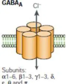
GABAA is the main GABA receptor and its structure was identified in
1990 (Olsen and Tobin, 1990). GABAA receptors belong to the
ligand-gated ion channel superfamily that includes nicotinic receptors for acetylcholine, glycine receptors and also serotonin receptors. GABAA
is a pentameric receptor composed by distinct polypeptides. At the moment, several distinct peptides have been identified, namely six α-subunits, three β-subunits, three γ-subunits, one δ-subunit, one ε-subunit, one π-subunit and one θ-subunit (Schofield, 1987; MacDonald and Olsen, 1994; Mehta and Ticku, 1999).
Figure 1.2 – Schematic representation of a GABAA receptor (adapted from
Owens and Kriegstein, 2002).
Although a functional GABAA receptor always includes one α, one β
and one γ subunit (Pritchett et al., 1989), the GABAA subunits can be
assembled in several possible combinations, with some of them being preferred. For example, GABAA receptors containing α1
subunits combined with β2 and γ2 subunits are the most abundant form in the brain (McKernan and Whiting, 1996). Furthermore, different subunits compositions of the receptors seem to be related with different cellular localizations, where they can mediate synaptic or extrasynaptic signaling (Mody, 2001).
The majority of GABAA receptors have affinity for benzodiazepines,
which, allosterically, exacerbate the effect of GABA. This property is the basis for therapeutic effect of benzodiazepines as anxiolytic agents (for review, Bowery and Smart, 2006). The GABAA receptors
contain other modulatory binding sites, sensitive to barbiturates, neurosteroids and ethanol (Macdonald and Olsen, 1994).
GABA binding to the receptor leads to a conformational change, which allows a net inward or outward flow through the channel, depending on the electrochemical gradient. GABAA receptors carry
bicarbonate (HCO3-), although with lower efficiency (Bormann et al.,
1987; Kaila, 1994). 1.1.1.2 GABAC receptors
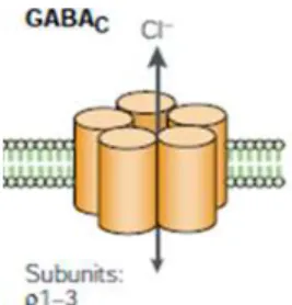
A related ionotropic GABA receptor was named GABAC by Drew and
colleagues, in 1984 (Drew et al., 1984). This receptor is a chloride-selective channel but is insensitive to the GABAA antagonist,
bicuculline. GABAC are homo or hetero-pentomeric receptors
composed by ρ subunits (Cutting et al., 1991, 1992).
Figure 1.3 – Schematic representation of GABAC receptor (adapted from
Owens and Kriegstein, 2002).
As the ρ subunits share extensive sequence homology with some GABAA subunits, GABAC receptors can be considered as
pharmacological variants of GABAA receptors. However different
studies involving GABAC receptors have strongly suggested that they
have distinct pharmacology, structure, function, genetics and cellular localization, and consequently should be considered as an independent class of GABA receptors (Bormann, 2000). Although GABAC receptors were first identified in the retina (Feigenspan et al.,
1993; Qian and Dowling, 1993), posterior data suggests that GABAC
receptor has a widespread distribution in the CNS (López-Chávez et al., 2005).
1.1.1.3GABAB receptors
In 1980, it was described the existence of a different GABA receptor (GABAB), that was insensitive to bicuculline, chloride independent
and responsible for the baclofen inhibitory effect upon neurotransmitter release in CNS (Bowery et al., 1980). The GABAB
receptor was insensitive for bicuculline and chloride independent and was termed GABAB receptor (Bowery et al., 1980). GABAB is a
metabotropic receptor which signals through Gi/0 proteins,
decreasing adenylate cyclase activity and modifying calcium and potassium conductances. GABAB receptors are expressed either at
pre-synaptic and pos-synaptic levels. Presynaptic activation of GABAB receptor results in inhibition of calcium channels, which leads
to a reduction in neurotransmitter release. At the pos-synaptic level, the effect of GABAB is mediated by enhancement of potassium
conductance, which leads to neuron hyperpolarization (Bormann, 1988).
Cloning of GABAB receptor showed a seven-transmembrane
receptor that exists as a heterodimer composed of GABAB1 and
GABAB2 subunits (White et al., 1998). Interestingly, the GABAB
receptors were the first G-protein coupled receptors to be described as heteromeric receptors (Kaupmann et al., 1998). However GABAB
subunits cannot work separately: the GABAB1 subunit binds to all
known GABAB ligands (Kaupmann et al., 1997), while GABAB2
involved in different roles, namely: (1) to mask the retention signal of the GABAB1 subunit, allowing that GABAB heteromer to reach the
Pagano et al., 2001), (2) to contain the molecular determinants required for G-protein coupling (Galvez et al., 2001); (3) to play a critical role in G-protein activation within the heteromer (Margeta-Mitrovic et al., 2001; Robbins et al., 2001; Duthey et al., 2002; Havlickova et al., 2002); and (4) to increase agonist affinity on GABAB1 (Kaupmann et al., 1998; White et al., 1998; Galvez et al.,
2001). Thus GABAB can be considered as a natural complementation
of two non-functional receptors: one subunit recognizing the ligand and the other activating the G protein (Pin et al., 2003).
Figure 1.4 – Schematic representation of GABAB receptor (adapted from
http://www.pharma.uzh.ch/research/neuropharmacology/researchareas/ signaltransduction/introduction.html).
1.1.2 GABA metabolism
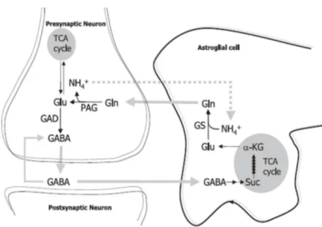
Neurons lack pyruvate carboxylase, so they are unable to de novo synthesis of glutamate and GABA (Patel, 1974; Shank et al., 1985), which suggests the occurrence of a metabolic crosstalk between neurons and glial cells. In fact, the discovery of glutamine and glutamate pools in neurons and astrocytes was indicative of a
glutamate/glutamine cycle, working between neurons and glial cells (van den Berg and Garfinkel, 1971; Benjamin and Quastel, 1972). Considering a GABAergic synapse, the majority of GABA is taken up into presynaptic terminals (see Schousboe, 2000), where without any metabolic changes, GABA can quickly be package into vesicles by a vesicular GABA transporter (vGAT), being available for dependent calcium exocytic release.
However, GABA can also be taken up into astrocytes (it has been estimated that 20% of released GABA is taken up into surrounding astrocytes, see Schousboe, 2000). By this path, GABA is firstly metabolized into glutamate and glutamine. Then, glutamine is redirected to the presynaptic terminal, where, GABA is resynthetized. This metabolic pathway is known as the aforementioned glutamate/GABA-glutamine cycle. Briefly, after being taken up into astrocytes, GABA is catabolised to succinate (a tricarboxylic acid (TCA) cycle intermediate via the concerned action of GABA transaminase and semialdehyde dehydrogenase. Succinate, through TCA cycle, will originate α-ketoglutarate, which is transaminated to glutamate by glutamate transaminase. Glutamate is then converted to glutamine by the glutamine synthetase (GS). Glutamine is then released to the extracellular space, from where is taken up into presynaptic neurons. After being taken up, glutamine is converted to glutamate by phosphate-activated glutaminase (PAG). Glutamate is finally converted into GABA, by glutamate decarboxylase (GAD) (see figure 1.5 and Bak et al., 2006).
Figure 1.5 – Schematic representation of glutamate/GABA-glutamine cycle
in a GABAergic synapse (adapted from Bak et al., 2006).
1.1.3 GABA transporters
Ambient levels of neurotransmitter in the synapse result from the balance between neurotransmitter release, diffusion within the synaptic cleft and the uptake through transporters. The main role attributed to neurotransmitter transporters is the shutdown of synaptic transmission. In the case of ionotropic receptors, both receptor desensitization and neurotransmitter uptake mediated by transporters contribute to the end of transmission, being unclear which is the principal phenomenon. Within these, extrasynaptic receptors desensitize slower than synaptic ones, so the relative contribution of transporters for shutting down GABA action at ionotropic receptors varies according to their location. In contrast, for metabotropic receptors, which desensitize slower than ionotropic receptors, neurotransmitter uptake is the predominant mechanism through which the synaptic transmission is finished (Borden, 1996).
Besides the temporal delimitation of synaptic transmission, transporters also contribute for synaptic spatial control by preventing the neurotransmitter spill over, assuring the precise location of transmission (see Krogsgaard-Larsen et al., 1987). It was indeed demonstrated that the maintenance of appropriate levels of transmitter in the synaptic cleft by transporters is crucial for normal brain function (Giros et al., 1996; Tanaka et al., 1997).
From a metabolic perspective, neurotransmitter uptake by transporters prevents the depletion of intracellular pools, preserving metabolic energy balance. The maintenance of these pools is critical for homeostasis, especially during periods of high activity. As mentioned above, neurotransmitter recycling can, however, be direct or indirect: the direct recycling occurs when the neurotransmitter is taken up directly to the pre-synaptic terminal, allowing the prompt replenishment of synaptic vesicles. The indirect recycling involves the surrounding astrocytes and the uptake by these cells implies metabolic conversion before the transmitter is available to be packaged into exocytic vesicles, as it was shown above.
GABA levels at the synapse are therefore mainly regulated by the specific transporters (GATs) expressed in neurons and astrocytes, which were identified in 1968 (Iversen and Neal, 1968). However, even before the recognition of GATs, Elliot and van Gelber (1958) had already demonstrated that GABA added to the incubation
medium can accumulate in slices of cerebral cortices (Elliot and van Gelber, 1958).
Figure 1.6 – Schematic representation of a GABAergic synapse. As
illustrated, GATs are expressed in both presynaptic terminals and astrocytes (adapted from Owens and Kriegstein, 2002).
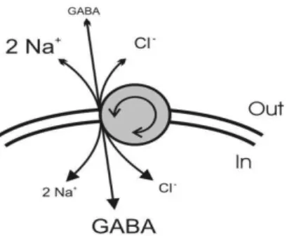
1.1.3.1GABA transporters family
GABA transporters belong to the family of Na+/Cl- coupled receptors. In each transporter cycle, one uncharged GABA molecule is co-transported with two Na+ ions and one Cl- ion (see figure 1.7 and Kanner and Schuldiner, 1987). Thus GABA is carried up its concentration gradient using energy from the inward Na+ gradient and the flow of one positive charge downs the electrical gradient (Richerson and Wu, 2003).
Figure 1.7 – Schematic representation of GABA transport. Each molecule of
GABA is co-transported with two sodium ions and one chloride ion (adapted from Richerson and Wu, 2003)
Shortly, GABA binds the transporter in its resting state, facing extracellular space. After the binding of GABA, Na+ and Cl- ions, the transporter undergoes a conformational change enabling GABA and ions to face intracellular space, where they are released. Then, the unfilled transporter reverts to the resting state, waiting for a new cycle of transport (Borden, 1996).
Furthermore, it was shown that the magnitude of GAT-1 mediated currents is altered in response to changes in Na+, Cl- and GABA gradients (Lu and Hilgemann, 1999). Although these experiments were performed using nonphysiological concentrations, they provided initial evidence that transport can be modified (and possibly reverted) in response to ionic concentration changes. So far, four distinct GABA transporters have been identified: GAT-1, GAT-2, GAT-3 and BGT-1. The first three are high affinity-transporters and the last is a low-affinity transporter.
1.1.3.2GAT-1
GAT-1 was the first neurotransmitter transporter to be cloned (Guastella et al., 1990). GAT-1 mRNA was found in all brain regions, being the highest levels of hybridization signal observed in brainstem nuclei, ventral tegmental area, pontine nuclei, cerebellum, thalamus, basal forebrain, hippocampus, olfactory bulb and neocortex. Concerning cellular localization, GAT-1 is expressed by both neurons and astrocytes. Although GAT-1 is mainly expressed at GABAergic neurons (which express concomitantly GAT-1 and GAD67), it can also be found in a small number of glutamatergic cells, suggesting that GABA uptake system mediated by GAT-1 is strongly expressed in the brain, being more widespread than the GABA synthesizing system (Minelli et al., 1995). GAT-1 is a 67 kDa protein with twelve transmembrane regions, with both N- and C- termini in the intracellular space. GAT-1 has four putative glycosylation sites, being three of them located on the large extracellular loop that connects membrane segments 3 and 4. GAT-1 has also eight putative phosphorylation sites: three putative protein kinase C phosphorylation sites are intracellular, two at the N-terminal and one at C-N-terminal. The other three sites for PKC and the single site for PKA are located externally or within membrane segments (Guastella et al., 1990), being therefore of questionable functional relevance.
Figure 1.8 – Schematic representation of the GABA transporter GAT-1.
The rectangles represent the twelve transmembrane domains, connected by extramembrane loops. The large extracellular loop, between membrane segments 3 and 4, contains three potential glycosylation sites, illustrated by the branched lines (adapted from Kanner, 1994).
1.1.3.3GAT-2
GAT-2 mRNA was initially identified only at cells of the leptomeninges (pia and arachnoid - Durkin et al., 1995). Later, a weak GAT-2 immunoreactivity was observed in the cortical parenchyma, where GAT-2 is expressed by neuronal and non-neuronal cells (Conti et al., 1999). However, the predominant localization of GAT-2 to leptomeninges and blood vessels suggests that GAT-2 function is mainly related to the regulation of GABA levels in the cerebrospinal fluid (Minelli et al., 2003).
1.1.3.4GAT-3
In the early 90’s, several studies indicated that GAT-3 was absent or very weakly expressed at the cerebral cortex (Clark et al., 1992; Durkin et al., 1995; Ikegaki et al., 1994). Then, in 1996, Minelli and colleagues showed that GAT-3 is expressed in cerebral cortex, exclusively at astrocytes, being GAT-3 the main GABA transporter
expressed in glial cells (Minelli et al., 1996). In addition, Durkin and colleagues showed that GAT-3 mRNA is present in many brain areas, though it is less abundant the GAT-1 mRNA (Durkin et al., 1995). 1.1.3.5BGT-1
BGT-1, the fourth GABA transporter, was known as a result from renal physiology studies, where a betaine transporter was initially identified. This transporter had the ability to concentrate urine through the uptake of the betaine (glycine betaine), that was co-transported with Na+ and Cl- (see Borden, 1996). Then, Yamauchi and colleagues isolated a clone encoding a Na+/Cl- dependent transporter, which was able to utilize both GABA and betaine as substrates. Hence, the clone was named BGT-1 (Betaine/GABA transporter). Surprisingly, BGT-1 has higher affinity for GABA (KM =93
µM) than for betaine (KM =398 µM) (Yamauchi et al., 1992).
The expression of BGT-1 as well as its role in CNS is still not clear although the presence of BGT-1 mRNA in human and mouse brain may suggest its involvement in GABAergic transmission. However, the distribution of BGT-1 mRNA does not correlate with GABAergic pathways, which corroborates the hypothesis that BGT-1 might not play a role in finishing GABA effects at synapses. Alternatively, betaine may be the primary substrate for BGT-1 in the brain (as it is in the kidney), which suggests an involvement of BGT-1 in osmoregulation, thus BGT-1 could contribute to the volume control in the CNS. Osmoregulation is particularly important for brain
function (see Borden, 1996), as the skull is a rigid and non-expandable container.
1.1.3.6GABA transporters modulation
Considering that the control of GABA concentration at synapse is strongly regulated by GABA transporters, their modulation is a key factor to assure adequate GABA levels for brain function. GAT modulation occurs mainly by changes in their expression at the membrane level. In fact, this expression is not static and transporters can cycle to and from membrane on the time scale of seconds to minutes, being this cycling regulated by different factors, including extracellular signals (Zahniser and Sorkin, 2004; Sattler and Rothsein, 2006), protein phosphorylation (Foster et al., 2006) and interactions with accessory proteins (Quick, 2006).
The average number of GAT-1 molecules in a cultured cortical inhibitory bouton is approximately 3000-4000 (Chiu et al., 2002) and the size of the recycling pool is about 30% of total GAT-1 (Wang and Quick, 2005). Then, about 1000 GAT-1 molecules traffic to and from the membrane, with GAT-1 being inserted into the membrane in a calcium-dependent manner and internalized via a clathrin-dependent process (Deken et al., 2003). The regulation of membrane expression levels can occur through changes in recycling pool size or modification of exocytosis and endocytosis rate. Changes at transporters surface membrane expression occur in a scale of minutes (Mennerick et al., 1999; Blakely and Bauman, 2000; Deken et al., 2000; Robinson, 2002).
Firstly, GAT-1 is modulated by its own substrate, GABA, being this a concentration dependent effect, involving changes in phosphorylation state, which consequently modify the transporter surface expression (Hu and Quick, 2008).
In fact, phosphorylation seems to be a critical cellular mechanism responsible for changes in the membrane expression of transporters. As aforementioned, GAT-1 has eight putative phosphorylation sites (Guastella et al., 1990) and it was described that PKC activation decreases membrane GAT-1 levels (Corey et al., 1994), in a way that is independent of protein synthesis. This action of PKC activators is mimicked by phosphatase inhibitors, suggesting that changes in membrane levels are dependent on protein phosphorylation. However, it was shown that the effect of PKC upon GAT-1 surface expression still occurs when consensus residues for PKC phosphorylation are removed (Corey et al., 1994). Thus, it seems that there are other cellular mechanisms through which PKC can influence GAT-1 levels. In fact, a subsequent study showed that PKC effect upon GAT-1 function occurs through the regulation of syntaxin A1 availability to interact with the transporter. Further, a mutant transporter that fails to interact with syntaxin A1 is not regulated by the PKC (Beckman et al., 1998).
Like PKC, Ca2+ depletion also decreases surface expression of GAT-1, while hypertonic sucrose treatment increases its surface expression (Beckman et al., 1999; Deken et al., 2003). Interestingly, these effects are not mediated by the same cellular mechanism: PKC and sucrose application modifies the endocytosis rate but does not
change the recycling pool size, which is dramatically diminished by extracellular calcium depletion (Wang and Quick, 2005).
1.1.3.7Reversal of GABA transporters
It is known for a long time that neurotransmitters transporters can revert (Schwartz, 1987; Pin and Bockaert, 1989; O’Malley et al., 1992; Attwell et al., 1993; Levi and Reiteri, 1993; Cammack et al., 1994; Lu and Hilgemann, 1999). For instance, after extracellular calcium removal, GABA is still released from cortical slices in response to: (1) an increase in K+ to 50mM; (2) an exposure to veratridine (50 µM); (3) a high-frequency electrical stimulation (Szerb, 1979). GABA is also released from cultured striatal neurons, in the absence of calcium (Pin and Bockaert, 1989) and from cortical neurons in response to 55-56 mM of K+ or 100 µM of glutamate (Belhage et al., 1993). Neurotransmitter release in the absence of extracellular calcium occurs by the reversal of transporters since it is prevented by the blockade of transporters (Pin and Bockaert, 1989; Belhage et al., 1993). As these studies were performed using non-physiological stimuli, the transporters reversal was initially associated with pathological conditions such as ischemia (Nicholls and Attwell, 1990; Levi and Raiteri, 1993). However, transporter-mediated GABA release can also occur in response to presynaptic stimulation (Schwartz, 1987; Bernath and Zigmond, 1988) or in response to an elevation in extracellular K+ concentration from 3 to 12 mM (Gaspary et al., 1998), a level of K+ that is reached in vivo during neuronal firing (Krnjevic et al., 1980; Somjen and Giacchino, 1985) and during seizures (Fisher et al., 1976). In 2001, a study by
Wu and colleagues corroborate the hypothesis that GABA transporters reverse more easily than previously recognized: GABA reversal was induced by a small depolarization of membrane potential caused by 6mM K+ in hippocampal cultures (Wu et al., 2001. This potassium increase is physiologically relevant as extracellular potassium concentration increases during neuronal firing in vivo. For example, an increase in [K+] to > 12mM occurs in the rat hippocampus in vivo in response to electrical stimulation of the angular bundle (Somjen and Giacchino, 1985). Then, instead of being associated with some pathological state, the reversal of GABA transporters seems to be related with modifications in the ratio between intra and extracellular concentrations of GABA. In fact, the rate of GABA uptake is governed by a driving force, which decreases as extracellular GABA concentration reduces. Then, the GABA uptake by transporters occurs until the equilibrium between intra and extracellular concentrations is achieved. Thus, GABA transporters are unable to uptake all the GABA, and extracellular GABA concentration fall around 0.1-0.8 µM (Hagberg et al., 1985; Lerma et al., 1986; Tossman et al., 1986; Phillis et al., 1994; Cavelier et al., 2005).
1.1.3.8GABA transport impair and neurological disorders
As GATs are the main responsible for GABA clearance from synapse, a transporter dysfunction will consequently originate an unbalance in GABA concentrations, which has been associated with several pathologies, especially in epilepsy (e.g. Richerson and Wu, 2004), in which some therapeutic options are related with GABA transporter.
A possible therapeutic option in epilepsy is to increase the inhibitory tone in order to prevent seizures. This can be achieved through the blockade of GABA uptake or by the reversal of GABA transporters. Interestingly, in animal models, specific blockers of GAT-1 and blanket GABA transporter inhibitors have been shown to be selectively effective against different types of seizures. This can be due to a differential distribution of GABA transporters among brain regions. GAT-1 is predominantly expressed in the forebrain and limbic areas while GAT-3 is mainly presented in the brainstem. The kindled focal and generalised secondary seizures, and clonic convulsions, induced by chemoconvulsants and originated in limbic system or forebrain, are abolished by GAT-1 blockers. On the other hand, convulsions induced by electro shocks (originated in the basal ganglia and brainstem) are inhibited by non-selective GATs inhibitors (Nielsen et al., 1991; Swinyard et al., 1991; Dalby et al., 1997). Actually, the discovery of a potential use of GABA transporter inhibitors in epilepsy occurred thirty years ago (Krogsgaards-Larsen et al., 1981) and since then, several non-selective inhibitors have been experimentally demonstrated to possess anticonvulsant activity (Dalby et al., 1997; Dalby, 2003; White et al., 2005). Nowadays, tiagabine, a specific GAT-1 inhibitor (Borden et al., 1994), is used as an adjuvant therapy for patients with focal seizures, namely with complex partial temporal lobe epilepsy and secondary generalised tonic-clonic seizures (Uthman et al., 1998).
Other therapeutic approach involving the GATs reversal is the use of vigabatrine, which blocks GABA transaminase, the enzyme
responsible for GABA catabolism. The inhibition of GABA transaminase will increase GABA intracellular levels, favouring the reversal of GABA transporters (Wu et al., 2001, 2003).
The reversal of GABA transporters was also observed during ischemic conditions, where the increase of extracellular K+, by the inactivation of Na+/K+ ATPase, is enough to reverse GATs. In fact, GAT-1 inhibition reduced the amount of GABA released (and subsequently, cell swelling) during energy deprivation (Zeevalk and Nicklas, 1996). So, although it has been considered that the potentiation of GABAergic transmission contributes to reduce ischemia-induced damage (Green et al., 2000), the neuroprotective effect mediated by GATs inhibition can be related with a decrease in the GABA released by the reversal of transporters (and consequently to a decrease in extracellular GABA levels) instead of being associated with a enhancement in extracellular GABA, caused by the blockade of GABA uptake, as some authors had suggested (Phillis, 1995; Green et al., 2000). The reversal of GATs associated with an increase in intracellular GABA concentrations, which is also observed during ischemia (Erecinska et al., 1984), contributes to an overactivation of GABAA receptors, which leads to an increased
influx of Cl- ions, and subsequently, to cell swelling and eventually cell death.
The enhancement in intracellular GABA levels is the result of three different aspects: first, the reduced ATP levels found under ischemia, stimulate GABA synthesis by GABA decarboxylase (Madl and Royer, 2000); second, the reduction of pH, that occurs under ischemia,
inhibits GABA transaminase, leading to an accumulation of intracellular GABA (Madl and Royer, 2000); third, the disruption of vesicular H+-dependent GABA storage during ischemia rises cytoplasmic GABA, promoting GABA transporters reversal under ischemic conditions (see Allen et al., 2004). Recently, Clarkson and colleagues identified an excessive tonic inhibition after stroke which was associated to the observed decrease in GAT-3 expression in peri-infarct cortex (Clarkson et al., 2010). Thus, besides the reversal of the transporters, changes in transporters expression are also associated with ischemic conditions, which make GATs potential targets in treatment of stroke.
1.1.3.9Influence of GATs upon tonic and phasic GABAergic transmission.
GABA acts through GABAAR and mediates both phasic and tonic
inhibition. Phasic inhibition is a transient and point-to-point phenomena, as a consequence of the release of GABA following nerve terminal depolarization, leading to inhibitory postsynaptic currents (IPSCs). Tonic inhibition is associated with the spillover of GABA outside the synaptic areas leading to lower but sustained extracellular GABA levels. The main feature of phasic GABAA
R-mediated inhibition is the rapid synchronous opening of a relatively small number of channels that are clustered at the synaptic junction, whereas tonic inhibition results from random, temporally dispersed activation of receptors that are distributed over the neuronal surface. This distinction involves a deep difference in the control of
neuronal network activity by phasic and tonic forms of inhibition (see Farrant and Nusser, 2005).
At the central nervous system, the extracellular GABA concentration ranges from 0.2 to 2.5µM (Lerma et al., 1986; Tossman et al., 1986; Ding et al., 1998; Kuntz et al., 2004). At ambient GABA concentrations, the extra- and peri-synaptic GABAAR are
preferentially activated as they have higher affinity for GABA, as compared to synaptic GABAAR. These extra-synaptic receptors are
then responsible for tonic inhibition (Brickley et al., 1996; Farrant and Nusser, 2005), which is highly related with sustained changes in extracellular GABA concentration, therefore to changes in the activity of GABA transporters. In fact, GABA transporters are responsible for the maintenance of homeostatic GABA levels. This control can be achieved by changes in uptake rates- which are related with protein surface expression – but also by the reversal of GABA transporters (Gaspary et al., 1998; Richerson and Wu, 2004). Some electrophysiological studies have reported the influence of GATs in GABAergic transmission. For instance, in rat neocortex and in the hippocampus, GAT-1 blockers increase the decay time of evoked inhibitory post-synaptic currents (eIPSCs) without modifying the spontaneous IPSCs (sIPSCs) (Thompson and Gahwiler, 1992; Engel et al., 1998; Overstreet and Westbrook, 2003; Keros and Hablitz, 2005), as well as enhance the amplitude of tonic GABAAR
mediated currents (Nusser and Mody, 2002; Semyanov et al., 2003; Keros and Hablitz, 2005; Marchionni et al., 2007;), thus indicating that GAT-1 affects both tonic and phasic GABAergic transmission.
This influence of GAT-1 upon phasic and tonic transmission was further confirmed by Bragina and colleagues, using GAT-1 KO mice (first characterized by Jensen in 2003), who showed that a chronic impairment in GAT-1-mediated GABA uptake in the neocortex modifies both phasic and tonic GABAAR-mediated inhibition. GAT-1
KO mice showed an increase in glutamate decarboxylase (GAD65/67) expression but no changes in GAT-3 expression or distribution (Bragina et al., 2008).
The influence of GABA transporters upon tonic transmission is probably related with transporters features, since GATs just take up GABA until reach equilibrium, being not able to remove all the extracellular GABA. This implies that a small (few micromolar) amount of GABA remains extracellularly, being enough to activate extra-synaptic GABAA receptors, which are the responsible for tonic
inhibition.
The majority of studies about the influence of GABA transporters upon GABAergic transmission have been focused in the neuronal transporter, GAT-1. But, recently, a study had highlighted the role of glial transporter GAT-3 upon GABAergic transmission. It was observed that the GAT-3 blockade (just when GAT-1 was previously blocked) reduces the frequency of sIPSC (Kirmse et al., 2009), attributing to GAT-3, a role in modulation of phasic GABAergic transmission, at least when GAT-1 is blocked, which means that the contribution of GAT-3 under basal conditions is still unclear.
1.2
The tripartite synapse and glial cells
For many years, glial cells were considered as supportive cells and unable to communicate with neurons or other glial cells. But, in the 90’s years, several studies revealed the existence of bidirectional communication between neurons and astrocytes. These different studies led Araque and colleagues to propose that synapses are tripartite, consisting of synaptically associated glia as well as the presynaptic and the postsynaptic terminals (Araque et al., 1999). It was probably the discovery that astrocytes can be excited non-electrically that expanded the brain communication to non-synaptic elements and changed the paradigm of brain function based on neuronal activity to a function dependent on neuron-glia network. There are four different types of glial cells: astrocytes, oligodendrocytes, microglia and Schwann cells. The first three are present in the central nervous system while the last one can be only found in the peripheral nervous system. Oligodendrocytes and Schwann cells are responsible for myelin production while microglia is related with immune defense of nervous system.
1.2.1 Astrocytes
The central nervous system is mainly composed of glia, in a proportion (in humans) of 1 neuron to 10 glial cells. These cells were identified by Rudolf Virchow in 1858. In his lecture, he described astrocytes as connective tissue, a kind of as “nervenkitt” or nerve-cement. Virchow named these cells as neuroglia (Virchow, 1858, quoted by Kettenmann and Verkhratsky, 2008). Although for him
neuroglia was a connective tissue, he admitted that it also contained a certain number of cellular elements. But Virschow was not the first to identify glial elements, Robert Remak had previously described, in 1838, a sheath around single nerve cells (R. Remak, PhD Thesis, University of Berlin, 1838, quoted by Kettenmann and Verkhratsky, 2008) and Heinrich Müller had previously discovered the radial fibers in the retina in 1851 (these cells were latter called as Müller glial cells).
During the following years, many different forms of glial cells were described and images were published (Deiters, 1865; Henle and Merkel, 1869; Golgi, 1903, quoted by Kettenmann and Verkhratsky, 2008). Later, in 1893, Michael von Lenhossek proposed the name astrocyte (von Lenhossek, 1893, quoted by Kettenmann and Verkhratsky, 2008). Astrocytes were then classified as fibrous or protoplasmic astrocytes by Kölliker (Kölliker, 1889, quoted by Kettenmann and Verkhratsky, 2008) and Andriezen (Andriezen, 1893, quoted by Kettenmann and Verkhratsky, 2008). Thus, although they are known since the 19th century, astrocytes were neglected for a long time, probably because they are not able to generate action potentials. They used to be considered as silent and supportive cells with relevant roles in metabolic pathways and also neurotransmitters uptake from synapse. In fact, it was early suggested that glial cells may remove and inactivate the products of neuronal activity (Lugaro, 1907, quoted by Henn and Hamberger, 1971). Later, it was shown that glial cells have the ability to accumulate amino acids (aspartic acid, threonine, serine, proline;