Full paper published online: May 30, 2009 ISSN 1678-9199.
NEUROTRANSMITTER EVALUATION IN THE HIPPOCAMPUS OF RATS AFTER INTRACEREBRAL INJECTION OF TsTX SCORPION TOXIN
Nencioni ALA (1), Barreto SA (2), Lebrun I (2), Florio JC (3), Lourenço GA (1), Dorce VAC (1)
(1) Laboratory of Pharmacology, Butantan Institute, São Paulo, São Paulo State,
Brazil; (2) Laboratory of Biochemistry and Biophysics, Butantan Institute, São Paulo,
São Paulo State, Brazil; (3) Laboratory of Pharmacology, School of Veterinary
Medicine and Zootechny, University of São Paulo, USP, São Paulo, São Paulo State,
Brazil.
ABSTRACT: TsTX is an α-type sodium channel toxin that stimulates the discharge of neurotransmitters from neurons. In the present study we investigated which neurotransmitters are released in the hippocampus after TsTX injection and if they are responsible for electrographic or histopathological effects. Microdialysis revealed that the toxin increased glutamate extracellular levels in the hippocampus; however, levels of gamma-aminobutyric acid (GABA), glycine, 5-hydroxyindoleacetic acid (5-HIAA), homovanillic acid (HVA) and 3,4-dihydroxyphenylacetic acid (DOPAC) were not significantly altered. Neurodegeneration in pyramidal cells of hippocampus and electroencephalographic alterations caused by the toxin were blocked by pretreatment with riluzole, a glutamate release inhibitor. The present results suggest a specific activity of TsTX in the hippocampus which affects only glutamate release.
KEY WORDS: scorpion toxin, microdialysis, hippocampus, neurotransmitters, neurodegeneration, riluzole.
CONFLICTS OF INTEREST: There is no conflict.
CORRESPONDENCE TO:
VALQUIRIA ABRÃO CORONADO DORCE, Laboratório de Farmacologia, Instituto
Butantan, Av. Dr. Vital Brasil, 1500, São Paulo, SP, 05503-900, Brasil. Phone: +55
INTRODUCTION
The toxicity of scorpion venoms is due to neurotoxins that are composed of basic
polypeptides with molecular weights of approximately 7,000 Da. These toxins affect
cell permeability to ions including sodium, potassium, chloride and calcium (1-4).
Sodium channel neurotoxins affect sodium conductance in various excitable tissues,
thus serving as important pharmacological tools for the study of excitability and
sodium channel structure (5).
Scorpion neurotoxins that act on sodium channels have been divided into two
groups, α and β toxins, according to the ligand-binding sites in the channel. The α
-scorpion toxins were the first to be studied. Their primary effect is interference in
channel inactivation upon prolonged depolarization, increasing sodium permeability
and consequently extending the duration of the action potential (6). One of the main
consequences is the augmented release of neurotransmitters from neuronal endings
(7).
Gomez and Diniz (8) were the first to isolate toxic polypeptides from the venom of the
Brazilian scorpion Tityus serrulatus. According to Arantes et al. (9), TsTX was first
isolated by Coutinho Netto in 1975. This α-type sodium channel toxin increases the
release of glutamate, acetylcholine and GABA from different preparations of nervous
tissue (10-13). When injected in the dorsal hippocampus of rats, it induces behavioral
alterations and epileptic discharges besides neurodegeneration (14).
The aim of the present study was to investigate which neurotransmitters are released
after TsTX intrahippocampal injection. To do this, microdialysis analysis was
performed to evaluate extracellular levels of some cerebral neurotransmitters.
In a previous study, we observed an increase in extracellular levels of glutamate only
until three hours after toxin injection (11). Based on the toxin kinetic and on the
glutamate release profile observed in that study, we decided to lengthen the time of
collection (15).
Moreover, since the previous work by Nencioni et al. (11) had indicated the
involvement of glutamate in the convulsive/neurodegenerative effect of the toxin; in
the present study, we examined the result of pretreatment with riluzole
(2-amino-6-trifluoromethoxy benzothiazole, RP54274), a drug that inhibits glutamate release and
MATERIALS AND METHODS Subjects
Male Wistar rats (200 to 250 g), obtained from an established colony kept by the
Central Animal House Service at the Butantan Institute, were used. Upon their arrival
in the laboratory (seven days before the experiments), the animals were individually
housed in wire mesh cages and kept at constant temperature (22 ± 1°C), in a
12-hour light/12-12-hour dark cycle (lights on at 0700 h), with food and water provided ad
libitum.
The animals employed in the current study were maintained in accordance with the
policies of the Ethics Committee on Use of Laboratory Animals, Butantan Institute,
Brazil.
TsTX Purification
Tityus serrulatus toxins were obtained according to the method described by
Carvalho et al. (14). Briefly, the venom was extracted with 0.05 M ammonium
bicarbonate buffer, pH 8.0, and eluted in Sephadex® G-50 M (Pharmacia Fine
Chemicals, USA). Active fractions 30 to 63 (pool IV) were eluted with ammonium
bicarbonate (0.05 M, pH 8.0) in Sephadex® G-25 M column (Pharmacia Fine
Chemicals, USA). The peptides purified by gel filtration chromatography were
separated by high performance liquid chromatography (HPLC) and the peak 8 was
used (TsTX).
Chemicals
• Ringer solution (Aster, Brazil)
• TsTX scorpion toxin (Butantan Institute, Brazil) dissolved in Ringer solution • L-glutamic acid HCl (Research Biochemicals International, USA)
• L-aspartic acid (Research Biochemicals International, USA) • Glycine (Sigma, USA)
• γ-aminobutyric acid (Sigma, USA)
• Riluzole hydrochloride (Tocris, USA) dissolved in 10% dimethyl sulfoxide (DMSO – Sigma, USA)
• Triethylamine (Sigma, USA)
• Ethylenediaminetetraacetic acid (EDTA – Merck, Germany) • Acetonitrile (Malinkrodt, USA)
• 3,4-dihydroxybenzylamine (DHBA – Sigma, USA) • Perchloric acid (Merck, Germany)
• Sodium metabisulfit (Merck, Germany)
Surgery
Rats were anesthetized with an intraperitoneal (IP) injection of 3 mL/kg of a mixture
of pentobarbitone (1 g) and chloral hydrate (4 g) diluted in 100 mL of 0.9% NaCl.
Animals were placed in a stereotaxic frame and a microdialysis guide cannula was
implanted unilaterally in their dorsal hippocampus. Guide cannulas were fixed to the
skulls with stainless steel jeweler screws and dental acrylate. The guide cannulas for
microdialysis were implanted in rats according to the following coordinates: 5.3 mm
posterior to bregma, 3.2 mm lateral to midline, and 2 mm below dura mater (17). The
cannulas were used for both toxin injection and dialysate collection. In rats
undergoing behavioral studies and EEG recording, the coordinates were 4.8 mm
posterior to bregma, 3.2 mm lateral to midline, and 2.8 mm below dura mater. In
these rats, bipolar twisted electrodes for depth recordings were implanted
contralaterally at the same coordinates and anchored to the skull as were the
cannulas. Jeweler screws were inserted bilaterally in the skull over the occipital
cortex for surface recordings. A screw placed in the frontal sinus was employed as
reference (indifferent electrode). The surgery was conducted using aseptic
techniques and animals with surgical complications were excluded. Subsequently,
rats were individually housed and allowed to recover for one or two days.
Experimental Groups
A control group of six rats, used for EEG recording and histology, received a 2-µL
intrahippocampal injection of Ringer solution. A second group composed of 11 rats
received an intrahippocampal injection of 2 µg of TsTX diluted in 2 µL of Ringer
solution. These animals were used for microdialysis experiments (five for amino acid
test and six for analysis of monoamine) and histology. A third group of 11 rats
2-µg intrahippocampal injection of TsTX. Six rats from this latter group were employed
for EEG recording and histology, while the remainder were utilized for amino acid
analysis.
Microdialysis
Microdialysis was performed in freely-moving rats between 24 and 48 hours after
implantation of guide cannulas. The probe (CMA/11 microdialysis probe, membrane
length 2.0 mm – CMA Microdialysis Instruments, Sweden) was introduced into the
guide cannula of each animal while the inlet was connected through 70-cm
polyethylene tubing to a 2.5-mL Hamilton® syringe (Hamilton Company, USA). The
outlet was connected to a 15-cm polyethylene tubing. Ringer solution was perfused
at a flow rate of 2.13 µL/min and the final volume of samples was approximately 60
µL. After 30 to 45 minutes of equilibration period, the outflow was collected every 30
minutes during two hours. Subsequently, the probe was disconnected without flux
interruption and the toxin was injected through the cannula. After that, the probe was
connected again to each rat and twelve additional microdialysis samples were
collected until the end of the experiment. Pretreated animals received an IP injection
of riluzole 15 minutes before the toxin administration, without disconnecting probes.
At the end of the collection, samples were dried and frozen (–80°C) until the analysis.
For biogenic amine determination, samples were collected in a tube with 5.0 μL of a
solution composed of EDTA, sodium metabisulfite and perchloric acid.
Measurement of Neurotransmitter Concentration
Extracellular neurotransmitter levels in the dialysate were expressed as μg/μL or μg/mL. The first four samples from each animal were used as control and compared with post-treatment samples of the same individual.
Statistical significance was evaluated by one-way repeated measures analysis of
variance and Tukey’s post-hoc test. Values of p < 0.05 were considered significant.
Monoamines
Levels of dopamine (DA) and its metabolites (DOPAC and HVA), 5-HT and its
metabolite (5-HIAA), and norepinephrine (NE) were measured by a HPLC system
(model 6A, Shimadzu, Japan) with a C-18 Shim pack-ODS® column (Shimadzu,
(valve for 20 #1), and an integrator (model 6A Chromatopac®, Shimadzu, Japan).
Dihydroxybenzylamine (DHBA) was used as the internal standard. The limit of
detection was 0.002 ng for DA, DOPAC, NE, 5-HT and 5-HIAA, and 0.02 ng for HVA.
Amino Acids
All samples were analyzed for amino acid content through HPLC along with
ultraviolet (UV) detection after precolumn phenylisothiocyanate derivatization. The
mobile phase consisted of solution A (19 g sodium acetate, 0.5 mL triethylamine and
200 µL of 1 M EDTA; for 470 mL of this solution, 30 mL acetonitrile was added), and
solution B (400 mL acetonitrile, 100 mL water and 100 µL of 1 M EDTA). The
gradient profile was determined to clearly identify each amino acid sample by
comparison with the retention time of an amino acid standard solution containing 1
mM glutamate, 1 mM γ-aminobutyric acid (GABA) and 1 mM glycine.
The sample was mixed with 50 µL phenylisothiocyanate, and distilled water was
added to a 350-µL volume. Levels of glutamate, glycine and GABA from the dialysate
were determined by means of HPLC with UV detection at 254 nm.
Electroencephalographic Recordings and Behavioral Observations
Electroencephalographic recordings and behavioral observations were carried out in
a glass compartment placed in a Faraday cage. Animals were connected to a
PowerLab® recording apparatus (ADInstruments, Canada) and allowed to settle
down for 15 minutes. Subsequently, basal electroencephalographic trace was
recorded for 15 minutes. In rats treated with riluzole, the drug basal trace was
analyzed for additional 15 minutes after its injection. Afterwards, an intracerebral
injection of the toxin was administered through a 5 µL Hamilton® microsyringe
(Hamilton Company, USA) connected with polyethylene tubing to an injection needle.
Then, EEG was recorded and the behavior was observed for extra four hours.
Histology
The location of implanted electrodes and guide cannulas was histologically
examined. Animals that presented missing target areas were excluded. Seven days
after the injection, animals were completely anesthetized with CO2 and received a
cardiac perfusion (left ventricle) with phosphate-buffered saline (PBS) solution
least one week, and embedded in Paraplast® (Oxford Labware, USA). Coronal brain
sections of 10 µm were cut on microtome from a 700 µm brain block containing the
cannula track. Every sixth tissue slice was mounted on a glass slide and stained with
cresyl violet. And the other five slices from each animal had their hippocampal field
analyzed. The number of cells in the CA1, CA3 and CA4 hippocampal areas was
examined through a light microscope at 400x magnification. A two-dimensional cell
counting was performed using a 100 x 100 µm reticulum. Only pyramidal neurons
located in the area of the reticulum that had a visible nucleus and nucleolus were
considered intact. ANOVA followed by Tukey’s test were employed for statistical
analysis (p < 0.05).
RESULTS
Monoamine Levels
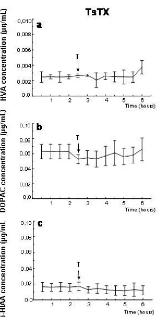
TsTX did not significantly affect levels of homovanillic acid (HVA),
3,4-dihydroxyphenylacetic acid (DOPAC) and 5-hydroxyindoleacetic acid (5-HIAA)
(Figure 1). Levels of norepinephrine and its metabolites, serotonin and dopamine
Figure 1. Extracellular levels of hippocampal HVA (a), DOPAC (b) and 5-HIAA (c). Levels determined by microdialysis in conscious rats before and after ipsilateral
intrahippocampal injection of 2 µg TsTX. Arrows indicate time of toxin (T) injection.
Error bars show standard deviation p < 0.05 compared to pre-injection level, one-way
repeated measures ANOVA and Tukey’s test. All groups n = 5.
Extracellular Amino Acid Levels
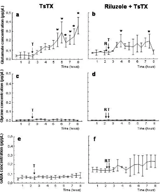
Injection of TsTX toxin into the hippocampus augmented glutamate concentration in
extracellular fluid (Figure 2), which reached statistical significance three hours after
Pretreatment with riluzole (5 mg/kg body weight, IP at t = –15 min) prevented TsTX
effect on hippocampal glutamate level (Figure 2).
Figure 2. Extracellular levels of hippocampal glutamate (a), glycine (c), and GABA
(e) determined by microdialysis in conscious rats before and after ipsilateral intrahippocampal injection of 2 µg TsTX. Panels on the right (b, d, f) show the effect of pretreatment with riluzole (5 mg/kg IP) on the response to toxin intrahippocampal
injection. Arrows indicate time of riluzole (R) and toxin (T) injection. Error bars show
standard deviation p < 0.05 compared to pre-injection level, one-way repeated
Electroencephalographic Recordings and Behavioral Observations
Ringer solution injection into CA1 hippocampal area caused no alterations in
behavior or in electroencephalographic records. Results of TsTX intrahippocampal
injection were previously described and included long electroencephalographic
epileptic-like discharges, immobility, orofacial movements and “wet dog shakes” in all
animals (14). The effects started five to ten minutes after the injection and persisted
for the entire recording period.
Rats treated with riluzole or with riluzole and toxin did not show epileptiform activity in
electroencephalographic records or behavioral signs of convulsion.
Histology
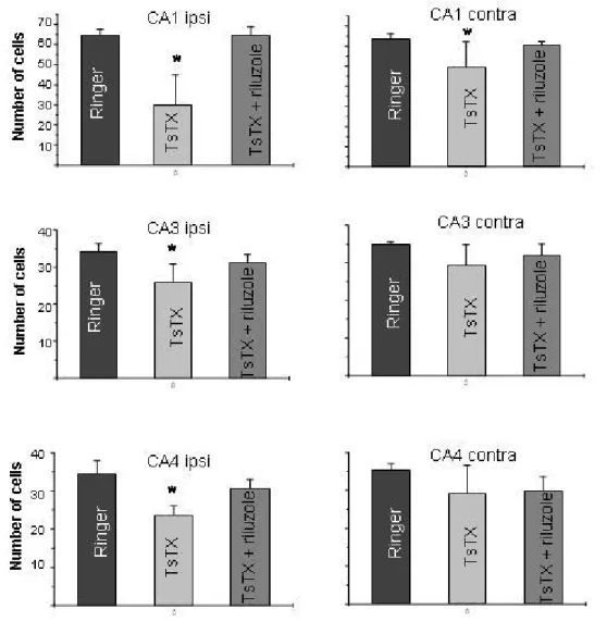
Injection of Ringer solution into the hippocampus did not influence the number of
cells in this area neither in the ipsilateral or contralateral regions (Figure 3).
According to previous descriptions, outcomes provoked by intracerebral injection of
the toxin were degeneration of pyramidal cells in ipsilateral and contralateral CA1
regions, and in CA3 and CA4 fields of the ipsilateral hippocampus (11, 14). Many
dark-stained neurons were observed, which indicated the degeneration. Pretreatment
with riluzole completely prevented toxin-induced cell loss. The number of cells in the
ipsilateral and contralateral areas in these animals was similar to the control group
Figure 3. Number of cells in the ipsilateral and contralateral CA1, CA3 and CA4 areas of the hippocampus after intrahippocampal injection of 2 µg TsTX, both in the
presence and absence of riluzole (5 mg/kg). Rats were killed seven days after the
inoculation. Error bars indicate standard deviation p < 0.05 compared to control
group (Ringer) and Tukey’s test. All groups n = 6.
DISCUSSION
Among scorpion neurotoxins, the α-group is the most studied and the more useful in
the functional mapping of the sodium channel structure (18).
TsTX is an α-toxin that acts by enhancing neurotransmitter release in the central and
peripheral nervous system (19, 20). Some studies have demonstrated that this toxin
releases glutamate, acetylcholine and adenosine triphosphate (ATP) from rat cortical
Moreover, in vivo studies reveal that it induces electroencephalographic and
histopathological alterations (14).
The present work aimed to assess the profile of in vivo neurotransmitter release in
the hippocampus after TsTX injection. Based on the toxin kinetics and on the
glutamate release profile registered in a previous study, in the current study the
collection period was prolonged up to six hours after the injection and other
neurotransmitters were analyzed (11, 15). It was observed that the toxin injected into
the hippocampus did not modify extracellular levels of 5-HIAA, HVA, DOPAC, glycine
or GABA. Nevertheless, extracellular levels of glutamate were significantly increased.
These findings agree with Nencioni et al. (11) who observed enhanced levels of
glutamate for three hours after the toxin injection and no alterations in GABA or
glycine levels.
According to Nunan et al. (15), TsTX can be found in rat brain after subcutaneous
(SC) injections. In adult animals the toxin increase is smaller and slower than in
young ones. The highest brain concentration of the toxin was observed one hour
after the inoculation in young rats and three hours later in adult ones, in which the
levels were augmented up to 12 hours. These results were in accordance with ours,
which revealed continuous enhancing in glutamate release that persisted until the
end of the collection and probably after (six hours or more). Moreover, considering
that in our study the toxin was injected directly into the brain, there was a large
quantity of glutamate in the hippocampus.
Similar results had been obtained by Dorce and Sandoval (25) with crude venom,
when DA, HVA, NE and 5-HIAA levels as well as glutamic acid decarboxylase (GAD)
activity were determined after intravenous (IV) or intrastriatal injection of Tityus
serrulatus crude venom. Subsequently, an increase in HVA levels and a decrease in
GAD activity in the striatum and hypothalamus were observed. On the other hand, no
alteration was registered in neurotransmitter levels in the hippocampus.
Previous data provided evidences that glutamate is the main responsible for TsTX
neurotoxic effects since glutamate receptor antagonists totally or partially block the
toxin effects (11). This hypothesis was corroborated by the utilization of riluzole, a
substance that belongs to a class of anticonvulsant and neuroprotective agents
which selectively inhibits glutamate release over release of other neurotransmitters
(26, 27). Zona et al. (28) concluded that riluzole modulates Na+ currents and the late
anticonvulsant and neuroprotective properties of this compound. A decrease of the
voltage-activated Na+ current certainly reduces neuronal excitability and enhances
excitatory amino acid release (28). Under experimental conditions, the drug
prevented audiogenic convulsion in DBA/2 mice, maximal electroshock and
amygdala-kindled seizures in rats, as well as delayed the appearing of seizures and
reduced the duration of afterdischarges (29, 30).
Riluzole injection 15 minutes before the toxin inoculation abolished all electrographic
alterations as well as neuronal losses. Riluzole, when intraperitoneally injected,
achieves the entire brain, including ipsi and contralateral areas of hippocampus,
preventing the excitotoxic effects of the toxin. Animals did not show behavioral
alterations either. They slept throughout the collection period, which is consistent with
the finding that riluzole easily crosses the blood brain barrier and enhances
slow-wave and rapid eye movement sleep (31). The level of extracellular glutamate
remained elevated and reached statistical significance in 2 points in rats pretreated
with riluzole; however, it is not enough to provoke epileptiform activity or neuronal
loss.
According to Meldrum (32), enlarged release of glutamate may worsen or prolong
preexisting seizure activity, while GABA release may be a compensatory inhibitory
mechanism that limits the progression and spread of seizure activity (33). However,
hippocampal augmentation of extracellular glutamate and GABA levels are directly
related to seizure activity rather than to the convulsant agent (34). Meurs et al. (34)
described that maximal glutamate increases occurred in the early stages of seizure
development, and preceded maximal GABA elevation. Nevertheless, in our results,
the electrographic convulsive activity did not keep a relation with the maximal
extracellular glutamate level. Seizure activity onset occurred (approximately 15
minutes after toxin injection) before glutamate extracellular level reach a significant
increase (approximately three hours after the injection), indicating that it is not
necessary the maximal increase in glutamate extracellular levels to initiate
convulsions. The convulsive activity enhanced by TsTX is not so intense to increase
GABA levels. Rats treated with riluzole and TsTX maintained elevated levels of
glutamate, although, they did not present behavioral or electrographic signs of
convulsion neither neuronal loss. The glutamate level in these animals was not as
convulsions and neurotoxicity elicited by TsTX without completely revert this
increase.
In hippocampal neurons, voltage-gated sodium channels appear to be the primary, if
not the only, target responsible for synaptic effects of riluzole at low micromolar
concentrations. Riluzole’s voltage-dependent blockade of sodium channels enhances
the depression of excitatory postsynaptic currents, which does not apply to inhibitory
postsynaptic currents determined by GABA. Inhibition of sodium channels account
for the preferentially anti-glutamate effect of this drug. Therefore, the preferential
depression of glutamate release is explained by a direct effect of riluzole on
glutamatergic cells (35).
These findings constitute an evidence of a putative specific activity of TsTX, at least
in the hippocampus, that affects only glutamate release.
In our opinion, outcomes of TsTX in the hippocampus may be due to its interaction
with a specific subtype of sodium channel. Different actions on sodium channels
produce diverse consequences on neurotransmitter release that involve distinct
presynaptic calcium channels, which supports the idea that sodium channels may
modulate neurotransmitter release (22). Depending on the rate of increase in channel
conductance, the outcome in terms of neurotransmitter release and calcium channel
type coupled to that event is different (36).
A large number of biological toxins exert their effects by modifying sodium channels
properties (37). Gilles et al. (38) elucidated, for the first time, how different toxins
affect mammalian central and peripheral excitable cells. They interact selectively with
sodium channel subtypes in a discrete subcellular region. The study by Gilles et al.
(39) revealed unexpected subtype specificity of toxins that interact with receptor site
3 (α-type toxins). Additionally, it was found that multiple sodium channel subtypes in
mammalian brain can be pharmacologically distinguished by their sensitivity into
certain toxins, such as ScαTxs (scorpion α-toxins) and αLTxs (spider α-latrotoxins)
(39).
Numerous different isoforms of mammalian voltage-gated sodium channels have
been identified (37). These multiple subtypes of sodium channels are the products of
at least six distinct genes that differ in subcellular localization, developmental pattern
of expression, and abundance in different brain regions (40-48).
Scorpion α-toxins provide a unique instrument for the identification of sodium channel
an important tool to study the functional role and distribution of channel subtypes in
the hippocampus.
REFERENCES
1. Rodriguez de la Vega RC, Possani LD. Overview of scorpion toxins specific for
Na+ channels and related peptides: biodiversity, structure-function relationships and
evolution. Toxicon. 2005;46(8):831-44
2. Rodriguez de la Vega RC, Possani LD. Current views on scorpion toxins specific
for K+-channels. Toxicon. 2004;43(8):865-75.
3. Debin JA, Maggio JE, Strichartz GR. Purification and characterization of
chlorotoxin, a chloride channel ligand from the venom of the scorpion. Am J Physiol.
1993;264(2):361-9.
4. Valdivia HH, Kirby MS, Lederer WJ, Coronado R. Scorpion toxins targeted against
the sarcoplasmic reticulum calcium-release channel of skeletal and cardiac muscle.
Proc Natl Acad Sci USA. 1992;89(24):12185-9.
5. Cestele S, Catterall WA. Molecular mechanisms of neurotoxin action on
voltage-gated sodium channels.Biochimie. 2000;82(9-10):883-92.
6. Kirsch GE, Skattebol A, Possani LD, Brown AM. Modification of Na+ channel
gating by a α-scorpion toxin from Tityus serrulatus. J Gen Physiol. 1989;93(1):67-83.
7. Massensini AR, Romano-Silva MA, Gomez MV. Sodium channel toxins and
neurotransmitter release. Neurochem Res. 2003;28(10):1607-11.
8. Gomez MV, Diniz CR. Separation of toxic components from the Brazilian scorpion
Tityus serrulatus venom. Mem Inst Butantan. 1966;33(3):899-902.
9. Arantes EC, Sampaio SV, Vieira CA, Giglio JR. What is tityustoxin? Toxicon.
1992;30(7):786-9.
10. Massensini AR, Moraes-Santos T, Gomez MV, Romano-Silva MA. Alpha- and
beta-scorpion toxins evoke glutamate release from rat cortical synaptosomes with
different effects on [Na+]i and [Ca2+]i. Neuropharmacology. 1998;37(3):289-97.
11. Nencioni AL, Lebrun I, Dorce VA. A microdialysis study of glutamate
concentration in the hippocampus of rats after TsTX toxin injection and blockade of
toxin effects by glutamate receptor antagonists. Pharmacol Biochem Behav.
12. Casali TAA, Gomez RS, Moraes-Santos T, Gomez MV. Differential effects of
calcium channel antagonists on tityustoxin and ouabain-induced release of [3H]acethylcholine from brain cortical slices. Neuropharmacology.
1995;34(6):599-603.
13. Nicolato R, Fernandes VMV, Moraes-Santos T, Gomez RS, Prado MAM,
Romano-Silva MA, Gomez MV. Release of γ[3H]aminobutiric acid in rat brain cortical
slices by α-scorpion toxin. Neurosci Lett. 2002;325(3):155-8.
14. Carvalho FF, Nencioni AL, Lebrun I, Sandoval MR, Dorce VA. Behavioral,
electroencephalographic and histopathologic effects of a neuropeptide isolated from
Tityus serrulatus scorpion venom in rats. Pharmacol Biochem Behav.
1998;60(1):7-14.
15. Nunan EA, Moraes MF, Cardoso VN, Moraes-Santos T. Effects of age on body
distribution of tityustoxin from Tityus serrulatus scorpion venom in rats. Life Sci.
2003;73(3):319-25.
16. Kretschmer BD, Kratzer U, Schmidt WJ. Riluzole, a glutamate release inhibitor,
and motor behavior. Naunyn Schmiedebergs Arch Pharmacol. 1998;358(2):181-90.
17. Paxinos G, Watson C. The rat brain in stereotaxic coordinates. 4th ed. San Diego:
Academic Press; 1998.
18. Bosmans F, Tytgat J. Voltage-gated sodium channel modulation by scorpion
alpha-toxins. Toxicon. 2007;49(2):142-58.
19. Gomez MV, Romano-Silva MA, Prado MAM. Effects of tityustoxin on central
nervous system. Toxin Rev. 1995;14(3):437-56.
20. Lima EG, Freire-Maia L. Cardiovascular and respiratory effects induced by
intracerebroventricular injection of scorpion toxin (tityustoxin) in the rat. Toxicon.
1977;15(3):225-34.
21. Fletcher PL, Fletcher M, Fainter LK, Terrian DM. Action of new world scorpion
venom and its neurotoxins in secretion. Toxicon. 1996;34(11-12):1399-411.
22. Falqueto EB, Massensini AR, Moraes-Santos T, Gomez MV, Romano-Silva MA.
Modulation of Na+-channels by neurotoxins produces different effects on [3H]ACh
release with mobilization of distinct Ca2+-channels. Cell Mol Neurobiol.
23. Salgado AH, Prado MA, Moraes-Santos T, Romano-Silva MA, Gomez MV.
Tityustoxin-induced release of ATP from rat brain cortical synaptosomes. Neurosci
Lett. 1997;229(2):113-6.
24. Fernandes VM, Massensini AR, Prado MA, Silva MA, Moraes-Santos T, Gomez
MV. Effects of α-scorpion toxin, tityustoxin on the release of [3H]dopamine of rat
brain prefrontal cortical slices. Neurochem Int. 2004;44(2):91-7.
25. Dorce VA, Sandoval MR. Effects of Tityus serrulatus crude venom on the
GABAergic and dopaminergic systems on the rat brain. Toxicon.
1994;32(12):1641-7.
26. Maciver MB, Amagasu SM, Mikulec AA, Monroe FA. Riluzole anesthesia:
use-dependent block of presynaptic glutamate fibers. Anesthesiology. 1996;85(3):626-34.
27. Martin D, Thompson MA, Nadler JV. The neuroprotective agent riluzole inhibits
release of glutamate and aspartate from slices of hippocampal area CA1. Eur J
Pharmacol. 1993;250(3):473-6.
28. Zona C, Siniscalchi A, Mercuri NB, Bernardi G. Riluzole interacts with
voltage-activated sodium and potassium currents in cultured rat cortical neurons.
Neuroscience. 1998;85(3):931-8.
29. De Sarro G, Siniscalchi A, Ferreri G, Gallelli L, De Sarro A. NMDA and
AMPA/kainate receptors are involved in the anticonvulsant activity of riluzole in
DBA/2 mice. Eur J Pharmacol. 2000;408(1):25-34.
30. Yoshida M, Noguchi E, Tsuru N, Ohkoshi N. Effect of riluzole on the acquisition
and expression of amygdala kindling. Epilepsy Res. 2001;46(2):101-9.
31. Wokke J. Riluzole. Lancet. 1996;348(9030):795-9.
32. Meldrum BS. The role of glutamate in epilepsy and other CNS disorders.
Neurology. 1994;44(11 Suppl 8):14-23.
33. Treiman DM. GABAergic mechanisms in epilepsy. Epilepsia. 2001;42(Suppl
3):8-12.
34. Meurs A, Clinckers R, Ebinger G, Michotte Y, Smolders I. Seizure activity and
changes in hippocampal extracellular glutamate, GABA, dopamine and serotonin.
Epilepsy Res. 2008;78(1):50-9.
35. Prakriya M, Mennerick S. Selective depression of low-release probability
36. Bicalho AF, Guatimosim C, Prado MA, Gomez MV, Romano-Silva MA.
Investigation of the modulation of glutamate release by sodium channels using
neurotoxins. Neuroscience. 2002;113(1):115-23.
37. Catterall WA, Cestele S, Yarov-Yarovoy V, Yu FH, Konoki K, Scheuer T.
Voltage-gated ion channels and gating modifier toxins. Toxicon. 2007;49(2):124-41.
38. Gilles N, Chen H, Wilson H, Le Gall F, Montoya G, Molgo J, Schönherr R,
Nicholson G, Heinemann SH, Gordon D. Scorpion α and α-like toxins differentially
interact with sodium channels in mammalian CNS and periphery. Eur J Neurosci.
2000;12(8):2823-32.
39. Gilles N, Blanchet C, Shichor I, Zaninetti M, Lotan I, Bertrand D, Gordon D. A
scorpion α-like toxin that is active on insects and mammals reveals an unexpected
specificity and distribution of sodium channel subtypes in rat brain neurons. J.
Neurosci. 1999;19(20):8730-9.
40. Schaller KL, Krzemien DM, Yarowsky PJ, Krueger BK, Caldwell JH. A novel,
abundant sodium channel expressed in neurons and glia. J Neurosci. 1995;15(5 Pt
1):3231-42.
41. Sangameswaran L, Fish LM, Koch BD, Rabert DK, Delgado SG, Ilnicka M,
Jakeman LB, Novakovic S, Wong K, Sze P, Tzoumaka E, Stewart GR, Herman RC,
Chan H, Eglen RM, Hunter JC. A novel tetrodotoxin-sensitive voltage-gated sodium
channel expressed in rat and human dorsal root ganglia. J Biol Chem.
1997;272(23):14805-9.
42. Toledo-Aral JJ, Moss BL, He ZJ, Koszowski AG, Whisenand T, Levinson SR,
Wolf JJ, Silos-Santiago I, Halegoua S, Mandel G. Identification of PN1, a
predominant voltage-dependent sodium channel expressed principally in peripheral
neurons. ProcNatl Acad Sci USA. 1997;94(4):1527-32.
43. Westenbroeck RE, Merrick DK, Catterall WA. Differential subcellular localization
of the RI and RII Na+ channel subtypes in the central neurons. Neuron.
1989;3(6):695-704.
44. Black JA, Yokoyama S, Higashida H, Ransom Br, Waxman Sg. Sodium channel
mRNAs I, II and III in the CNS: cell-specific expression. Brain Res Mol Brain Res.
1994;22(1-4):275-89.
45. Denac H, Mevissen M, Scholtysik G. Structure, function and pharmacology of
voltage-gated sodium channels. Naunyn Schmiedebergs Arch Pharmacol.
46. Felts PA, Yokoyama S, Dib-Hajj S, Black JA, Waxman SG. Sodium channel α
-subunit mRNAs I, II, III, NaG, Na6 and hNE (PNI): different expression patterns in
developing rat nervous system. Mol Brain Res. 1997;45(1):71-82.
47. Gordon D, Gurevitz M. The selectivity of scorpion α-toxins for sodium channel
subtypes is determined by subtle variations at the interacting surface. Toxicon. 2003;
41(2):125-8.
48. Gordon D, Merrick D, Auld V, Dunn R, Goldin AL, Davidson N, Catterall W.
Tissue-specific expression of RI and RII sodium channel subtypes. Proc Natl Acad Sci