UNIVERSIDADE DE LISBOA
FACULDADE DE CIÊNCIAS
DEPARTAMENTO DE BIOLOGIA ANIMAL
The role of Cadherin11 (Cdh11) in the establishment of laterality
in the zebrafish
José Maria Lage de Sousa Leitão
Mestrado em Biologia Evolutiva e do Desenvolvimento
Dissertação orientada por:
Leonor Saúde
Solveig Thorsteinsdóttir
3 Acknowledgements
Queria agradecer a todos os que me acompanharam ao longo deste ano pelo apoio e por todo o tipo ajuda que ofereceram. Desde aqueles que me motivaram com palavras de incentivo, até aos que acom-panharam o avançar desta tese diariamente. Todos foram uma ajuda e espero ter a oportunidade de agradecer pessoalmente a cada um, mas é especialmente àqueles que estiveram lá todos os dias que gostava de agradecer aqui.
Tenho de agradecer acima de tudo e muito especialmente à Leonor. Obrigado por me ter recebido no estágio e depois para fazer a tese. Nunca poderia imaginar um ano tão bom como este. Já é um privilégio poder trabalhar num local onde posso aprender com investigadores de alto nível, e ao mesmo tempo ser tão bem recebido e com este ambiente fantástico. É um privilégio ainda maior ter uma chefe com estas mesmas qualidades. Obrigado por me levar a ser um melhor investigador, tanto pelo seu exemplo como pelas suas orientações e desculpe lá qualquer coisinha.
Muito obrigado à professora Solveig pela preocupação e atenção que teve comigo e com todos nós no mestrado. Obrigado por estar sempre disponível ao longo do primeiro ano para aturar com os nossos falhanços e desesperos, e por toda a ajuda neste ano. Obrigado à professora Gabriela por todo o entusiasmo e atenção ao longo destes anos, e pelas primeiras aulas de desenvolvimento no 3º ano, que me mostraram como o desenvolvimento é extraordinário. Obrigado ao professor Élio por sempre se preocupar com a exigência e qualidade do mestrado, nas suas aulas e nos testes. Agora olhando para trás vejo como isso foi importante.
Dalila, não sei o que posso escrever que seja suficiente para agradecer tudo o que fizeste por mim este ano. Obrigado por me ensinares praticamente tudo o que sei, pelas dicas e ajudas, e pelo teu trabalho incansável no ministério da propaganda. Tive a sorte enorme de ter um exemplo de uma grande cientista que pude seguir de perto todo este tempo. Obrigado pelas luzes e motivação nos momentos mais caóticos desta tese. Queria agradecer também muito especialmente a Sara porque sem ela este projecto não aconteceria, literalmente. Obrigado por estares sempre disponível para qualquer dúvida, mesmo quando já estavas a escrever a tua tese, e também pelo teu exemplo como investigadora.
Guida, obrigado por seres a mãe deste grupo, sempre preocupada com os teus filhos desorien-tados. Ajudaste-me em tantas coisas ao longo da tese que seria preciso um capítulo só para agradecer cada uma dessas coisas. Os nossos projectos no laboratório não seriam possíveis sem ti, não só pelo teu trabalho mas também pela alegria e atenção que tens com cada um. Boa sorte para o teu mestrado! Queria agradecer a Fish Facility e obviamente a quem a faz! À Lara, por todo o trabalho em tornar a facility de alta qualidade, e o cuidado com cada um de nós. Obrigado pelo ânimo quando as experiências encravavam, ou quando os peixes não estavam interessados no progresso científico. Obrigado Aida por trazeres um entusiasmo contagiante em todos os momentos, e pelas caixas de cruzamentos personaliza-das. Obrigado também por te preocupares em ajudar o mais possível sempre que precisei. Queria agra-decer às duas por irem além dos aspectos técnicos e se preocuparem a fundo com cada pessoa e pela união do grupo todo. Obrigado à Isaura por toda a alegria e entusiasmo no laboratório, e o bocadinho de loucura saudável nos tempos mais críticos. Obrigado pelo teu exemplo de alguém que verdadeira-mente gosta de fazer ciência, por estares sempre disponível para ajudar e pelo teu relógio biológico apuradíssimo.
Obrigado à Ana e à Rita por toda a ajuda que me deram ao longo deste ano, sempre disponíveis para responder a dúvidas e grandes exemplo de investigadoras que pude acompanhar. Obrigado ao Di-ogo pela companhia e pela ajuda em equilibrar a balança para o lado masculino neste grupo. Força! Obrigado à Mariana Ferreira, pela caridade de me relembrar em todos os momentos que devia estar a trabalhar e à Mariana Costa pelo croissant (a vingança serve-se fresca). Queria gradecer ao Domingos, ao João e ao Gonçalo pela companhia neste ano, por me ajudarem seja com reagentes ou com dúvidas, e muito especialmente pelos bolos e queijos.
Queria agradecer à Bioimaging, Flow, Histology Facilities pela ajuda ao longo do projecto e por toda a atenção que recebi de cada um dos técnicos sempre que precisei.
Obrigado à Susana Lopes e todo ao seu grupo pela ajuda com protocolos, reagentes e dúvidas no geral. Obrigado à Catarina Certal e especialmente à Joana Monteiro por todo o trabalho com os gRNA, e pela ajuda técnica que ofereceram
4
Obrigado muito especialmente aos meus pais por me terem recebido como o seu primeiro projecto de desenvolvimento e por tudo, mesmo. Aos meus irmãos, à minha família e a todos os amigos que sempre se perguntaram sobre o que era exactamente a minha tese, “para que serve isso?”, e “peixes fluorescen-tes? Fixe!”. Para aqueles que ainda estão confusos, boas leituras.
5 Resumo
O plano corporal aparentemente simétrico dos vertebrados esconde assimetrias no seu interior. O esta-belecimento consiste em 4 momentos conservado nos vertebrados: começa com a quebra de simetria no organizador de Esquerda-Direita (ED), que é transmitida para Placa Lateral Mesodérmica (PLM). Aqui desencadeia a cascata de Nodal, restrita ao lado esquerdo deste tecido que finalmente informa a forma-ção dos órgãos.
Neste trabalho identificámos uma molécula de adesão celular, a caderina11 (Cdh11) que aparentemente influencia o estabelecimento da ED no peixe zebra.
Através de ensaios de perda de função deste gene, usando Morpholinos (MO), observámos que a late-ralidade do coração e do tracto digestivo se alteravam. No entanto, enquanto no tracto digestivo a sime-tria era invertida, a maior parte dos embriões afectados tinham o coração simétrico. Esta observação indica que há um desemparelhamento do estabelecimento da simetria na formação destes dois órgãos. Para além disso, após injecção com MO, a expressão dos dois genes da cascata de Nodal na PLM (spaw e pitx2), inverte-se e fica restrito ao lado direito da PLM. Consequentemente, a expressão anormal destes genes pode explicar a inversão do tracto digestivo mas não a simetria do coração.
A expressão de cdh11 foi detectada na endoderme e na Mesoderme Intermédia (MI) nos estadios em que o sinal de ED passa do organizador, chamado Vesícula de Kupffer (VK) no peixe-zebra, para a PLM, entre os estadios de 8 e 12 somitos. Este resultado sugere que a Cadh11 pode estar envolvida na passagem do sinal da VK para PLM. Por um lado, no rato e no Xenopus a endoderme tem um papel nesta transmissão, e por outro lado podemos observar que a MI localiza-se entre a VK e a PLM.
A cdh11 também foi detectada nos Pronefros (PN), que deriva da MI, nos estadios em que o tracto digestivo adquire a sua torção., entre as 24 e 30 horas pós-fertilização (hpf). Nós observámos que a porção anterior dos PN coincide dorso-ventralmente com a região de migração da PLM que provoca a torção do tracto digestivo. Deste modo é possível que os PN sirvam como estrutura de suporte para esta migração.
Cdh11 também foi observado na PLM anterior ao estadio de 20 sómitos. Esta expressão coin-cide espacial e temporalmente com a migração dos percursores do coração.
Tendo em conta estes resultados, sugerimos que a Cdh11 tem um papel no desenvolvimento do coração e tracto digestivo. Quanto ao desenvolvimento do tracto digestivo, a Cdh11 nos PN pode ser importante para a estabilidade desta estrutura e consequentemente da migração assimétrica da PLM. Por outro lado, a Cdh11 na endoderme e na MI podem afectar a passagem do sinal assimétrico da VK para PLM. A Cdh11 pode estar presente nas junções comunicantes da endoderme atravessadas pela onda de cálcio que transporta a informação assimétrica para PLM, ou nas junções aderentes da IM quando o próprio Nodal passar da VK para a PLM. Quanto ao coração, a Cdh11 pode afectar a migração dos percursores do coração e consequentemente a formação deste órgão.
Como uma abordagem complementar analisámos um mutante para cdh11 que no entanto não reproduziu os fenótipos obervados com o MO. Para entender melhor os efeitos da inactivação deste gene produzimos um novo mutante através da técnica de CRISPR-Cas9.
6 Abstract
Symmetric body plans in vertebrates hide asymmetrical organs on the inside. The establishment of this asymmetry is generally conserved in vertebrates. It starts in the Left-Right Organizer (LRO), is then translated to the left Lateral Plate Mesoderm (LPM) and ultimately informs organogenesis.
Using a morpholino (MO) loss-of-function approach for the cell adhesion molecule, Cadherin 11 (Cdh11), we observed that the establishment of LR in organogenesis was disrupted. However, while the majority of the affected embryos exhibited reversed laterality phenotypes in the gut, the predominant disorder of the heart was the absence of asymmetric looping. This is an indication of some kind of uncoupling of laterality between these organs. Additionally, in a high number of the cdh11MO injected embryos the conserved LR genes (spaw and pitx2) were expressed on the right LPM instead of the left LPM. Thus, we proposed that the abnormal expression of the conserved LR genes in the right LPM might underlie the reversed gut loop phenotype but not of the heart.
Transcript of cdh11 was found to be present in the Endoderm at 8-somite stage (ss), and in the Intermediate Mesoderm (IM) from 8ss to 12ss. These stages coincide with the transfer of the laterality signal from the zebrafish LRO, the Kupffer’s Vesicle (KV) to the LPM. Recent findings in other model vertebrates suggested a model for this relay: the LR information might be transferred either by calcium waves going from the LRO to the LPM through gap junctions in the endoderm, or Nodal itself might cross the extracellular matrix towards the LPM, or a combination of both. This suggests that, in the zebrafish, Cdh11 could play a role in the relay of the LR signal. Calcium waves starting from the KV have been described. These might travel from the KV through gap junctions composed of Ca2+ sensitive Cdh11 molecules, in the endoderm towards the LPM. Alternatively, the Nodal protein (Spaw) might travel directly along the extracellular matrix, crossing the IM, which is located between the KV and the LPM.
cdh11 was also detected in the Pronephros (PN), an IM derivative, at the same stages when the looping of the gut occurs, between 24 and 30 hours-post-fertilization (hpf). We confirmed that the ante-rior portion of the PN coincides dorsoventrally with the gut looping region at the level of the third somite pair. This supports the hypothesis that the PN could affect the asymmetric migration of the LPM that is essential to promote the displacement of the gut.
Furthermore, cdh11 was also detected at 20ss in the anterior LPM. This region is located anteriorly to the LPM asymmetric migration area, however, it coincides both spatially and temporally with the mi-grating heart primordia territory.
Taken together, these results suggest that Cdh11 might play a role in the LR development of both the gut and heart:
Gut - Cdh11 in the PN might be important for the stability of this structure, and consequently to the asymmetric migration of the LPM; Alternatively, Cdh11 might affect the transfer of the asym-metric signal from the KV to the LPM, between 8ss and 12ss, upstream of the Nodal cascade in the LPM. It participates either through the gap junctions in the endoderm relaying a calcium wave, or at the adherens junctions of the cells of the IM, affecting the passage of Spaw in the extracellular matrix. Absence of Cdh11 disrupts the Nodal cascade in the LPM and the asymmetric migration of this tissue over the gut endoderm that ultimately leads to the displacement of the gut.
Heart: At 20ss, in the anterior LPM, Cdh11 might affect the migration of the heart primordia and consequently the LR development of the heart.
As a complementary approach, we analysed a cdh11 mutant, which, however, did not reproduce the phenotypes observed in the MO-injected embryos. This discrepancy raised questions about the spec-ificity of our MO and the quality of the null cdh11 mutant. To better understand the effect of a cdh11 knockout, we produced a new cdh11 mutant, resorting to the CRISPR-Cas9 genome editing system.
In this work, we have detected the expression of cdh11 in tissues where it had not been observed before. This raised new hypotheses for the function of Cdh11 in the establishment of LR more in line with what has been described in other vertebrate models. Nevertheless, the specific mechanism of action of Cdh11 during the establishment is still not clear and needs further investigation.
7 Abbreviations
AP – Anterior-Posterior; DV – Dorsal-Ventral; LR – Left-Right;
LRO – Left-Right Organizer; KV – Kupffer's vesicle;
DFC – Dorsal Forerunner Cells; hpf - hours post-fertilization;
TGF- β - transforming growth factor-β; Spaw – Southpaw;
LPM - lateral plate mesoderm; aPKC - atypical protein kinase C; ZO-1 - zonula occludens 1; IM - Intermediate Mesoderm; PN – Pronephros;
i.e – That is; Cdh – Cadherin;
EC – Extracellular Domain; TM - Transmembrane Domain; IC – Intracellular Domain; Cdh2 – N-cadherin;
HH - Hamburger and Hamilton; DM – Dorsal Mesentery; Cdh11 – Cadherin-11; WT – Wild Type;
EZRC - European Zebrafish Resource Center; gDNA – genomic Deoxyribonucleic acid; PK – Proteinase K;
RT – room temperature;
PCR – Polymerase Chain Reaction; Fw – Forward;
Rv – Reverse;
MO - Morpholino oligonucleotides; cdh11MO - cdh11 splice blocking MO; ControlMO - Standard Control MO; DIG – Dioxigenin;
WISH - Whole mount in situ hybridization; s – seconds;
h – hours;
Pre-Hybmix - pre-Hybridization Mix; PBS - Phosphate-buffered saline; PFA – Paraformaldehyde; MetOH – Methanol;
BSA - Bovine Serum Albumin; qPCR – quantitative PCR;
CRISPR - Clustered Regularly Interspaced Short Palindromic Repeat; gRNA - single-guide RNA;
Cas9 - CRISPR associated protein 9; NTR – Nitroreductase;
Mtz – Metronidazole.
8 Index Acknowledgements III Resumo V Abstract VI Abbreviations VII Chapter 1 – Introduction 2
1.1 – Left Right Development 2
1.1.1 – Phase 1: Breaking of symmetry 3
1.1.2 – Phase 2: nodal expression in the KV and transfer of the asymmetric signal to the left lateral plate mesoderm (LPM)
4
1.1.3 – Intermediate Mesoderm and Pronephros 6
1.1.4 – Phase 3: Nodal Cascade in the LPM 6
1.1.5 – Phase 4: Left-right asymmetric organ morphogenesis 7
1.2 – Cadherins in development 9
1.2.1 – Cadherin 11 10
1.3 - Objectives 13
Chapter 2 – Experimental procedures 14
2.1 - Zebrafish maintenance 14 2.2 – cdh11 Knockdown 14 2.2.1 – Embryo microinjections 14 2.2.2 – Cdh11 mRNA synthesis 14 2.3 – CRISPR/Cas9 15 2.3.1 – gRNA design 15 2.3.2 – gRNA/Cas9 microinjection 15 2.3.3 – DNA Extraction 16 2.3.4 – T7 Endonuclease 16
2.4 – Fluorescence activated cell sorting (FACS) 16
2.4.1 – RNA extraction (DNAse I) 17
2.4.2 – Reverse trancriptase 17
2.4.3 - cDNA Purification 17
2.4.4 – Polymerase Chain Reaction (PCR) 17
2.5 – Cdh17 ablation line plasmid 18
2.5.1 – Plasmid cloning 18
2.5.2 – Transformation of competent Echerichia coli bacteria 18
2.6 – Whole mount in situ hybridization (WISH) 19
2.6.1 – WISH protocol 19
2.6.2 – Double WISH protocol 19
2.6.3 – Embryo embedding 20
2.6.4 - Cryosectioning 20
2.7 – Agarose gel electrophoresis Chapter 3 - Results
20
3.1 - Independent cdh11 knockdown assays produce similar heart laterality phenotypes
21
3.2 - Generation of cdh11 mutations using the CRISPR-Cas9 genome editing system 22 3.2.1 - T7 Endonuclease I assay reveals the occurrence of mutagenesis 24 3.2.2 - Left-Right phenotypes in the heart and gut are not observed in the F0 population of cdh11 CRISPR-Cas9 mutants
26
3.3 - Where is cdh11 expressed at the breaking of symmetry? 27 3.3.1 - New in situ hybridization assays suggest that cdh11 is expressed around the Kup-ffer’s Vesicle.
9
3.3.2 - FACS profiling identifies three GFP-expressing sub-populations of cells in sox17:EGFP embryos at 8ss.
28
3.3.3 - PCR assays indicate that cdh11 is expressed in the endodermal cells. 30 3.4 - Expression of cdh11 in the Intermediate Mesoderm and Pronephros
3.4.1 - The Pronephros could have a role in the asymmetric migration of LPM during the displacement of the gut
30 31
3.4.2 - Developing a new method to study the role of the Intermediate Mesoderm in Left-Right asymmetry
32
3.5 - Is Cdh11 expressed in the LPM? 33
Chapter 4 – Discussion 35
4.1 - Determining the specificity of the knockdown LR phenotypes 35 4.2 – Confirming the morpholino assays with a new mutant line
4.2.1 – Should we observe the mutant phenotype in CRISPR-Cas9 injected founder embryos?
35 36
4.3 - Where is cdh11 expressed at 8ss? 37
4.4 – Cdh11 involved in both pathways of the relay of the LR information to the LPM
38
4.5 – Is Cdh11 in the anterior LPM affecting organogenesis? 41 4.5.1 – Cdh11 might affect the heart primordia, disrupting the intrinsic chirality of the
heart
41
4.5.2 – Gut looping affected by the disruption of the Nodal cascade 42
Chapter 5 – Bibliography 43
10 Chapter 1 - Introduction
1.1 – Left Right Development
The external bilateral symmetry in vertebrates hides internal organ asymmetries. Organs such as the heart, liver, spleen, gall bladder, among others, are consistently asymmetrically distributed regarding the Left-Right (LR) body plan. This normal distribution of the internal organs is referred to as situs solitus and is largely conserved among a population of a given species (Grimes & Burdine, 2017).
Of all of the organs, the asymmetry of the digestive tract was probably the first to evolve. In all vertebrates, its length is greater than the main body axis and in all kinds of species we can observe the functional specialization of each module: mouth, oesophagus, stomach and gut. On the other hand, in evolutionary terms, a primitive heart was nothing more than a linear contractile muscle that facilitated the distribution of nutrients throughout the body (Blum et al., 2014). This morphology can be seen in the Drosophila. Additionally, the asymmetries of the lungs might reflect spatial constraints in the thorax resulting from asymmetric heart placement rather than a specific function (Blum et al., 2014).
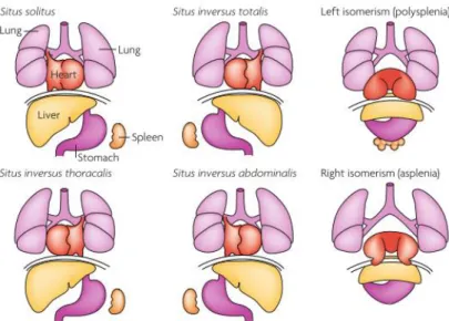
For human individuals it is crucial to understand how consistent LR asymmetry is established in embryogenesis. These patients might face difficulties due to the lack of proper connections between the heart and the different organs. Disorders of the organization of the LR axis include the complete reversal of the internal organs (situs inversus totalis), partial asymmetries (situs inversus abdominalis or thoracalis) and symmetry, leading to duplication or complete loss of single organs such as the spleen (isomerism) (Figure 1) (Vandenberg & Levin, 2013). Situs inversus totalis occurs in 1 of 20.000 cases in humans and is the only non-life-threatening condition (Vandenberg & Levin, 2013).
Figure 1 – Human Laterality disorders - Schematic illustration of normal left–right body asymmetry (situs
solitus) and five laterality defects that affect the lungs, heart, liver, stomach and spleen. Taken from Fliegauf et al. 2007.
The mechanisms that direct the establishment of LR asymmetry are highly conserved across the different vertebrate model organisms (Blum et al., 2014). In zebrafish, the process of establishment of asymmetry throughout development can be divided in four phases: 1 - Breaking of symmetry; 2 - Nodal expression in the KV and transfer of the asymmetric signal to the left lateral plate mesoderm (LPM); 3 - Nodal Cascade in the LPM; 4 - Left-right asymmetric organ morphogenesis (Collins & Ryan, 2014; Hamada et al., 2002; Shiratori, 2006).
11 1.1.1 – Phase 1: Breaking of symmetry
The establishment of LR asymmetries results from a series of molecular and morphogenetic events. It has been shown that, before gastrulation, ion transporters that are asymmetrically distributed in the embryo, generate differences in pH and membrane voltage potential between the left and right sides (Kawakami et al., 2003). It is believed that this asymmetric membrane polarization promotes the accumulation of LR determinants, such as serotonin, through directional transport involving gap-junction channels (Fukumoto et al., 2005a; Fukumoto et al., 2005b). LR asymmetry is further established during early somite stages in the conserved ciliated organ of asymmetry, the LR organizer (LRO), known as the Kupffer’s vesicle (KV) in zebrafish (Matsui et al., 2015).
The KV originates from a cluster of 20-30 cells, the dorsal forerunner cells (DFCs), which is maintained by cadherin- based adherens junctions (D’Amico & Cooper, 1997; Matsui et al., 2015; Oteiza et al., 2010). These cells are formed via a Nodal signalling-dependent ingression of surface enveloping layer cells from the dorsal blastoderm margin. The initial group migrates ahead of the dorsal margin and proliferates during epiboly stage. At the end of epiboly, by 10hpf, these cells undergo a mesenchymal- to-epithelial transition (MET) to form the KV in a vesicle-like structure with a mono-ciliated epithelium (Essner et al., 2005; Gokey et al., 2017; Oteiza et al., 2008). KV cells create a fluid filled lumen arising from the apical membrane that rapidly expands during early somite stages (G. Wang et al., 2011). At the same time, a single cilium forms and elongates from the apical surface of each KV cell to extend into the lumen (Oteiza et al., 2008; Smith et al., 2014; Wang et al., 2012) Directional ciliary beating in the LRO generates leftward flow of extraembryonic fluid which is essential to LR development. (Kramer-Zucker, 2005; Nonaka et al., 1998; Okabe et al., 2008). This directional fluid flow is induced by a combination of planar cell polarity (PCP) and rotational movement of cilia in the organizer (Shinohara & Hamada, 2017). In order for the KV to produce a robust fluid flow it requires a minimum of 30 motile cilia and an anterior-dorsal cluster of motile cilia (Sampaio et al., 2014; Smith et al., 2014). The number of motile cilia in the KV seems to be modulated by Notch signalling, through a mechanism that involves the activity of Her12 (hairy- related 12) (Sampaio et al., 2014; Tavares et al., 2017). Changes in the ratio of motile to immotile cilia mediated by Her12 impacts both the intensity of the flow and the distribution of cilia (Tavares et al., 2017).
Two alternative and nonexclusive theories have been proposed to explain how the LR flow is translated to asymmetric information:
1 – The chemosensing hypothesis states that asymmetric flow creates a LR concentration gradient of morphogens that is detected by receptors on the left side, triggering LR asymmetric gene expression; (Okada et al., 2005; Tanaka et al., 2005).
2 – The mechanosensing (or two-cilia) hypothesis states that cells of the KV can mechanically sense flow due to a particular type of non-motile sensory cilia, activating an asymmetric response on the periphery of the KV.(McGrath et al., 2003; Sampaio et al., 2014; Tabin & Vogan, 2003, Yoshiba et al., 2012).
A recent study has showed that in the mechanosensing mechanism the number of immotile cilia in the KV is insufficient, however, motile cilia could sense their own motion. Additionally they showed that a chemosensory mechanism could explain the observed robust LR asymmetry establishment, provided that the particle size is above the lower limit of about 2 nm (Ferreira et al., 2017)
The LR information of the flow has been shown to lead to the activation of cation channel PKD2 (Yoshiba et al., 2012). This in turn is necessary for the asymmetric release of calcium around the KV, which is initiated within the cilia (Yuan et al., 2015). This leads to an increase in cytoplasmic calcium at the left side of the KV (McGrath et al., 2003; Yoshiba et al., 2012; Yuan et al., 2015).
12
1.1.2 – Phase 2: nodal expression in the KV and transfer of the asymmetric signal to the left lateral plate mesoderm (LPM)
The Nodal related gene spaw is expressed bilaterally in the cells surrounding the KV at the 4ss to 6ss (Long et al., 2003). Dand5 (member of the Cerberus/Dan family) is a negative regulator of Nodal related genes. Dand5 binds to Spaw, a Nodal related gene, inhibiting its binding to receptors in adjacent region on the right side of the KV (Hashimoto et al., 2004). The expression of dand5 in the KV is initially symmetric but by the 8ss it has become restricted to right-side. It has been shown that the transcription of dand5 in zebrafish is sensitive to fluid flow, given that in the absence of flow in the KV its expression is no longer biased (Lopes et al., 2010). The LR information of the flow leads to the activation of cation channel PKD2 (Yoshiba et al., 2012). This activates an asymmetric calcium release, initiated within the cilia, which in turn leads to repression of Dand5 on the left side of the KV (Yoshiba et al., 2012; Yuan et al., 2015). How calcium affects Dand5 is, therefore, the next challenge to understand mechanisms of symmetry breakage driven by flow (Blum & Vick, 2015).
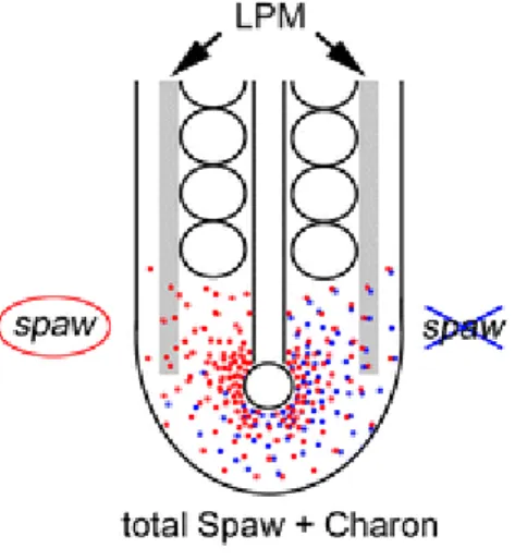
Figure 2 – Dand5 antagonism of Spaw – Dand5 antagonizes Spaw by attaching to it. Spaw signal reaches the
left LPM after repression of Dand5 on this side. Taken from Matsui & Bessho 2012.
Consequently, due to the repression of Dand5, a left-sided spaw restricted signal starts from the KV towards the left LPM, becoming delimited to this tissue at the 10 to 12-somite stage (Figure 2). Here the Nodal Cascade is activated on the left but not on the right side. (Marques et al., 2004; Matsui & Bessho, 2012).
The mechanism of transfer of information from the LR organizer to the LPM in vertebrates is still relatively unknown but previous findings in mouse and frog development have raised two complementing hypotheses for this process (Grimes & Burdine, 2017; Norris, 2012; Saijoh et al., 2014). In mouse embryos, Ca2+ signals have been observed to spread laterally beyond the node and reaching as far as the LPM (McGrath et al., 2003). Previous experiments in Xenopus (Beyer et al., 2012) and mouse, (Saund et al., 2012; Viotti et al., 2012), have described that this Ca2+, or other signals, might travel intracellularly through endodermal cells, which are connected by gap junctions, towards the LPM. The expression of spaw would then be activated in the LPM by this Ca2+ signal (Figure 3) (Viotti et al., 2012; Saund et al., 2012; Beyer et al., 2012).
13
Figure 3- Model for the transfer of LR information in the mouse. After LR symmetry is broken in the node by
rotating cilia, the resulting nodal flow induces left-biased asymmetries around the node. These asymmetries are transmitted via gap junctions comprised of Cx43 within the gut endoderm to the left LPM, where the Nodal cascade is activated. Adapted from Viotti 2012
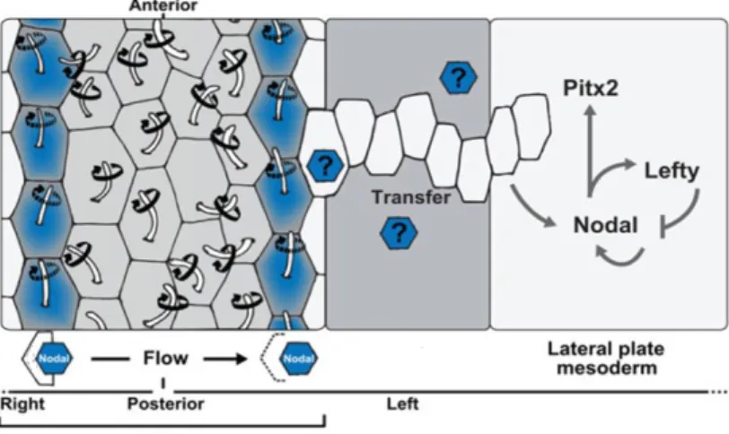
Conversely, Nodal itself, which is produced in greater amounts at the left side of LROs, might directly travel to the LPM through the extracellular matrix, and activate its own expression (Figure 4). In mouse embryos, sulfated glycosaminoglycans (sGAGs) are located in the basement membrane between the endoderm and the mesoderm (Oki et al., 2007). In fact, Nodal expression at the LRO is required for Nodal activation in the LPM (Brennan et al., 2002; Saijoh et al., 2003) and Nodal is able to activate its own expression (Adachi et al., 1999; Norris & Robertson, 1999). However, this may not be the case in zebrafish since mutants lacking either expression of spaw in the KV, or spaw mutants themselves still express spaw in the LPM (Burdine et al., 2016; Noel et al., 2013). Some experiments have proposed that Dvr1/Gdf3 (a member of the TGFβ family) facilitates the transfer of the LR signal from KV to the LPM (Peterson et al., 2013). Knockdown of gdf3 prevents the expression of spaw from occurring in the LPM even in the absence of Nodal inhibitors Dand5 and Lefty1 (Pelliccia et al., 2017).
Figure 4- Representation of the left sided flow in the KV and later relay of the LR signal to the LPM. Motile
and polarized cilia (positioned at the posterior pole of cells) rotate in a clockwise fashion to produce a leftward fluid flow in the extracellular space. Dand5is repressed on the right side, freeing Spaw (Nodal) on the left side. Spaw crosses across the paraxial and intermediate mesoderm towards the left LPM triggering the Nodal cascade. Adapted from Blum 2014
14
A combination of these two hypotheses suggests that Ca2+ spreading through gap junctions in endodermal cells may enhance the secretion of sGAGs, assisting the transfer of Nodal protein from the organizer to the LPM (Beyer et al., 2012; Norris, 2012).
1.1.3 - Intermediate Mesoderm and Pronephros
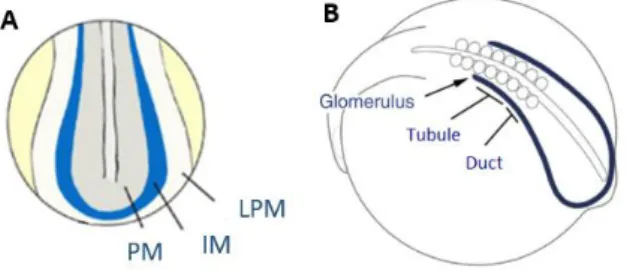
In zebrafish, between the KV and the LPM stands a stretch of mesodermal cells called the Intermediate Mesoderm (IM), which could play a role in the transfer of Spaw to the LPM. Shortly after epiboly, these IM cells emerge as a band of tissue at the ventrolateral edge of the paraxial mesoderm (Figure 5A) (Drummond et al., 2016). In zebrafish the IM gives rise to both kidney and blood cells, and, as development proceeds, the nephrogenic component of the IM is determined by the expression of renal markers such as the transcription factors hnf1ba, pax2a, pax8, and lhx1a. Later, this tissue develops into the Pronephros (PN). In teleost fish, such as the zebrafish, the PN is the functional kidney of early larval life (Drummond et al., 2016). This structure is composed by three segments: anteriorly the glomerulus, then the tubule and posteriorly the duct(Figure 5B). Each of these segments is determined by the anterior to posterior interaction of wt1, pax2a and sim1.Anteriorly the domain expressing only wt1 will give rise to the glomerulus, the region expressing wt1 and pax2a will originate the tubule and the tissue expressing pax2a and sim1 will develop into the duct. (Drummond et al., 2016; Serluca & Fishman, 2001).
Figure 5 – Development of the Intermediate Mesoderm and the Pronephros. A – At 10 hpf, the Intermediate Mesoderm (IM) is located between the Paraxial Mesoderm (PM) and the Lateral Pate Mesoderm (LPM). Adapted from Marra and Wingert et al., 2014. B –The IM later gives rise to the Pronephros (PN) with its three distinct segments: Glomerulus, Tubule and Duct. Adapted from Drummond et al., 2016
1.1.4 –Phase 3: Nodal Cascade in the LPM
Expression of spaw in the left LPM can be seen by 10ss. In the LPM Spaw activates itself and pitx2, and the expression of both spreads from the posterior to the anterior end of the LPM, eventually covering the whole left side of the LPM (Shiratori et al., 2006). The left-sided expression of pitx2 remains for many hours after Nodal signalling has stopped (Shiratori et al., 2006). Before reaching the LPM, Spaw from the KV activates lefty1 expression in the posterior notochord (Grimes et al., 2016). It has been proposed that lefty1 acts to repress Spaw signalling to the LPM, preventing the activation of Nodal target genes in the LPM before the asymmetric signal from the KV (Grimes et al., 2016). The expression of lefty1 in the notochord is driven by Spaw as it propagates anteriorly within the LPM (X. Wang & Yost, 2008) and acts as a molecular midline barrier preventing spaw activation on the right LPM (Lenhart et al., 2011). The expression of lefty2 is induced within the left side of the heart field once Spaw reaches the anterior left and also prevents Spaw from spreading to the right LPM (Lenhart et al., 2011). Therefore, both lefty1 and lefty2 play a critical role in confining the expression of spaw to the left side of the LPM (Zinski et al., 2017).
15
1.1.5 – Phase 4: Left-right asymmetric organ morphogenesis
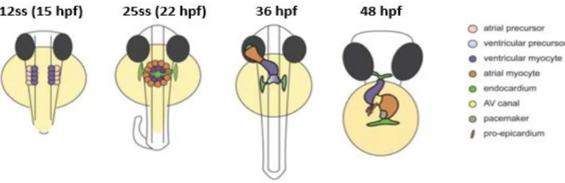
The asymmetric information in the LPM is interpreted by individual organ primordia, resulting in the asymmetric morphogenesis of several organs. The heart is the first organ to form and function during vertebrate embryo development (Bakkers et al., 2009). At 12 hpf (15ss), myocardial progenitors are found in the anterior LPM, with ventricular progenitors more medial than atrial progenitors. Endocardial progenitors lie anteriorly. Then, myocardial and endocardial progenitors migrate to the midline and fuse by 19 hpf (20ss) to create the cardiac cone. The endocardium covers the inner lining of the myocardial tube forming the linear heart tube by 24 hpf (Bakkers, 2011; Grimes & Burdine, 2017; Staudt & Stainier, 2012).
The first asymmetric displacement is called the heart jogging. It occurs when the cells on the left side of the cone migrate anteriorly more quickly than cells on the right side, resulting in a clockwise rotation and the movement of the cone to the left. It then involutes and extends to produce a leftward pointing cardiac tube by 24–26 hpf. At 36 hpf, the second asymmetric displacement, called cardiac looping, starts with a shift of the ventricle towards the mid-line, and the constriction at the position of the atrioventricular canal is first visible. The heart tube continuous to loop and by 48 hpf, it has formed a right-sided ventricle and left-sided atrium in a D-loop shape (Figure 6) (Bakkers, 2011; Grimes & Burdine, 2017; Staudt & Stainier, 2012).
Asymmetric heart morphogenesis seems to be directed by Spaw asymmetrically expressed in the anterior LPM (Bakkers et al., 2011). However, it has been observed that after loss of spaw, normal looping is reduced but still occurs in most of the embryos (Baker et al., 2008). Additionally, zebrafish heart tubes, isolated and cultivated cultured ex vivo, still undergo D-looping most of the times (Noel et al., 2013). This movement could be prevented by blocking actin polymerization, or the activity of non-muscle myosin II (Noel et al., 2013). Altogether, these data indicate that emerging cardiomyocytes have an intrinsic bias to laterality, requiring actomyosin activity, which could be amplified by the action of the Nodal pathway (Campione & Franco, 2016)
Figure 6 - Stages of heart development. At the 12-somite stage (ss) cardiogenic differentiation starts by the expression of cardiac myosins (purple). By 25ss, the cardiac disc is formed, with the endocardial cells within the hole at the centre, ventricular myocytes at the circumference and atrial myocytes at the periphery of the disc. Cardiac jogging then forms the cardiac tube with the endocardium forming the inner lining of the myocardial tube. At 36 hpf, cardiac looping has started, with a displacement of the ventricle towards the mid-line. The heart tube continuous to loop and forms an S-shaped loop by 48 hpf. Adapted from Bakkers et al., 2011
16
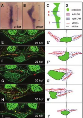
It is assumed that the establishment of laterality of the heart, gut, liver and pancreas is a consequence of the events that happen in the LPM (Davis et al., 2008). However, while the heart derives from the LPM tissue, the digestive tract organs originate from the underlying solid rod of endodermal cells that forms at the ventral midline between 24 and 30 hpf (Davis et al., 2008). Their development starts at a particular position along the anterior–posterior axis by a process known as gut looping. This displacement is mediated by the asymmetric migration of the LPM. It occurs specifically within the gut looping region and requires functional LR gene expression and establishment of epithelial polarity within the LPM (Horne-Badovinac et al., 2003). At 20 hpf the endodermal rod lies in the midline and epithelial cells of the LPM flank the endoderm at the same dorsoventral level (Figure 7 A, E, E’). Starting at 26 hpf, both sides of the LPM migrate towards the midline. The left LPM migrates dorsally to gut and the right LPM migrates ventrolaterally (Figure 7 F, F’). At 30 hpf the developing gut has shifted to the left and the LR position of the LPM is highly asymmetric (Figure G-I’). (Horne-Badovinac et al., 2003). It has been shown that bidirectional signalling between EphrinB1, in the liver progenitors, and EphB3b, in the LPM, coordinates the movements of the hepatic endoderm and adjacent LPM, resulting in asymmetric positioning of the zebrafish liver (Cayuso et al., 2016). Remodelling of the extracellular matrix (ECM) is also required. During gut looping, Laminin is reduced by the activity of matrix metalloproteinases (MMP) along the LPM/gut boundary, and the activity of the MMP is regulated by transcription factor Hand2. In hand2 mutants there is no asymmetric migration of the LPM nor gut looping, showing that Laminin depletion is necessary for LPM migration (Hochgreb-Hägele et al., 2013; Yin et al., 2010).
Figure 7 - The LPM undergoes asymmetric migration in the gut-looping region – (A/B) Whole mount in situ
hybridization reveals digestive tract morphology. (C to I) – Transverse section of the endoderm and LPM and respective diagrams (E’ to I’). Taken from Horne-Badovinac et al 2003.
Interestingly, Noel et al. observed that heart looping was mostly normal in spaw mutants, where asymmetric pitx2 expression is lost in the LPM (Noel et al., 2013). However, Ji et al. described that gut laterality was completely randomized after loss of spaw, but was normal in pitx2 mutants. This indicates
17
that LR signalling downstream of Spaw is mediated by molecules other than Pitx2 during zebrafish gut LR development (Ji et al., 2015).
1.2 - Cadherins in development
We have seen so far that during embryonic development, individual cells participate in multicellular processes to co-ordinately remodel tissue (Oteiza et al., 2008; Bakkers et al., 2011; Horne-Badovinac et al., 2003). This means that cells sense and adapt to each other via physical contacts between them. A principal intercellular structure that links cells together is the adherens junction. These junctions consist of cadherin adhesion receptors that can interact with the actin cytoskeleton via catenin adaptor proteins that link cadherins to the actin cytoskeleton, cell signalling and regulatory proteins. They regulate the adhesive interaction between adjacent cells in a polarized epithelium (Takeichi et al., 2014, Malinova et al., 2017).
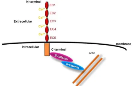
Cadherins, a key component in adherens junctions, represent one class of CAMs (cell adhesion molecules) that mediate Ca2+ dependent interactions between cells. In general, classical cadherins are transmembrane glycoproteins that have a common cytoplasmic domain and an extracellular domain containing five tandem extracellular cadherin domains (Figure 8). These domains are highly homologous to each other and hold Ca2+ binding sites. (Alimperti et al., 2015). Cadherin-mediated cell– cell homophilic junctions are formed as a result of interaction between extracellular domains of identical cadherins, located on the membranes of the neighbouring cells (Ivanov et al., 2001). Even though some cadherins can mediate weak heterophilic interactions, highly specific homophilic adhesions play a key role in tissue and organ development during embryogenesis and in maintenance of normal tissue structure in the adult organism. (Ivanov et al. 2001; Alimperti et al., 2015; Collins et al., 2014)
Figure 8 - Structure of classical cadherins and their interaction with cytoplasmic proteins. Adapted from
Alimperti et al. 2015.
Experiments with chick embryos have shown that cadherins, especially N-Cadherin (Cdh2), have been implicated in the establishment of LR asymmetry both early and later in development (Davis et al., 2008; García-Castro et al., 2000; Mendes et al., 2014; Plageman, et al., 2011, Gonzalez-Morales et al., 2015). Some have established a link between actin dynamics and cadherin-based junctions, which culminate in the asymmetric cell behaviours seen during gut morphogenesis (Davis et al., 2008; Kurpios et al., 2008; Welsh et al., 2013). Work from our lab showed that N-cadherin plays a role in LR patterning in the chick
18
(Mendes et al., 2014). It is a key molecule responsible for finishing the leftward cell movements at the node. Stopping these movements at the right time is crucial to stabilize the molecular asymmetries generated in the node, so that the correct asymmetric information is conveyed to the LPM and the proper looping of the heart is achieved (Mendes et al., 2014)
1.2.1 – Cadherin 11
In zebrafish, Cadherins are required to maintain the adhesive interactions of the dorsal forerunner cells (DFC) during their migration and subsequently for lumen formation when they differentiate to form the KV (Matsui et al., 2011; Oteiza et al., 2008; Tay et al., 2013). One specific cadherin, Cadherin-11 (Cdh11) cadherin has been studied in the mouse and Xenopus. It is involved in the cell differentiation and migration of neural crest (Pegoraro et al., 2013; Simonneau et al., 1995; Vallin et al., 1998) and metastatic tumour cells (Chu et al., 2009; Huang et al., 2011). In zebrafish, this cadherin was initially detected during epiboly, later in the neural keel, and at 20ss in the ventral neural tube, otic vesicle, midbrain and diencephalon (Franklin & Sargent, 1996). Experiments have found that Cdh11 is present in membrane structures important for otolith formation (Clendenon et al., 2009), and that it is also required for the development of the visual system of the zebrafish (Clendenon et al., 2012). However, Cdh11 had not been described in the establishment of LR asymmetry.
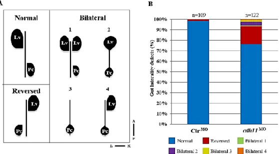
At the start of this project, in order to understand the role of Cdh11 we injected embryos with a cdh11 morpholino. We saw that at 48 hpf both the heart and the gut had laterality defects (Figures 9 and 10). By injecting into a sox17:EGFP transgenic line, we observed that approximately 20% of the embryos showed an inverted gut conformation. In some cases we also saw bilateral disturbances, such as an extra liver (Figure 9). Using a myl7 probe, we saw that cdh11 morphants often showed reversed or symmetric hearts (50%) (Figure 10).
Figure 9 – Cdh11 knockdown causes gut laterality defects at 50 hpf. A- Schematic representation of normal
gut loop (Normal) and five gut laterality defects (Reversed, Bilateral 1, 2, 3 and 4); B- Percentages of normal (blue), reversed (red), bilateral 1 (green), bilateral 2 (purple), bilateral 3 (yellow) and bilateral 4 (Orange) in Ctr MO (n=109) and cdh11 MO (n=122) injected embryos analyzed at 50 hpf. Lv-liver, Pc-Pancreas, L-Left, R-Right, A-Anterior, P-Posterior. Unpublished data from Sara Fernandes
19
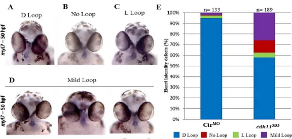
Figure 10 – Morphants embryos exhibit heart laterality defects at 50 hpf. A-D - Ventral view of a ControlMO
or cdh11MOinjected embryos at 50 hpf after myl7 hybridization (A- embryo with a WT conformation (D-loop); B-
Embryo with a symmetric heart (No-Loop); C- embryo with inverted heart (L-loop); D– Example of three embryos with Mild heart loops ); E – Percentages of D loop (blue), No loop (red), L loop (green) and Mild loop hearts (purple) in ControlMO (n=133) and cdh11MO (n=189) injected embryos. R-Right, L-Left. Data from Silva 2017.
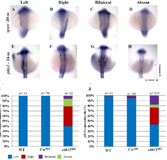
Furthermore, after cdh11 knockdown, the expression patterns of the LR markers spaw and pitx2 were altered in the LPM. For both genes around 60% of the embryos displayed an alternative pattern, in which 30% of the morphant embryos showed spaw and pitx2 expression restricted to the right LPM (Figure 11).
20
Figure 11 – Left-Right markers spaw and pitx2 are affected in morphants at late somite stages. A to H -
Whole-mount in situ hybridization in ControlMO or cdh11MO injected embryo using spaw probe at 20-somite stage
(A to D) and pitx2 probe at 24-somite stage (E to H) (spaw and pitx2 expression on the left side (WT) (A, E), right (B, F), bilateral (C, G) or absent (D, H); I-J - Percentages of Left (blue), Right (red), Bilateral (purple) and Absent (green) expression of spaw (I) and pitx2 (J) in WT (spaw n=24; pitx2 n=63), Ctr MO (spaw n=70; pitx2 n=88) and cdh11MO (spaw n=62; pitx2 n=145) injected embryos analyzed at 20- and 24-somite stage respectively. L-Left,
R-Right, A-Anterior, P-Posterior, ss- somite stage. Data from Silva 2017
Interestingly, the laterality phenotypes of the heart and gut were not concordant after cdh11MO injection. The predominant heart defect was absence of loop, but the majority of the affected embryos showed a reversed gut (Figures 9 and 10). This indicates an uncoupling in the development of these organs, which had been observed before (Lopes et al., 2010; Noel et al., 2013; Ji et al., 2015). Additionally, we noticed that the percentage of bilateral expression of spaw and pitx2 after MO injection also showed a lack of concordance (Figure 11). Therefore, we proposed that the abnormal expression of the conserved LR genes in the right LPM might account for the reversed gut loop phenotype, and the absence of these genes in the prospective heart territory would prevent the breakage of symmetry in the heart.
In order to understand what role Cdh11 might play throughout development, we searched for cdh11 transcripts and we detected cdh11 mRNA in the IM at 8ss (Figure 12A). Curiously, this corresponds to the stage where the laterality signal (spaw) is being relayed to the LPM (Shiratori et al., 2006). Additionally, Cdh11 had been detected in the PN (which derives from the IM) at 48 hpf (Figure 12B) (Clark et al., 2011). We know that by 24 hpf the PN has developed (Drummond et al., 2016), and at this stage the LPM begins its asymmetric migration to the midline (Horne-Badovinac et al., 2003). This led
21
us to suggest a new, not previously described role for these tissues in the establishment of symmetry in zebrafish. We proposed that that the IM could be used as mediator of the transfer of Spaw from the KV towards the LPM at the 10 to 12-somite stages, and that the PN can provide a stable structure for the LPM asymmetric migration between 26-30 hpf, that ultimately leads to the shift of the gut to the left.
Figure 12 - cdh11 expression pattern in WT embryos. A - WISH for cdh11 in 8-somite stage (A- whole-mount
embryo (dorsal view), A’- transversal section through KV)
WISH done by Sara Fernandes, Photos taken by Dalila and Sara. B – cdh11 expression at 48 hpf using a protein-trap system. This produces a RFP transcript where and when the cdh11 mRNA is transcribed. Adapted from Clark 2011
Additionally, we also analysed a mutant line for cdh11, which displayed a premature stop codon in one of its Cdh-Cdh interaction domains, producing presumably a truncated protein (Supplementary Figure 1). In contrast to what happened the morphants, the cdh11 mutant did not show any of the heart and gut phenotypes that we previously characterized (Figure 9). This means that the cdh11MO is not specific and we are observing off-target effects, that the mutant might not be functioning as a null mutation or that a genetic compensation mechanism is activated in the mutant.
1.3 – Objectives
In this project we propose to investigate the role of Cdh11 in the establishment of LR. We propose that Cdh11 could be affecting LR asymmetries either at 10-12ss during the transfer of the Nodal signal from the KV to the LPM, and also at 24 hpf during the asymmetric migration of the LPM. Additionally we want to understand how this dual role of Cdh11 might explain the lack of concordance between the heart and gut phenotypes after cdh11 knockdown.
In this project we aim to:
- Test the specificity of the cdh11MO
- Confirm whether cdh11 is expressed in LR associated tissues
- Understand the role of Cdh11 in the transfer of asymmetric signal from the KV to the LPM - Evaluate the putative role of the PN during the migration of the LPM and looping of the gut
22 Chapter 2 – Experimental procedures
2.1. Zebrafish maintenance
Adult zebrafish (Danio rerio) and embryos used in this project were maintained and bred under standard laboratory conditions (Westerfield et al. 2000). During this project, the embryonic stages were confirmed according to Kimmel et al., 1995.
The cadherin-11 (cdh11) knockdown characterization was performed using embryos from wild-type (WT) AB strains and transgenic Tg(sox17:EGFP) line, maintained at Instituto de Medicina Molecular (IMM) (Sakaguchi et al., 2006). The mutant line cdh11sa14413 was obtained from the European Zebrafish Resource Center (EZRC)1 that was generated within the TILLING project context (Moens, Donn, Wolf-Saxon, & Ma, 2008).
This line produces a truncated protein in the Cdh-Cdh interaction domain (fourth EC) due to a single nucleotide nonsense mutation (T to A) in the 454th amino acid (aa). This change leads to the formation of a premature STOP codon (TTA) instead of a Leucine (Leu) aa (TTT).
2.2. cdh11 Knockdown
To knockdown the cdh11, we used a cdh11 specific splice blocking MO, the MO3-cdh11 (5' - TGTCACGCACCTCTGTTGTCCTTGA - 3') (cdh11MO) (Clendenon et al., 2009), and a Standard Control MO (CtrMO) as a negative control (5' - CCTCTTACCTCAgTTACAATTTATA - 3') (GeneTools).
Stock solution of 3mM (Control MO) and 2,5mM (cdh11 MO) was stored at -20°C. Injection mixture was prepared by diluting the MOs in RNase-Free water to reach a 0,2mM injection concentration.
2.2.1 - Embryo microinjections
Adult zebrafish of interest (lines WT AB) were kept overnight in a breeding cage. In the morning, a loaded injection needle was clipped using forceps, using a micrometer and mineral oil, and calibrated each time to produce a consistent injection volume. The embryos were then collected, aligned to a microscope slide in a Petri dish with a pipette
Each one-cell stage embryo was injected in the cell cytoplasm with 1,4nL of 0,2mM MO solution (0,23ng).
For each experiment, both conditions (control and cdh11 MO) were injected into sibling embryos from two to three independent batches, and incubated in 1x Embryo Medium at 28°C until the desired developmental stage was reached.
2.2.2 - Cdh11 mRNA synthesis
cdh11 sense and anti-sense transcripts were obtained through the respective DNA plasmids, PCS2+ cdh11 sense and PCS2+ cdh11 anti-sense, available in our lab. The linearization reaction was composed of 5μg of DNA plasmid, 1μL of NotI restriction enzyme, 5μL of the respective buffer (10x) and water mixed together for a final volume of 50μL. The reaction mixture was incubated 1h at 37°C.
After 1% agarose gel electrophoresis the corresponding linearized fragments were extracted and purified with the Gel DNA Recovery kit (Zymo Research).
23
The anti-sense transcripts were produced through the mMessage mMachine kit (Thermo Fisher) by adding approximately 1μg of the purified linearized plasmid DNA with the SP6 RNA Polymerase and following the provided protocol. 1μL of TURBO DNAse was also added. This produces the capped mRNA.
The sample was purified according to the manufacturer's instructions of the illustra™ Probe Quant™G-50 Micro Columns (GE Healthcare Life Sciences). The resulting mixture was observed in a denaturing gel and its concentration measured in the Nanodrop 2000 Spectrophotometer (Thermo Fisher).
2.3. CRISPR/Cas9 2.3.1 - gRNA design
Three different gRNAs were designed to target three regions of the Cdh11 protein: signal peptide, extracellular domain and transmembrane domain (gRNA 1, 2 and 3 respectively).
- gRNA1 was designed to recognize the signal peptide, at the N-terminal, which directs the newly synthesized protein to its destination. Targeting this region should prevent the protein from localizing to the membrane.
- gRNA2 targets the extracellular domain of the cadherin, downstream of the signal peptide. While mutations in this region will not prevent alternative ATG usage, if a frameshifting mutation occurs, it will give rise to two outcomes: on the one hand, transcripts using the correct ATG will produce a truncated or altered protein, thus lacking the appropriate transmembrane domain. On the other hand, transcripts that use an in frame alternative ATG, located downstream of the mutation site, will produce proteins lacking both the signal peptide and the upstream extracellular domain. This means that they probably won’t localize to the membrane, but if they do, they won’t be functional.
- gRNA3 targets the transmembrane domain, stopping the protein from attaching to the membrane. The gRNAs were genearated as described in Talbot et al., 2014. The gRNA synthesis was done as described in Ribeiro et al., 2017.
Cas9 protein was produced by the Weizmann Institute of Science, Israel as a purified batch at 1mg/mL in 20 mM Tris pH 8.0, 10mM MgCl2, 0.2M KCl.
2.3.2 – gRNA/Cas9 microinjections
All gRNA combinations were tested, from injecting each single guide, two at the time and also all three of them together. Several gRNA and Cas9 concentrations were analysed, from 107 ng/μL to 880 ng/μL of gRNA and either 666,7 ng/μL or 800 ng/μL of Cas9 protein (Table 1). The analyses present in this work derives from injecting gRNAs 1 and 2 combined, both at 293,3 ng/μL, with the Cas9 protein at 800 ng/μL. The mixture was incubated at R.T. for 5 minutes to allow for the gRNA/Cas9 complexes to form. Embryos at one-cell stage were injected with 1.4nL of this solution, as described above.
24
Table 1
Conditions Attempts Analysed
gRNA combinations
1, 2
1+2, 1+3, 2+3 1+2+3
1+2
gRNA concentration (ng/uL) 107 to 880 293,3 each Cas9 concentration (ng/uL) 667,7 to 800 800
Table 1 – Different concentrations and combinations of cdh11 gRNAs and Cas9 protein were injected in 1-cell stage embryos.
2.3.3 - DNA extraction
At 24 hours, embryos were collected and placed into microcentrifuge tubes in pairs (two embryos per tube). Following the protocol as described in Meeker et al., 2007.The Embryo Medium was replaced with 100μL of 50 mM NaOH and incubated at 95°C for 20 minutes. The tubes were cooled to 4°C, and then 10μL of 1 M Tris-HCl, pH 7.5, was added. The sample was centrifuged at 670g for 10 minutes at R.T., and the supernatant transferred to a new tube. The resulting mixture was purified through the DNA clean & concentrator™- 5 kit (Zymo Research)
2.3.4 - T7 endonuclease
Forward (Fw) and reverse (Rv) primers for each gRNA were designed using NCBI primer blast2 and synthesized by STABVida. The annealing temperature was calculated using Tm Calculator by Ther-moFisher Scientific™3.
The samples were amplified by PCR and then 200ng of the purified PCR product was added to 2μL of NEBuffer 2 (adjusting with water up to 19μL) and denatured and reannealed using a thermocycler with the respective protocol (Supplementary Table 5).
After this, 1μL was added to the samples and incubated at 37 oC for 90 minutes in the thermocycler. The samples were then run in a 2.5% agarose gel electrophoresis and gene modification levels were estimated using the ImageJ software.
2.4. Fluorescence activated cell sorting (FACS)
Approximately 100 embryos at 8-somite stage, i.e. 13 hour-post-fertilization (hpf). were dechorionated and washed in Daniaeu’s buffer These cells were transferred to a CO2 independent medium (Gibco) complemented with 0.5 mM EDTA. These cells were then dissociated through pipetting. After this, the embryos were centrifuged at 700g and re-suspended in 5 mL of the same medium (this step repeated
2https://www.ncbi.nlm.nih.gov/tools/primer-blast/
3 https://www.thermofisher.com/pt/en/home/brands/thermo-scientific/molecular-biology/molecular-
25
three times). Afterwards, the cells were re-suspended in 1 mL of medium and filtered with a 70μm filter
(Falcon) directly into a 5mL round bottom tube (Falcon).
FACS was performed with a FACSAria bench top High Speed Cell Sorter (Becton Dickinson), with the 100μm nozzle with 0-16-0 mask, and the sheath fluid pressure at 20psi.
GFP excitation was made through a 488nm (Blue) laser and the detection using 502LP e 530/30nm filtres. GFP positive cells in WT and and in sox17:GFP transgenic embryos were selected and collected into TRIzol buffer.
2.4.1 - RNA extraction (DNAse I)
After sorting, TRIzol was added up to 1 mL. The cells were vortexed for 30 seconds and incubated 5 minutes at room temperature. 200μL of chloroform was added and the tubes were shaken vigorously by hand for 15 seconds, incubated for 5 minutes at room temperature and centrifuged at 12000g, for 15 minutes at 4oC. The aqueous phase was collected into a new RNAse-free tube and mixed with 0.5μL of 20μg/μL RNAse-free glycogen (ROCHE). 500μL of isopropanol was also added and mixed by hand. The samples were incubated for 10 minutes at room temperature and centrifuged at 12000g, for 10 minutes, at 4oC. The supernatant was removed, and the pellet washed with 1 mL of cold 75% Ethanol and centrifuged at 8000g for 10 minutes at 4oC. The wash was discarded and this step repeated. After the second wash, ethanol was removed from the tubes by pipetting and for 10 minutes the tubes were left to dry to remove all traces of ethanol. The pellet was re-suspended in 14μL of RNase free water. The samples were warmed at 60oC for 10 minutes and cooled on ice. The samples were treated with DNase I (Promega), purified with the RNA clean and concentrator kit (Zymo Research) and stored at -80 oC.
2.4.2 - Reverse transcriptase
First strand synthesis of cDNA was carried out using ProtoScript® II Reverse Transcriptase kit (NEB), through random primers, following the manufacturer protocol. Approximately 120 ng of the total RNA was added in a 20 µl reaction. This was initially denatured through heating (65°C for 5 minutes), followed by a 5 minutes incubation at 25°C, then 1 hour at 42°C and for 20 minutes at 65°C.
2.4.3 - cDNA Purification
100μL of Phenol:Chloroform:Isoamyl alcohol (25:24:1) (Sigma) was added into each tube and the samples were then vortexed for 30 seconds and centrifuged at 13200rpm for 5 minutes at R.T. The upper phase was transferred into a new tube, and mixed with 200μL of chloroform per 100μL of supernatant. The mixture was vortexed for 30s and centrifuged at 13200rpm for 5 minutes at R.T. Once more, the upper phase was transferred to a new tube, and mixed with 1/10 of the volume transferred of ammonium acetate 4,5M, and 2,5x the volume transferred of absolute ethanol. The samples were homogenized and incubated overnight at -20°C. On the following day, the tubes were centrifuged at 14000rpm, for 45minutes, at 4°C.
The supernatant was discarded and the pellet was washed with previously cooled 70% ethanol. The tubes were centrifuged for 15 minutes at 14000rpm, at 4°C and the supernatant was discarded. The pellet was air dried and then re-suspended in 20μL of water. The samples were quantified using the NanoDrop 2000 spectrophotometer and stored at -20°C.
2.4.4 Polymerase Chain Reaction (PCR)
All the reaction specifications were performed according with Thermo Scientific Phusion High-Fidelity DNA Polymerase product information. For a final volume of 50μL, 150ng/50μL of gDNA template was added, 10μL of 5x Phusion HF Buffer (ThermoFisher Scientific™), 1μL of 10mM dNTP mix (ThermoFisher Scientific™), 2,5μL of 10μM primer mix, 0,5μL Phusion DNA polymerase
26
(ThermoFisher Scientific™) and water. Cycle sequencing was performed with conditions described in Supplementary Table 2 and 3. Then the samples were analysed by agarose gel electrophoresis.
In order to amplify the cdh11 gene region, we used the primer pair previously designed for cdh11 gRNA 1 (see above) using the corresponding thermocycler protocol (Supplementary Table 2). Then the samples were analysed in an agarose gel electrophoresis. Primer pair for amplification of dand5 was kindly provided by Susana Lopes laboratory.
2.5. Cdh17 ablation line plasmid
In order to produce a cdh17 specific ablation line plasmid, two plasmids were used: the Osx:mCherry-NTRO plasmid (Renn and Winkler 2009) and the pSceI-cdh17prom-eGFP-cdh17intron plasmid (Sanker et al., 2013), kindly provided by the Didier Stainier and Neil Huckriede repectively (Supplementary figures 4 and 5)
2.5.1 - Plasmid cloning
The plasmids were initially digested with KpnI initially, later replaced by Acc65I (NTRO plasmid) and XhoI (cdh17 plasmid) restriction enzymes. To linearize each plasmid DNA, 5μg of DNA plasmid, 1μL
of the appropriated restriction enzyme, 5μL of the respective buffer (10x) and water were mixed together to a final volume of 50μL. The reaction mixture was incubated for 1h at 37°C. The efficiency of digestion was visualized on an agarose gel and later purified using the DNA clean & concentrator™- 5 kit (Zymo Research).
The 5’ ends of the fragments were blunted by Klenow fragment or T4 DNA polymerase filling. With the Klenow, the reaction was composed of 15μL of the DNA sample, 2μL of the respective buffer, 0,5μL
of the dNTP mix (10μM), 1μL of the Klenow fragment and water up to 20μL.
The T4 polymerase reaction was composed of the purified samples, plus 1μL of T4 DNA polymerase, 3μL of the respective buffer (10x), 1.5μL of dNTPs (10μM) and water up to 30μL. The tubes were incubated at 12°C for 15 minutes.Both samples were then cut with BamHI enzyme. This was done by adding all of the sample DNA, 10μL of the respective buffer (10x), 1μL and water up to 100μL. The
mixture was incubated for 1h at 37°C.The samples were analysed in an agarose gel and the required fragments extracted and purified, using the ZymocleanTM Gel DNA Recovery kit (Zymo).The fragments
were ligated using the T4 DNA Ligase (NEB) protocol, with different insert:vector ratios, for 2h at R.T. or ON at 12°C.
2.5.2 - Transformation of competent Escherichia coli bacteria
Frozen aliquots of competent Escherichia coli bacteria previously prepared in our lab (DH5α strain, kept at -80°C), were thawed on ice. 5μL of the cloned plasmid was added to 100μL of cells and incubated on ice for 30 minutes. This was followed by 40s heat shock at 42°C and a 2 minutes cooldown on ice. 900μL of SOB solution was added to the mixture and left for incubation, for 45 to 90 minutes, at 37°C with agitation. After this, 100μL of this mixture was plated on LB agar medium (containing ampicillin 100μg/mL of LB agar medium) and left at 37°C overnight. This was also done for a positive control (the original Osx:mCherry-NTRO plasmid) and a negative one (the AmpR expressing fragment, after ligation protocol).
On the next day, an isolated colony was inoculated on a 15mL falcon with LB media and left overnight at 37°C with agitation. DNA was purified according to the manufacturer's instructions of the GeneJET Plasmid Miniprep Kit (ThermoFisher Scientific™). DNA concentration was determined by spectrophotometry using the NanoDrop 2000 spectrophotometer.
27
2.6. Whole mount in situ hybridization (WISH)
In these experiments, several probes were used, previously synthesized in our lab: DIG-labelled cdh11,
cmlc2, cdh6, cdh17, foxa3 and FITC labelled myoD.
Zebrafish embryos were collected at specific developmental stages and fixed in a 4% paraformaldehyde solution prepared in 1x Phosphate-buffered saline (1x PBS) (4% PFA) during 4 to 5 hours at R.T. or overnight at 4°C. The embryos were then stored at -20°C after dehydrate washes performed with increasing concentrations of methanol (MetOH) diluted in 0,1% Tween 20 in 1x PBS (0,1% PBT) (two washes with 0,1% PBT, 50% MetOH and 100% MetOH).
2.6.1 - WISH protocol
Day 1: The stored embryos were rehydrated by successive washes with 75%, 50%, 25% MetOH in 0,1% PBT, and four times in 0,1% PBT, for 5 minutes each. Chorions were removed using forceps. Each set of embryos was incubated with PK (Roche) (10μg/mL) in 0,1% PBT (incubation period according to embryo stage - see Table below) and immediately re-fixed in 4% PFA for 20 minutes at R.T. and washed five times with 0,1% PBT (5 minutes each). For 2 to 5 hours, the samples were incubated at 70°C in 500μL of pre-Hybmix, followed by an overnight incubation with 200μL of probe at 70°C, having this one been previously heated for 10 minutes at 70°C.
Stage Incubation time
Early somitogenesis (until 8 somites) 1 minute 18 – 20 hpf (19-23 somites) 5 minutes
24 hpf 15 minutes
36-48 hpf 30 minutes
Day 2: The probe solution was recovered in the next day and the embryos washed at 70°C with pre-heated solutions of 100% pre-Hybmix for 10 minutes, 25%, 50% and 75% 2x SSC in pre-Hybmix and 100% 2x SSC for 15 minutes. After that, the embryos were washed at R.T. twice in 0,2x SSC for 15 minutes each, once in 50% 0,2x SSC in 0,1% PBT and two times in 0,1% PBT, each one lasting 10 minutes. Embryos were incubated in 500μL of blocking solution for in situ at least for 1 hour at R.T. and after that incubated with Anti-DIG-AP (Roche Life Science) in blocking solution (1:5000) overnight at 4°C.
Day 3: In the last day, the embryos were washed six times with 0,1% PBT for 15 minutes each, and three times in Staining Buffer for 5 minutes each. To reveal the probe, the embryos were incubated with 500μL of purple AP substrate (Roche Life Science), or 500μl of NBT/BCIP in the dark at R.T..
The colorimetric reaction was monitored with a dissecting microscope, and stopped by changing the substrate for 0,1% PBT followed by a fixation in 4% PFA for 20 minutes at R.T. and a wash in 0,1% PBT. To store the embryos at 4°C in 100% glycerol, a series of washes in glycerol and 0,1% PBT (20%, 50% glycerol in 0,1% PBT) were performed.
2.6.2- Double WISH protocol
Double ISH was done by combining each of the DIG labelled cdh6, cdh11, and cdh17 probes with the FITC labelled myoD probe.
At the end of Day 2, after being incubated with blocking, the embryos were incubated with Anti-Fluo AP (Roche Life Science) in blocking solution (1:10000) overnight at 4°C.
Day 3: the embryos were washed six times with 0,1% PBT for 15 minutes each, and three times in Tris 0,1M for 10 minutes each. Fast Red substrate was prepared according to the provided protocol (SIGMAFAST™ Fast Red TR/Naphthol AS-MX Tablets) and 500μL was added to the embryos. The
28
revelation took place in the dark. When the revelation process was finished 0,1% PBT was added followed by a fixation in 4% PFA for 20 minutes at R.T. The embryos were one more washed with 0,1% PBT. The acid digestion took place with Glycine for 15minutes, in agitation. The embryos were then washed in tBST twice for 5 minutes, incubated in tBST at 70°C for 30 minutes and later washed with 0,1% PBT twice. Embryos were incubated in 500μL of blocking solution for at least for 1 hour at R.T. The following protocol proceeded as described in the normal ISH protocol from this step on.
2.6.3 Embryo embedding
After the ISH protocol, the embryos were washed in 1x PBS solution until the glycerol was completely removed and then transferred to a 5% sucrose in 1x PBS solution.
They were then fixed in 4% PFA at 4°C. They were washed in 1x PBS and incubated at 4°C for approximately 15 to 30 minutes in 15% sucrose in 1x PBS solution. Later, the samples were incubated for 1h at 42°C in a previously heated 7.5% gelatine and 15% sucrose in 1x PBS solution. The bottom of a plastic mould was filled with the same solution and allowed to harden at R.T. Finally, the embryos were disposed with the correct orientation for sectioning and embedded. The gelatine cubes were fast freezed using 2-Methylbutane (Isopentane) (Sigma) solution, previously cooled at -40°C, using dry ice, and stored at -80°C.
2.6.4 - Cryosectioning
20μm thick transversal sections cut with a Cryostat LEICA CM 3050S were mounted into microscope slides by the Histology and Comparative Pathology Laboratory at iMM and stored at -20°C.The frozen microscope slides were thawed for 30 minutes at R.T. and washed 4 times with a pre-heated 1x PBS in a water bath at 42°C for 5 minutes. After removing the maximum of solution as possible, 100μL of
Mowiol was added to the slide and the cover slip was set on top. The slides were left to dry at R.T. The samples were sealed using nail polish. The slides were stored at 4°C.
2.7. Agarose gel electrophoresis
To access the linearization of the plasmid DNA, PCR product size and probe synthesis, gels were prepared by heating agarose dissolved in 1x TAE buffer mixed with RedSafe™ Nucleic Acid Staining Solution (Intron) (5μL per 100mL TAE 1x). The samples mixed with Orange G (Loading Buffer) at a minimum of 1μL per 5μL of DNA sample and applied to the gel. A 1Kb Plus DNA ladder (Invitrogene) was used to evaluate samples size. The electrophoresis was performed in TAE 1x buffer for 15 minutes at 100V.
For a RNA Denaturing Gel 1,25g of agarose was dissolved by heating in 37mL in water milliQ and left ON at 70°C. On the following day, in a 15mL Falcon tube, the rest of the solution was prepared by adding 5mL of MOPS 10x, 8mL of formaldehyde 37% and 2,5μL of GelRed. This was then added
to the agarose solution. The electrophoresis was performed in a buffer solution composed of 50mL MOPS x10 in 450mL of water MIlliQ