Leonor Ribeiro Duarte Margalha
Dissertation presented to obtain the Ph.D degree in Molecular Biology
Instituto de Tecnologia Química e Biológica António Xavier | Universidade Nova de Lisboa
energy signaling by sumoylation
Leonor Ribeiro Duarte Margalha
Dissertation presented to obtain the Ph.D degree in Molecular Biology
Instituto de Tecnologia Química e Biológica | Universidade Nova de LisboaOeiras,
!"#$%&'()*(+*,)!-./0"1")0")2*
")"3#4*56#)&%6)#*74*5$8(4%&'()*
Margalha, L.
Regulation of SnRK1-dependent energy signaling by sumoylation
Ph.D thesis, Instituto Gulbenkian de Ciência, Universidade Nova de Lisboa, 2015
In English, with abstract in Portuguese
This thesis has been scanned for plagiarism and there was no conflict with published works
Elena Baena-González, Ph.D Plant Stress Signaling
Instituto Gulbenkian de Ciência Oeiras, Portugal
Examiners:
Jürgen Dohmen, Ph.D
Institute for Genetics, University of Cologne Cologne, Germany
Filip Rolland, Ph.D
Molecular Plant Biology, KU Leuven Leuven, Belgium
Isabel Abreu, Ph.D GPlantS Unit,
Instituto de Tecnologia Química e Biológica António Xavier Universidade Nova de Lisboa
Oeiras, Portugal
Alekos Athanasiadis, Ph.D Protein-nucleic acids interactions Instituto Gulbenkian de Ciência Oeiras, Portugal
" We shall not cease from exploration And the end of all our exploring Will be to arrive where we started And know the place for the first time."
T.S.Eliot in Little Gidding
"— ... ¿Qué le parece? — Posible, pero no interesante - respondió Lonnrot-. Usted replicará que la realidad no tiene la menor obligación de ser interesante. Yo le replicaré que la realidad puede prescindir de esa obligación, pero no las hipótesis." Jorge Luis Borges "La muerte y la brújula" in Ficciones
"Hoje vomitei um líquido esverdeado Eram as primeiras folhas Estou prestes a florir " Jorge de Sousa Braga "O último girassol" - in O Poeta Nú
I declare that this dissertation is a result of my own research jointly developed with Dr. Pierre Crozet and carried out between May 2011 and March 2015 in the laboratory of Dr. Elena Baena-González, Instituto Gulbenkian de Ciência in Oeiras, Portugal. Part of chapter 1 has been published in Frontiers in Plant Sciences entitled “Mechanisms of regulation of SNF1/AMPK/SnRK1 protein kinases”, Pierre Crozet, Leonor Margalha, Ana Confraria, Américo Rodrigues, Cláudia Martinho, Mattia Adamo, Carlos A. Elias, Elena Baena-González (2014). Chapters 2, 3, 4 and 5 are part of a manuscript in preparation to be submitted for publication, authored by Pierre Crozet, Leonor Margalha, Rafal Butowt, Noémia Fernandes, Alexandre Elias, Beatriz Orosa, Konstantin Tomanov, Markus Teige, Andreas Bachmair, Ari Sadanandom, Elena Baena-Gonzalez.
Declaro que esta dissertação é o resultado do meu próprio trabalho desenvolvido conjuntamente com o Dr. Pierre Crozet entre Maio de 2011 e Março de 2015 no laboratório da Dra. Elena Baena-González, Instituto Gulbenkian de Ciência em Oeiras, Portugal. Este doutoramento foi realizado no âmbito do Programa Gulbenkian de Doutoramento (edição 2010-2011). Parte do capítulo 1 foi publicado no Frontiers in Plant Sciences como “Mechanisms of regulation of SNF1/AMPK/SnRK1 protein kinases”, Pierre Crozet, Leonor Margalha, Ana Confraria, Américo Rodrigues, Cláudia Martinho, Mattia Adamo, Carlos A. Elias, Elena Baena-González (2014). Os capítulos 2, 3, 4 e 5 estão integrados num manuscrito em preparação para publicação como "SUMOylation represses energy signaling in Arabidopsis" com autoria de Pierre Crozet, Leonor Margalha, Rafal Butowt, Noémia Fernandes, Carlos A. Elias, Beatriz Orosa, Konstantin Tomanov, Markus Teige, Andreas Bachmair, Ari Sadanandom, Elena Baena-Gonzalez.
This dissertation had the financial support from FCT, doctoral fellowship SFRH/BD/51627/2011 and Fundação Calouste Gulbenkian.
Esta dissertação teve o apoio financeiro da FCT, bolsa de doutoramento SFRH/BD/51627/2011 e da Fundação Calouste Gulbenkian.
Turning the last page in my PhD is a special moment of fulfillment and achievement, and I am filled with gratitude for all those who were present and contributed in several ways to make this an amazing journey.
To my supervisor, Elena Baena-Gonzaléz, I am deeply grateful and honored for being your student. You have opened one way to my happiness and provided the most nurturing environment that allowed me to grow scientifically, engaged in your enthusiasm for science and in your demand for knowledge and excellence. You were always very inspiring, very supportive and kind. I feel fortunate with what I have learned, and will preserve it throughout my life.
It was a privilege to share with Pierre Crozet most of my bench time, a lot of work and scientific discussions. I value your dedication to science and to people, your broad interests and culture. Thank you for the scientific support and great company along these years.
Several people contributed to the work presented in this thesis, and to which I am most grateful: Pierre Crozet, Rafal Butowt, Noémia Fernandes, Alexandre Elias, Konstantin Tomanov, Andreas Bachmair, Ari Sadanandom and Elena Baena-González.
I will always be thankful to Paula Duque for being key in guiding me to science and to what became my scientific home in these last years, the Plant Stress Signaling Laboratory at Instituto Gulbenkian de Ciência.
My lab is made of the amazing people that work and worked there, my colleagues and friends: Elena, our top model boss, thank you for everything. Cláudia Martinho, Rafal Butowt and Américo Rodrigues, my companions at the Big Bang of PSS, thank you for scientific training and laughs. Ana Confraria, thank you for being a scientific "older sister", and for sharing so much knowledge and energy (also via delicious food). Pierre Crozet, Mattia Adamo and Alexandre Elias, thank you for extensive experimental support and for making the lab a joy, with music, gardens, pears and gadgets. Titti Valério,
adorable moments in the lab.
The unifying element amongst all Plant Groups at IGC is Vera Nunes, to whom I am deeply grateful for the amazing plant care, for hilarious moments and for her sweet presence. I thank all the Groups at IGC for sharing reagents, protocols and scientific insights. I am especially grateful to the Plant Molecular Biology Laboratory and to Raquel Carvalho, for providing the nicest atmosphere amongst neighboring groups.
It was an unique opportunity to be part of the IGC PhD program in Integrative Biomedical Sciences (PIBS) 2010, and I much value the gain that such experience brought. My acknowledgements go to Thiago Carvalho and Manuela Cordeiro, and extend to all people involved (from PhD colleagues to scientists, for sharing time and knowledge). I would also like to acknowledge my Thesis Committee, José Feijó and Vasco Barreto, for encouraging me to be critical and independent regarding my research work.
IGC has been a fantastic place to be, and I am grateful to each and everyone that contribute to this rich environment.
I am also very thankful to the scientists that accepted to be part of my PhD jury: Jürgen Dohmen, Filip Rolland, Alekos Athanasiadis and Isabel Abreu.
Other key people in my education and training should be acknowledged in this moment, for their dedication and affection: Prof. Clarisse Borralho, Prof. David Mendes, Dra. Teresa Correia and Dra. Sara Silva Alexandre.
Finally, I am very grateful to my family.
To my dear parents, my adorable brother and sisters, and my precious grandparents, for being part of what I am and for their unlimited support.
To my husband and son, Nuno and Sebastião, for offering me the ground and the sunlight that make me live happily.
Table of
Contents ... XV
List of Figures ... XVIII
Main abbreviations ... XX
Resumo ... XXI
Abstract ... XXV
CHAPTER 1 | ... 1
General Introduction ... 1
1.1 | Posttranslational regulation by members of the Ubiquitin family .... 2
1.2 | SUMO ... 4
1.2.1 | SUMO structure ... 4
1.2.2 | SUMO isoforms ... 5
1.2.3 | SUMO pathway ... 9
1.2.4 | SUMO Enzymology ... 13
1.2.5 | Molecular consequences of sumoylation ... 26
1.2.6 | SUMO Enigma ... 31
1.2.7 | Biological consequences of sumoylation ... 32
1.2.8 | Crosstalk with other posttranslational modifications ... 33
1.2.9 | Challenges and strategies for the identification of sumoylation substrates and SUMO interactors in Arabidopsis ... 39
1.3 | Energy sensing and signaling in eukaryotes ... 41
1.4 | SnRK1 in the core of plant energy homeostasis ... 42
1.4.1 | Stress ... 47
1.4.2 | Hormones ... 49
1.4.3 | Metabolism ... 51
1.4.4 | Growth and Development ... 55
1.5 | Conserved Structures of SNF1/AMPK/SnRK1 complexes ... 58
1.5.1 | !-catalytic subunit ... 59
1.5.2 | "-regulatory subunit ... 60
1.5.3 | #-regulatory subunit ... 61
1.6 | Regulation of SNF1/AMPK/SnRK1 kinases ... 62
1.6.1 | Complex composition ... 64
1.6.2 | Scaffold proteins ... 66
1.6.3 | Oligomerization ... 67
1.6.4 | Subcellular localization ... 68
1.6.5 | Adenylates, sugars and hormones ... 70
1.6.6 | Posttranslational modifications ... 71
1.7 | Aims and thesis scope ... 101
2.1 | Introduction ... 105
2.2 | Results ... 107
2.2.1 | SnRK1!1 bears two high probability SUMO attachment sites ... 108
2.2.2 | SnRK1!1 catalytic subunit interacts with SCE1 in Yeast two-hybrid assays ... 109
2.2.3 | Multiple SnRK1 subunits are sumoylated in a heterologous system in E. coli ... 111
2.2.4 | SnRK1 is sumoylated in planta ... 117
2.2.5 | SnRK1 sumoylation is mediated by the E3 ligase SIZ1 in planta . 121 2.2.6 | Alternative strategies employed for determining SnRK1 sumoylation in Arabidopsis ... 123
2.3 | Discussion ... 132
2.4 | Materials and Methods ... 137
Acknowledgements ... 146
References ... 147
CHAPTER 3 | ... 151
Biochemical and functional outcomes of SnRK1 sumoylation ... 151
3.1 | Introduction ... 154
3.2 | Results ... 155
3.2.1 | Sumoylation inhibits SnRK1 signaling ... 155
3.2.2 | Sumoylation triggers SnRK1 degradation ... 163
3.2.3 | Sumoylated SnRK1 is ubiquitylated and degraded by the proteasome ... 166
3.2.4 | Sumoylation affects SnRK1 complex composition and possibly its oligomerization ... 168
3.2.5 | Preliminary studies on the genetic interaction between SIZ1 and SnRK1!1 in Arabidopsis thaliana, and other approaches to address the relevance of SnRK1 sumoylation at the whole plant level ... 171
3.3 | Discussion ... 179
3.4 | Materials and Methods ... 186
Acknowledgements ... 192
References ... 192
CHAPTER 4 | ... 197
SnRK1 pathway activation and signal termination ... 197
Abstract ... 199
4.1 | Introduction ... 200
4.2 | Results ... 202
4.2.1 | SnRK1 activity triggers its degradation ... 202 4.2.2 | Sumoylation restores degradation of inactive SnRK1!1 variants . 204
4.2.4 | SnRK1$1 phosphorylates SIZ1 ... 208
4.3 | Discussion ... 213
4.4 | Materials and Methods ... 218
Acknowledgements ... 224
References ... 224
CHAPTER 5 ... 229
Concluding remarks and future perspectives ... 229
References ... 244
Figure 1.1 | Schematic and three-dimensional ribbon diagrams of mature
Ubiquitin and SUMO, evidencing the Ub/UBL "-grasp fold. ... 5
Figure 1.2 | Primary sequence alignment of Arabidopsis thaliana (At) SUMO and Ubiquitin precursors. ... 6
Figure 1.3 | SUMO and Ubiquitin conjugation pathways. ... 10
Figure 1.4 | SUMO conjugation reactions. ... 12
Figure 1.5 | Non-covalent (NC) and covalent (C) interactions between SUMO and substrates. ... 28
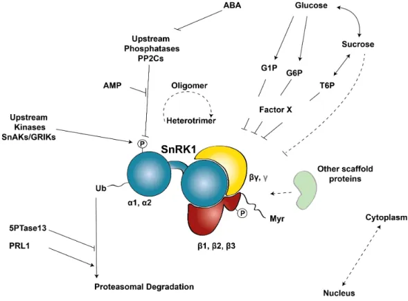
Figure 1.6. | SnRK1 in the interface of Stress, Metabolic, Hormonal, and Developmental signaling pathways. ... 47
Figure 1.7 | Heterotrimeric structure of the SnRK1 complex. ... 59
Figure 1.8 | Regulatory mechanisms controlling the SnRK1 kinase. ... 63
Figure 2.1 | Potential SUMO attachment sites on SnRK1!1 predicted using SUMOplotTM. ... 108
Figure 2.2 | Arabidopsis SnRK1!1 and Drosophila SNF1A interact with the E2 SUMO Conjugating Enzyme 1 (SCE1) in Y2H assays. ... 111
Figure 2.3 | Multiple SnRK1 subunits, as well as the Drosophila SNF1A catalytic subunit, are sumoylated in a heterologous E. coli system. ... 113
Figure 2.4 | SnRK1$1 residues sumoylated in the E. coli assay. ... 116
Figure 2.5 | Generation of SnRK1!1-GFP transgenic plant lines. ... 118
Figure 2.6 | The SnRK1 complex is sumoylated in planta. ... 121
Figure 2.7 | SIZ1 is required for SnRK1 sumoylation. ... 122
Figure 2.8 | Detection of SnRK1!1 high molecular weight bands in plant crude extracts (A-B), after nuclear enrichment (C) and in protoplast samples (D-F). ... 126
Figure 2.9 | Detection of SnRK1!1 high molecular weight forms in SUMO pathway mutants (A) and using the Ubc9 fusion-directed sumoylation (UFDS) methodology in protoplasts (B-C). ... 130
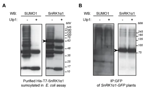
Figure 2.10 | Processing of SnRK1!1-SUMO conjugates by recombinant Ulp1 SUMO protease. ... 132
Figure 3.1 | SIZ1-mediated sumoylation of SnRK1$1 represses SnRK1 signaling. ... 157
Figure 3.2 | SnRK1 signaling overactivation in siz1-2 is due to lack of SnRK1 sumoylation. ... 161
Figure 3.3 | Salicylic acid (SA) has no effect on SnRK1 signaling. ... 162
Figure 3.4 | SnRK1 stability is increased in siz1-2. ... 166
Figure 3.5 | Sumoylation-dependent ubiquitylation of SnRK1 and its degradation by the proteasome. ... 168
Figure 3.6 | Sumoylation affects SnRK1 complex composition and oligomerization. ... 170
development of snrk1!1-3, siz1-2 snrk1!1-3, siz1-2,
SnRK1!1-OE, or Col-0 plants. ... 176 Figure 3.9 | Generation of SnRK1!1-SUMO-OE transgenic plants. ... 178 Figure 4.1 | SnRK1 activity triggers its degradation. ... 203 Figure 4.2 | Sumoylation restores degradation of inactive SnRK1!1 variants. 205 Figure 4.3 | SnRK1 activity triggers its sumoylation and turnover. ... 208 Figure 4.4 | SnRK1 phosphorylates SIZ1 in vitro. ... 210 Figure 4.5 | SnRK1-dependent phosphorylation of SUMO pathway
components and possible outcome. ... 212 Figure 5.1 | Model of SnRK1 turnover mediated by sumoylation. ... 242
!
ACC Acetyl-coA carboxylase AMPK AMP-activated protein kinase ASC Association with SNF1 complex bZIP Basic region/leucine zipper motif CBM Carbohydrate-binding motif
CIPK15 Calcineurin B–like (CBL)–interacting protein kinase 15 DUB Deubiquitylating enzyme
DUF Domain of unknown function ESD4 Early in short days 4
G1P Glucose-1-phosphate G6P Glucose-6-phosphate
GRIK1/2 Geminivirus Rep-interacting kinase 1/2
HMGR 3-Hydroxy-3-methylglutaryl CoA (HMG-CoA) reductase KA1 Kinase-associated 1 domain
LKB1 Liver kinase B1 (tumor supressor) MARD1 Mediator of ABA-regulated dormancy 1 NR Nitrate reductase
PCNA Proliferation cellular nuclear antigen
PIAL1/2 Protein inhibitor of activated STAT-like 1/ 2 PP Protein Phosphatase
PTM Posttranslational modifications PRL1 Pleiotropic Regulatory Locus 1 SAE SUMO activating enzyme SCE SUMO conjugating enzyme SIM SUMO interacting motif
SKIN SnRK1A-Interacting Negative Regulator SnAK1/2 SnRK1 activating kinase 1/2
Snf1 Sucrose non-fermenting 1 SnRK1 Snf1-Related protein Kinase 1 SPS Sucrose phosphate synthase STUbL SUMO-targeted ubiquitin ligase SUMO Small ubiquitin-like modifier TF Transcription factor
T6P Trehalose-6-phosphate
TPS Trehalose Phosphate Synthase UBA Ubiquitin-associated domain UPS Ubiquitin proteasome system
A capacidade de um organismo sentir e reagir a estímulos ambientais, mantendo a homeostasia num meio em constante mudança, é fundamental para a sua sobrevivência. Nas plantas, a SnRK1 (SNF1-related protein kinase 1) é um sensor energético central e um regulador metabólico que integra vários sinais para equilibrar os níveis de energia celular. Ao fazê-lo, a SnRK1 participa na resposta da plantas ao stresse (privação de energia), mas também no crescimento normal e decisões do desenvolvimento da planta. A SnRK1 está conservada em todos os eucariotas. Funciona como um complexo heterotrimérico constituído por uma subunidade catalítica (!) e duas subunidades reguladoras (" e !). No entanto, apesar da sua importância, a regulação da SnRK1 é ainda pouco compreendida. Em contraste com os homólogos em mamíferos e leveduras, a fosforilação/ desfosforilação da T-loop na subunidade catalítica SnRK1! é necessária, mas não suficiente, para determinar a sua activação/ inactivação, sob stress e uma vez restaurada a homeostasia. Com base em várias evidências, colocámos a hipótese de que outras modificações pós-tradução, nomeadamente sumolização, possam estar envolvidas na regulação da SnRK1.
Mostramos que a SnRK1 interage com a enzima E2 de conjugação do SUMO em ensaios de dois híbridos em levedura, e é sumolizada num sistema heterólogo em E. coli. Demonstramos, por imunoprecipitação, que a sumolização da SnRK1 também ocorre na planta, possivelmente como um evento coordenado ao nível do complexo da SnRK1, uma vez que tanto a subunidade catalítica como as reguladoras estão sujeitas a esta modificação. Além disso, identificamos SIZ1 como a enzima E3 de ligação de SUMO responsável pela sumolização da SnRK1 por SUMO1, em Arabidopsis.
A sumolização pode afectar a atividade, estabilidade e/ ou a localização subcelular dos substratos, principalmente através da modulação de interações
enzima E3 de ligação de SUMO, SIZ1, conduz a uma maior ativação da via da SnRK1, que pode ser revertida para níveis basais por complementação transiente com SIZ1, mas não com a variante cataliticamente inativa, SIZ1C379A. Além disso, a expressão de um constructo que mimetiza a sumolização, através da fusão de SUMO1 e SnRK1!1, restaura os níveis normais de sinalização da SnRK1, reforçando que a sobreativação observada em siz1-2 é causada pela ausência de sumolização da SnRK1. Observamos também uma maior estabilidade e acumulação de várias subunidades da SnRK1 em siz1-2, sugerindo que a sumolização da SnRK1 promove a sua degradação, restringindo assim a sinalização da SnRK1. Com efeito, a activação excessiva da via da SnRK1 em siz1-2 não é causada por alterações na atividade catalítica intrínseca da quinase, mas sim por uma acumulação da SnRK1 neste mutante.
Mostramos ainda que a SnRK1 é ubiquitinada na planta e encaminhada para degradação pelo proteosoma. Nos imunoprecipitados da SnRK1 são co-purificados conjugados de SUMO e de ubiquitina. No entanto, em siz1-2, os conjugados de ubiquitina associados à SnRK1 estão bastante reduzidos, sugerindo uma interação cooperativa entre a sumolização e a ubiquitinação no direcionamento da SnRK1 para degradação. Este processo poderá envolver a ação de ligases de ubiquitina dirigidas para SUMO (STUbLs), responsáveis pela ubiquitinação de substratos multi- ou polisumolizados. Adicionalmente, temos resultados preliminares que sugerem que a sumolização poderá afectar a composição do complexo da SnRK1 e, possivelmente, a organização das subunidades da SnRK1 em complexos heterotriméricos e putativamente oligoméricos.
Finalmente, mostramos que os variantes inativos da SnRK1, SnRK1K48M e SnRK1T175A, se acumulam em níveis mais elevados e são mais estáveis do que a proteína ativa, sugerindo uma ligação entre a atividade e a estabilidade da quinase. Para além disso, os variantes inativos SnRK1K48M e SnRK1T175A podem
é essencial para a sua degradação, e sugerindo que a deficiente degradação dos variantes inativos da SnRK1 é causada pela ausência de sumolização. O trabalho aqui descrito revela um mecanismo de feedback negativo pelo qual a atividade da SnRK1 desencadeia a sua própria degradação pelo proteosoma, mediada por sumolização e ubiquitinação, para evitar os efeitos nocivos resultantes de uma sinalização excessiva da SnRK1 na planta. Além disso, mostramos que a SnRK1 fosforila a enzima E3 de ligação de SUMO, SIZ1, in
vitro. Propomos que esse mecanismo poderá ser o elo de ligação entre o
stresse e consequente ativação da SnRK1 e a sua sumolização/ubiquitinação, que resulta na sua subsequente degradação na planta. A apertada regulação da ativação da via da SnRK1, e da acumulação da quinase em condições de privação de energia, poderá estar conservada evolutivamente e ser essencial para o equilíbrio entre respostas ao stresse e defesa, com as atividades biossintéticas, de crescimento e desenvolvimento da planta.
The ability of an organism to sense and react to environmental stimuli, maintaining homeostasis under ever-changing conditions, is key for survival. The plant SnRK1 (Snf1-Related protein Kinase 1) is a central energy sensor and metabolic regulator that integrates multiple inputs to balance cellular energy levels. In doing so, it takes part in the plant stress (energy deprivation) response but also on normal plant growth and developmental decisions. The SnRK1 kinase is conserved in all eukaryotes. It functions as a heterotrimer composed of !-catalytic and "- and #-regulatory subunits. However, despite its importance, SnRK1 regulation is still poorly understood. In contrast to its mammalian and yeast homologues, SnRK1! T-loop phosphorylation/dephosphorylation is required but insufficient to determine its activation under stress and subsequent inactivation once homeostasis is restored. Based on several lines of evidence, we hypothesized that additional posttranslational modifications, like SnRK1 sumoylation, may be involved.
We show that SnRK1 interacts with the E2 SUMO conjugating enzyme in yeast two-hybrid assays and undergoes sumoylation in a heterologous E. coli system. Importantly, immunoprecipitation experiments demonstrate that sumoylation of SnRK1 also occurs in planta, seemingly as a coordinated event at the whole-complex level, as both catalytic and regulatory subunits undergo this modification. Moreover, we identify SIZ1 as the E3 SUMO ligase responsible for SnRK1 sumoylation by SUMO1 in Arabidopsis.
Sumoylation can affect substrate activity, stability and/or subcellular localization, mainly through modulation of intra- and intermolecular protein interactions. We demonstrate that SnRK1 signaling is negatively regulated by sumoylation. In the siz1-2 mutant, the absence of the E3 SUMO ligase SIZ1 leads to enhanced SnRK1 pathway activation that can be reverted to wild-type (WT) levels by transient complementation with SIZ1, but not with the
SUMO1 to SnRK1!1 mimics sumoylation and restores normal SnRK1 signaling in siz1-2, supporting that the overactivation of the SnRK1 pathway in this mutant is caused by the lack of SnRK1 sumoylation. We observed increased stability and accumulation of several SnRK1 subunits in siz1-2, suggesting SnRK1 sumoylation promotes its degradation and thereby restrains SnRK1 signaling. Indeed, the higher activation of SnRK1 signaling in siz1-2 was not caused by changes in SnRK1 intrinsic activity, but rather by an enhanced accumulation of the kinase in this mutant background.
We further show that SnRK1 is ubiquitylated in planta and targeted for proteasomal degradation. SUMO and ubiquitin conjugates co-purify in SnRK1 immunoprecipitates. However, in the siz1-2 background, SnRK1-ubiquitin conjugates are greatly reduced, suggesting a cooperative interplay between sumoylation and ubiquitylation in targeting SnRK1 for degradation. This process is likely to involve the action of SUMO-targeted Ubiquitin Ligases (STUbLs), responsible for the ubiquitylation of multi- or polysumoylated substrates. Additionally, preliminary results suggest sumoylation may also affect SnRK1 complex composition, and possibly SnRK1 complex assembly and its putative oligomerization.
Finally, we show that the inactive SnRK1 variants SnRK1K48M and SnRK1T175A accumulate to a greater extent and are more stable than the active WT SnRK1 protein, underpinning a connection between kinase activity and stability. Importantly, inactive SnRK1K48M and SnRK1T175A can be destabilized if co-transfected with active SnRK1!1 or if expressed as translational protein fusions with SUMO1, further supporting that kinase activity is essential for its degradation and suggesting the impaired turnover of inactive SnRK1!1 variants is caused by lack of sumoylation. The work here described has uncovered a negative feedback loop by which SnRK1 activity triggers its own SUMO-mediated proteasomal degradation, to prevent deleterious effects derived from overly high SnRK1 signaling. Furthermore, we show that SnRK1 phosphorylates
subsequent turnover in planta. A tight regulation of SnRK1 pathway activation and kinase accumulation in energy deprivation conditions may be evolutionary conserved and essential for balancing stress and defense responses with biosynthetic activities, growth and development.
CHAPTER 1 |
!
!
!
Posttranslational modifications (PTMs) allow a sensitive, rapid and reversible response to internal or external stimuli, allowing cells to adapt to programmed or unpredicted changes (1, 2). The expansion in proteome diversity prompted by PTMs is crucial for an organism plasticity, adaptation and ultimately survival.
1.1 | Posttranslational regulation by members of the Ubiquitin
family
Ubiquitin (Ub) was discovered in the mid 1970s and is the founding member and the most prominent one (ubiquitous) of a broad family of protein modifiers (Ubiquitin-like, UBL). Ub an UBLs are transiently conjugated to other proteins, expanding the diversity and versatility of the proteome in eukaryotes (3). However, they can also modify prenyl groups or lipids, as it is the case for the membrane-anchored ubiquitin-fold protein (MUB) and the UBL-protein autophagy 8 (ATG8), respectively. The transient conjugation of ATG8 to the autophagosomal membrane is essential for autophagy (4).
All members of the ubiquitin family share a robust "-grasp structure, derived from the folding of a conserved ""!""" secondary motif, with some N- and C-terminal variations or extra helixes/folds, specific to each UBL member (3, 5, 6) (Figure 1.1). The compact structures of UBLs result in fast assembling and highly stable proteins resistant to harsh environmental conditions. Despite the reduced conservation of the primary structure amongst different UBLs, each UBL member shares a highly conserved sequence, structure, and possibly regulation and regulatory role across different organisms (3). UBL modifiers are encoded in the genome and therefore, have been targets of duplication and diversification events during evolution (7). Insight regarding the origins of the Ub/UBL family came from the prokaryotic ThiS and MoaD proteins, involved in the biosynthesis of several sulfur-containing enzyme cofactors. These proteins
are structurally related to the "-grasp structure and display surprising mechanistic parallels with the UBL conjugation pathway. Moreover, the eukaryotic UBL ubiquitin-related modifier 1 (URM1), implicated both in sulfur donation to transfer RNAs (tRNAs) and in covalent protein attachment to target proteins, could represent an ancestral bridge between sulfur chemistry and protein conjugation (1, 3, 5).
Several proteins have evolved as translational fusions with N-terminal Ub/UBL domains, in a stable association that overcomes the need for subsequent modification. These UBL domains have lost the characteristic C-terminal end and are thus resistant to deconjugation, providing enduring features in protein-protein interactions or folding properties to the whole protein (3, 5).
In plants, besides Ub, the UBL family includes the small ubiquitin-like modifier (SUMO), related to ubiquitin (RUB), ATG8 and ATG12, MUB, URM1, ubiquitin-fold modifier 1 (UFM1), and homology to ubiquitin1 (HUB1), whereas other UBL modifiers like the interferon-stimulated gene 15 (ISG15) or HLA-F adjacent transcript 10 (FAT10) are only present in metazoans (1, 3, 8).
The biological processes regulated by PTMs of the UBL family, and the specificities associated to each member, have been the focus of intense research and at present, Ub and UBLs are considered major regulators of growth, development, and survival in eukaryotes.
This thesis will focus on posttranslational modification by SUMO, given its prominent role in Snf1-Related protein Kinase 1 (SnRK1) regulation and due to the limitations of covering the entire UBL family here. SUMO regulation of SnRK1 signaling in plants will be addressed in detail in the experimental chapters, while the following introductory sections will provide an overview of the SUMO system and of the SnRK1 pathway.
1.2 | SUMO
SUMO stands for Small Ubiquitin-like Modifier and was discovered in 1996, when a peptide sequence analysis retrieved a protein with two N-termini but just one C-terminal end (9). The Ran-GTPase-activating protein 1 (RanGAP1) was the first SUMO substrate described, and the outcome of this modification was a change in the subcellular localization of RanGAP1 (10). Since then, a burst in the UBL research area allowed the identification of a myriad of SUMO targets and implicated SUMO in the regulation of crucial biological processes elevating it, together with Ub, as one of the most prominent UBL members.
Figure 1.1 | Schematic and three-dimensional ribbon diagrams of mature Ubiquitin and SUMO, evidencing the Ub/UBL "-grasp fold.
A. Schematic arrangement of the !-helix and strand secondary structures in the Ub/UBL "-grasp fold. B. Avena sativa Ub [PDB accession number 1UBQ; (11)]. C. Human SUMO1 [PDB accession number 1AR5; (12)]. D. Superimposed structures of Ubiquitin (orange, from B) and SUMO (yellow, from C). Arrows point to the hydrophobic groove between the "2-strand and !1-helix of Ub/SUMO. In A, B, and C, colors represent different secondary structures (red- !1-helix; yellow- sheet; green- loop). Adapted from (3).
SUMO and Ub share only ~18% amino acid (aa) sequence identity and their overall surface-charge distribution is different (6, 12-14). A specific feature of SUMO is an N-terminal unfolded extension that provides unique binding properties to this modifier and precedes the conserved hallmark "-grasp structure of the UBL class (Figure 1.1). SUMO is synthesized as a precursor (as most of the UBL members) and is processed to expose a di-Glycine (GG) C-terminal motif through peptide cleavage (6). Due to its N-C-terminal extension, mature SUMO has a few more amino acids (~100 aa) than Ub (76 aa). SUMO protein is around 12KDa but, when conjugated, it contributes to an increment of ~20KDa on the target protein molecular mass. The corresponding delay in migration in SDS-PAGE of the modified protein will also depend on where in the substrate the SUMO conjugation takes place and, for instance, on the occurrence of branching effects (15-18).
1.2.2 | SUMO isoforms
Whereas lower eukaryotes possess only one SUMO isoform (Smt3 in S.
cerevisae or Pmt3 in S. pombe), higher eukaryotes express several SUMO
variants possibly with distinct functional properties (19-21). In mammals, four SUMO paralogs (SUMO1 to SUMO4) exist, although SUMO4 has been suggested to be a pseudogene (only its mRNA is detected but not the protein),
and its ability to undergo maturation and conjugation even if expressed, has been questioned (20, 22).
In Arabidopsis thaliana, eight genes are putatively attributed to SUMO isoforms (6, 23, 24), but only 4 SUMO variants are highly, although differentially, expressed: SUMO1, SUMO2, SUMO3 and SUMO5 (6, 25, 26). SUMO1 and
SUMO2 are much more abundant (~7 fold) than SUMO3 and SUMO5
transcripts (25, 27). The transcriptional profile of SUMO isoforms is mostly unchanged in different tissues and developmental stages. Nevertheless,
SUMO3 and SUMO5 transcripts might be specifically enriched in roots and
flowers, respectively, reflecting isoform-specific roles in these tissues (27). However, at the protein level there is an enrichment of free SUMO1/2 and of the respective SUMO conjugates, in flowers and siliques (27). The SUMO1 and
SUMO2 genes are constitutively expressed, whereas SUMO3 can be strongly
and widely induced by salicylic acid (SA) and by the defense elicitor Flg22 (21). At the cellular level, most of the SUMOs are present in the nucleus as the majority of proteins involved in sumoylation, despite some may display distinct intracellular localizations (28).
Figure 1.2 | Primary sequence alignment of Arabidopsis thaliana (At) SUMO and Ubiquitin precursors.
(www.ebi.ac.uk/Tools/msa/clustalo/). A red box highlights the C-terminal di-glycine motif exposed during precursor maturation, and a black box evidences the variable amino acids important for deconjugation rate, at position -4 from the mature isoform C-terminus. Underlined residues represent identified sumoylation (black) and ubiquitylation (red) sites. Additional putative ubiquitylation (blue) and acetylation (green) sites are based on the detection of the respective modification in conserved residues of human SUMO1 and/or SUMO2. Below the alignment, asterisks (*) indicate amino acid residues conserved in all aligned sequences, colon (:) indicates conservation between groups of strongly similar properties, and period (.) indicates conservation between groups of weakly similar properties. The Delta (%) symbol in front of AtUbiquitin5 and AtSUMO3 indicates that further downstream amino acids of the precursor were omitted. Adapted from (7).
There is a high sequence similarity between SUMO1 and SUMO2 proteins (93%) (7) and probably functional equivalence (21, 23, 27). Accordingly, a single functional allele of SUMO1 or SUMO2 is sufficient for cell viability, whereas double mutants blocking the accumulation of both SUMO1 and SUMO2 are embryo lethal, underpinning an essential role for SUMO conjugation (21, 27). At the protein level, SUMO1 and SUMO3 share 53% sequence identity and the more divergent SUMO5 shares only 44% sequence identity with SUMO1 (7). Sequence alignment of the different isoforms revealed differences at a glutamine (Q) residue, conserved in most organisms, four amino acids distant from the mature C-terminus. SUMO1, SUMO3 and SUMO5 display Q, methionine (M) or leucine (L) at that position, respectively, and differences in deconjugation between SUMO isoforms in vitro were attributed to the identity of this amino acid (Q causing the fastest deconjugation) (Figure 1.2). Such type of regulation could result in a different half-life for the different SUMO-specific conjugates in vivo, and in diverse roles for the different paralogs (7, 25, 29). Besides SUMO proteases that can preferentially desumoylate specific SUMO conjugates (7), the SUMO paralog specificity also relies on E3 SUMO ligases and/or substrate interaction surfaces that may favor the binding of one SUMO isoform.
Whereas SUMO1 and SUMO2 can be further modified by SUMO in specific lysine (K) residues [K10, K23 and K42 in SUMO1 (30, 31) and K10 in SUMO2 (7, 25)], and feature chain assembly, SUMO3 does not share this property or is sumoylated to a much minor extent (Figure 1.2). SUMO1/2 chain formation is functionally important in plants, for instance in the crosstalk with the ubiquitin proteasome system (UPS), when Ub and SUMO cooperatively target a protein for degradation (32), as similarly reported in yeast and animals (33, 34). In plants, there is a free pool of SUMO1/2 protein that is rapidly diminished by conjugation to substrates upon exposure to a diversity of stresses like heat shock, H2O2, ethanol and to the aminoacid analog canavanine, whereas the SUMO3 profile does not seem to change in these conditions (23).
The differences observed in plant SUMO isoforms occur also in animals, although in this case SUMO2/3 are the highly similar and chain-forming isoforms, more abundant in vivo and massively conjugated as part of an early stress response (35, 36). Interestingly, mixed-chain formation between all SUMO family members was detected in mammals (37). SUMO1 mRNA levels increase in response to hypoxia in animals (38, 39), and higher SUMO2 transcripts are an indicator of malignancy in human hepatocellular carcinomas (40). In animals, a number of lysine residues (K11, K20, K32, K41 and K44) have been identified in SUMO2, as acceptor sites for ubiquitylation (41). Some of these residues are conserved in Arabidopsis SUMO isoforms (Figure 1.2). SUMO2 monoubiquitylation at its N-terminus primes its polyubiquitylation (42) through K63-linked ubiquitin chains that might target SUMO conjugates to insoluble protein inclusions (41). Ubiquitylation of AtSUMO1 on K23 was detected in heat stressed plants (30). SUMO can also be phosphorylated (43,
44) and acetylated (45) (Figure 1.2). Acetylation of K37 in SUMO1 and K33 in
SUMO2 neutralizes a positive charge on SUMO and inhibits its interaction with some SUMO interacting motifs (SIMs), whereas it promotes the interaction of SUMO2 with the bromodomain in p300, indicating that acetylation of SUMO can work as a molecular switch (28, 46).
!
1.2.3 | SUMO pathway
As for other UBLs, the SUMO pathway engages an E1-E2-E3 (and E4) enzymatic cascade that initiates with an inactive precursor processed by specific proteases, and terminates with the covalent attachment of the mature SUMO to a target substrate. The subsequent removal of the modifier by specific proteases confers a transient and versatile nature to the posttranslational modification of substrates by SUMO (Figure 1.3). SUMO conjugation requires energy and involves the formation of high-energy thioester bonds.
Figure 1.3 | SUMO and Ubiquitin conjugation pathways.
Both SUMO (S, bottom, yellow) and Ubiquitin (Ub, upper, orange) pathways share an E1 (activating), E2 (conjugating), E3 (ligase) and E4 (ligase, involved in chain formation) enzymatic cascade. After processing, the modifier is covalently attached through its C-terminal glycine (G) residues to the &-amino group of lysine (K) residues in the substrate (red). Modifiers can target single or multiple lysines in the substrate (mono-/multiubiquitylation/sumoylation) and subsequently form chains through different lysine residues of the modifiers (polyubiquitylation/sumoylation). There are diverse outcomes of these modifications that essentially modulate protein-protein interactions. SUMO-targeted ubiquitin ligases (STUbLs) can ubiquitylate SUMO conjugates, and ubiquitin chains with K48 topology target substrates for proteasomal degradation. Modifications can be reversed by Proteases, also involved in the processing of the respective precursors, which control the free pool of the modifier. The enzymes involved are specific for each pathway and there are striking differences in their number (the numbers of enzymes encoded in Arabidopsis are in parentheses). Adapted from (47).
1.2.3.1 | SUMO maturation
Most of the newly synthesized UBL isoforms need to be processed to expose a conserved di-glycine motif involved in the covalent attachment to the substrate. SUMO proteases fulfill this role through endopeptidase cleavage of carboxyl-terminal extensions from SUMO inactive precursors (6). SUMO proteases can perform both SUMO maturation and SUMO deconjugation, or be specifically engaged in only one of these functions (48).
The proteolytic activity of SUMO proteases can target SUMO-specific isoforms (7, 25, 29). In fact, in vitro pre-SUMO processing activity of the Arabidopsis SUMO proteases ESD4, OTS1 and OTS2, as well as yeast ULP1 and X. campestris XopD, was shown to be specific to AtSUMO1 and AtSUMO2 precursors. In this assay AtSUMO3 could only be processed by XopD, whereas AtSUMO5 was not processed at all, suggesting the enrollment of other SUMO proteases in precursor processing in Arabidopsis. The five cysteine proteases tested also showed SUMO-specific isopeptidase activity towards SUMO1 and
SUMO2 conjugates (7).
1.2.3.2 | SUMO conjugation/deconjugation
SUMO conjugation follows the same chemistry as Ub conjugation, however significant differences make E1, E2 and E3 enzymes specific for each UBL (3). One exception occurs for ATG8 and ATG12 modifiers that are activated by the same E1 (ATG7) but subsequently transferred as thioester intermediates to separate E2s (3, 5). E1 SUMO activating enzymes function as heterodimers (SAE1 and SAE2) and activate mature SUMO with the consumption of one ATP. Activated SUMO is transferred to the active site cysteine of SAE2 with the formation of a thioester bond and release of AMP (SAE2~SUMO). A transthiolation reaction allows the transfer of SUMO from SAE2 to the active site cysteine of E2 SCE1 (SCE1~SUMO). SUMO-loaded SCE1 will directly bind the target and conjugate SUMO through an amide (isopeptide) bond to a lysine residue on the substrate. It may modify a single lysine (monosumoylation) or multiple lysine residues (multisumoylation) on the substrate, and it may also result in the formation of SUMO chains (polysumoylation). E3 SUMO ligases facilitate SUMO conjugation and improve specificity either by bringing SCE1 and the substrate together, or by orienting SCE1~SUMO in a way that promotes SUMO transfer (32, 49). SUMO-substrate conjugates can be rapidly and specifically deconjugated through the isopeptidase activity of SUMO proteases that replenish the pool of free SUMO and release the substrate from its sumoylated state (Figure 1.4).
Figure 1.4 | SUMO conjugation reactions.
SUMO precursors are processed by SUMO proteases that expose a C-terminal glycine (Gly) in the SUMO modifier. Mature SUMO is activated with ATP by E1 SAE1, that adenylates the C-terminal carboxyl group of SUMO, forming a high-energy (~) AMP adduct. This intermediate is attacked (red arrow) and covalently bound by the sulfhydryl group of the E1 SAE2 catalytic cysteine, creating a thioester bond and releasing AMP. SUMO is subsequently transferred to the catalytic cysteine of the E2 SCE via a transthiolation reaction. SUMO can thereafter be ligated to a substrate with the help of an E3 SUMO ligase that can position the amino group of a substrate
lysine near the E2-SUMO thioester, catalyzing the transfer of SUMO to the substrate. Following this, SUMO proteases can remove SUMO from substrates, recycling the modifier. Adapted from (50).
1.2.4 | SUMO Enzymology
Unlike ubiquitylation, which in Arabidopsis involves 2 E1s, 37 E2s and over 1500 different E3s that provide substrate specificity, the SUMO machinery comprises only two different E1 heterodimers, a single E2, and just two E3s. Therefore, distinct features of the sumoylation process, such as the recognition of specific motifs by the E2 on the target, and a tight regulation must be key to attain specificity. The SUMO conjugation and deconjugation machinery is target of such regulation at the transcript level, protein amount and distribution, as well as through posttranslational modifications (3, 28).
1.2.4.1 | E1 SUMO activating enzymes (SAE1 and SAE2) The yeast Saccharomyces cerevisiae has a single E1 enzyme that coordinates the activation of Ub (Uba) while the SUMO activation in the same organism requires an E1 heterodimer (Aos1/Uba2). In Arabidopsis, the heterodimeric complex of E1 SAE is composed of a small subunit (SAE1a or SAE1b; encoded by two genes), and a big subunit (SAE2), that functionally resemble the N- and C-terminal domains of E1 in the Ub system, respectively (6, 7, 27). SAE1a and SAE1b share high amino acid sequence identity (81%) and have similar expression patterns in plants, suggesting functional redundancy. Accordingly, a potentially null mutant affecting SAE1a is viable, probably due to redundancy with SAE1b (27). SAE1 activates SUMO through the formation of a SUMO adenylate between the carboxyl group of SUMO C-terminal glycine (G) and the !-posphate group of ATP, with the subsequent release of pyrophosphate (PPi). The activated intermediate is then transferred
to the sulfhydryl group of the SAE2 active site cysteine, forming a highly energetic thioester linkage (SAE2~SUMO). Null T-DNA insertion mutants of the single gene encoding SAE2 in Arabidopsis are embryonic lethal, with arrest occurring early in embryo development (27).
In Arabidopsis, there is little variation in the mRNA profiles of SAE1a, SAE1b or SAE2 across different tissues and developmental stages, despite some differences at the protein level, for instance with SAE1 levels slightly elevated in flowers (27). Regulation at the transcriptional level was described in other organisms, with increased accumulation of SAE1/Aos1 transcripts in a variety of tissues (e.g., testis and thymus) (51, 52), in the S phase of the cell cycle (52), as well as during embryogenesis in Drosophila (53). On the other hand, high
SAE2/Uba2 and SUMO2 transcript levels were correlated with malignancy in
human hepatocellular carcinomas (40).
At the protein level, sumoylation of both SAE subunits has been observed in mammals. Sumoylation of SAE2 is thought to be more common and derived from auto-sumoylation or mediated by E2 SCE (54, 55). SUMO conjugation on the active site of SAE2 inhibits the transfer of SUMO from E1 to E2 (56), whereas sumoylation on the SAE2 C-terminus is important for SAE nuclear retention (57). AtSAE2 is also sumoylated in vivo (30). SAE1 and SAE2 are ubiquitylated and downregulated upon viral infection in the presence of the adenovirus protein Gam1 (58). Reversible oxidation, on the other hand, has also been shown to regulate sumoylation. In response to low concentrations of ROS, mammalian E1 SAE2 and E2 SCE1 form a disulfide bond between their catalytic cysteines, preventing SUMO thioester formation (59).
1.2.4.2 | E2 SUMO conjugating enzyme (SCE1)
In Arabidopsis, the SUMO machinery comprises a single E2 enzyme (SCE1), whereas more than 30 E2s exist in the Ub system. Therefore, SCE1 must have
relaxed substrate specificity to conjugate all SUMO substrates (and all SUMO isoforms) in vivo (7). SCE1 is crucial in SUMO conjugation since it binds directly the target on a consensus motif, through a patch surrounding the active site (60,
61). The SUMO moiety is then transferred from the thioester bond with SCE1 to
the &-amino group of a target lysine, establishing a covalent amide bond via the SUMO C-terminal glycine carboxyl group. This feature of SCE1 allows sumoylation in in vitro systems even in the absence of E3 SUMO ligases, despite co-expression of the latter might increase the efficiency of the reaction. SCE1 activity is modulated by the non-covalent binding of E1 (determinant for SCE1~SUMO formation) and of SUMO on a multifunctional binding site on SCE1 (6, 62). The consensus motif recognized by SCE, and ultimately the SUMO attachment site (SAS) in the target is 'KxE/D, where ' is a bulky hydrophobic amino acid (mainly valine (V), leucine (L), Isoleucine (I)), x is any amino acid, and where aspartate (D) can also replace the preferable glutamate (E) residue. There are several variations of this classic motif and also inverted motifs (63). The negatively charged amino acid-dependent sumoylation motif (NDSM) 'KxE(x)2-3(E/D)n comprises acidic residues downstream of the core motif that enhance the binding affinity with the positively charged patch of SCE1 (64). The same increase in affinity is seen in the phosphorylation-dependent sumoylation motif (PDSM) 'KxExxS/T, when phosphorylation of the serine (S)/ threonine (T) triggers sumoylation (65). A third sumoylation motif, hydrophobic cluster-containing sumoylation motif (HCSM), clusters N-terminal hydrophobic residues upstream of the core motif, 'nKxE. Inverted sumoylation motifs have also been described, E/DxK' (66).
The variation on SAS motifs contributes to the efficiency and specificity of SUMO conjugation at the substrate level in response to varied signaling cues (63). There are several algorithms that score the probability of a SAS to become modified by SUMO according to the hydrophobicity and acidity of the amino acids at given positions (e.g., SUMOplot, www.abgent.com; sumosp, http://sumosp.biocuckoo.org/; JASSA v4). Despite not all consensus motifs are
bona fide SUMO targets and sumoylation also occurs outside of canonical high
probability SUMO attachment sites (hpSAS), there seems to exist a correlation between hpSAS enrichment and SUMO conjugation. Proteomic analysis of putative SUMO targets reveals that 70% (25, 67) or even 80% (30) of these proteins contain hpSAS [defined with an arbitrary cutoff value of hpSAS (91% (67)], compared to the presence of hpSAS in ~40% of the Arabidopsis proteome. Interestingly, in a recent proteomic study, whereas more than half of the identified sumoylation sites matched the SUMO consensus motif in untreated cells, the adherence to the consensus motif dropped moderately for sumoylation sites mapped exclusively after heat-shock treatment, and more drastically after proteasome or SUMO/ubiquitin protease inhibitors treatment (37). However, besides the substrate primary sequence, SUMO attachment will also depend on the 3D context of the consensus motif, since it has to be exposed on the surface of the protein and thereby accessible to the sumoylation machinery (7). Furthermore, suitable modification sites are frequently outside of functional domains, within flexible regions not conserved, as described for protein group modification events (addressed in a later section) (47, 60).
SCE1 is essential not only in plants but in all organisms tested, with the exception of S. pombe, in which the mutant lacking the SUMO E2 grows poorly and has severe defects in genome maintenance (with the same phenotypes as
S. pombe mutants lacking SUMO or E1 subunits) (6, 27, 68-74). In Arabidopsis, SCE1 is the most highly expressed gene of all SUMO conjugation components
and there is little variation in its mRNA profile across different tissues and developmental stages, despite its relative enrichment at the protein level in flowers and siliques (27). The transcription of SCE1 can be modulated by temperature, as revealed by its induction by heat and repression by cold shock in rice (75). SCE is regulated by several posttranslational modifications with an impact on its efficiency and specificity towards different targets. Auto-sumoylation of the mammalian E2-conjugating enzyme Ubc9 at K14 leads to a gain in affinity for the transcriptional regulator Sp100 and to a decreased
interaction with RanGAP1, while not altering its activity towards other targets (76). On another report in budding yeast, E2 sumoylation at K153, and its subsequent interaction with the non-sumoylated SUMO-charged E2, promoted SUMO chain formation (77). Recently, SUMO conjugation to SCE1 at K15 was shown to be essential for SUMO chain formation in Arabidopsis (31). E2s can also be acetylated at K65 and this acetylation selectively reduces sumoylation of NDSM-containing proteins, such as the ETS-domain transcription factor ELK1 (28, 45). In response to oxidative stress, mammalian SAE2 (UBA2) and E2 SCE form a disulfide bond between their catalytic cysteines preventing SUMO thioester formation (59), whereas ubiquitylation and downregulation of E2 occurs in response to viral infections (58, 78, 79).
1.2.4.3 | E3 SUMO ligases
In the Ub system more than one thousand E3 Ub ligases were identified and coupled with specific substrates, whereas few E3 ligases exist in the SUMO pathway (3). E3 SUMO ligases are not required for SUMO conjugation in in vitro systems but increase the rate and substrate specificity of sumoylation in vivo (32). SCE1~SUMO and the target substrate can be brought together through binding interfaces in E3 ligases, that by this mean influence SUMO isoform and substrate specificity. This might be particularly relevant in cases when a consensus SAS motif is absent in the substrate, compromising a direct binding of SCE1 that needs to be recruited via an interaction with the ligase (47). On the other hand, E3 SUMO ligases may interact only with SCE1 and thus orient SCE1~SUMO in such a way to favor the SUMO transfer (49). A known SCE interaction domain present in most E3 ligases is the SP-RING domain (80), in which a zinc ion is coordinated via a set of conserved cysteine and histidine residues (32, 81, 82). The intracellular localization of E3 SUMO ligases is also critical for their function and substrate specificity, and since they possess SUMO
interacting motifs (SIMs) they can concentrate upon sumoylation and trigger a sumoylation chain reaction (47).
E3 SUMO ligases are grouped in several distinct types. The best characterized is the SIZ/PIAS (SAP and MIZ/ protein inhibitor of activated STAT) (83-86) that displays conserved SAP, PINIT, SP-RING, SUMO binding, and NLS domains (87). The PINIT and SP-RING domains are both required for SUMO ligase activity and the SP-RING is determinant for SCE binding (80, 87, 88). The SAP domain is composed of a helix-extended loop-helix structure and interacts with DNA (89). In the plant homologs of SIZ/PIAS, there is an additional specific Zn-finger domain, the plant homeodomain (PHD), associated with chromatin remodeling complexes (90) and contributing to SCE1 binding as well (91). Additional types of SUMO E3s include the non-SMC element/methyl methanesulfonate sensitive (NSE2/MMS21) that also belongs to the family of SP-RING ligases, and other structurally unrelated SUMO ligases like RanBP2 (92) or Pc2 (6, 93). In plants, SIZ1 (23, 24, 94) and HPY2/MMS21 (95, 96) are the only two E3 SUMO ligases identified and contain the hallmark SP-RING domain (32).
1.2.4.3.1 | SIZ1
Arabidopsis SIZ1 was the first SUMO E3 identified in plants, belonging to the SP-RING family, SIZ/PIAS type of SUMO ligases. AtSIZ1 is encoded by a single gene, and several orthologs are predicted in other plant species (32). In rice, two paralogs OsSIZ1 and OsSIZ2 have been described (97, 98). As determined for other components of the SUMO machinery in Arabidopsis, the relative expression of SIZ1 mRNA shows little variation across different tissues and developmental stages (27). Nevertheless, a relative enrichment of SIZ1 mRNAs was observed in root compared to shoot (94) and in female organs compared to anthers or pollen (99). At the cellular level, AtSIZ1 protein localizes predominantly to punctuate structures (speckles) in the nucleus, as described
for PIAS ligases in other organisms (94, 100). Despite its predominant role, null mutants of SIZ1 are still viable but have severe and pleiotropic phenotypes. On the other hand, the siz1-2 and hpy2-1 double mutant is embryonic lethal (82) showing that the combined role of the two ligases is essential for plant development, and questioning the physiological relevance of direct sumoylation by SCE1 via E3-independent mechanisms (27, 82).
SIZ1 is implicated in plant vegetative growth and developmental processes, as well as in the response to several biotic and abiotic stresses. Under control conditions, SIZ1 regulates the expression of important sets of genes (101). The
siz1 mutants accumulate salicylic acid (SA) to a higher extent (~28 fold) than
wild-type (WT) plants (102, 103), and many of the phenotypes caused by the absence of SIZ1 are SA-dependent. The siz1 mutations cause dwarfism and reduced leaf size (101) due to a decrease in cell size and number dependent) (104), an early flowering phenotype in short days (SD) (SA-dependent), and reduced fertility, probably due to defects in the female gametophyte (99), but also due to anther dehiscence (98). SIZ1 sumoylates and represses Flowering Locus D (FLD) activity, required for proper expression of the floral repressor Flowering Locus C (FLC) (105, 106). SIZ1 also modulates abscisic acid (ABA) signaling through sumoylation of the transcription factors ABI5 (107, 108) and MYB30 (109) that operate in parallel pathways to coordinate germination in response to ABA. Consequently, siz1 mutations cause ABA hypersensitivity with an inhibition of germination and of seedling primary root growth. Plants cope with nutrient imbalance through several molecular mechanisms in which SIZ1 is key. These include the response to Pi starvation, through sumoylation of the transcriptional activator PHR1 and the control of auxin distribution-induced root architecture (94, 110), as well as the regulation of copper homeostasis and tolerance (111), and sodium sensitivity (94, 112). In addition, SIZ1 sumoylates the nitrate reductases NIA1 and NIA2, stimulating their activity and favoring nitrogen assimilation (113) and an increase in alternative respiratory bypass pathways (114). SIZ1 mediates the
accumulation of SUMO1/2 conjugates required for basal thermotolerance (94,
115) and for cold acclimation. Sumoylation-dependent stabilization of the
transcription factor ICE1 inhibits MYB15 and activates CBF3/DREB1A downstream responses to low temperature (in a SA-dependent process)
(116-118). SIZ1 accumulates in response to drought stress, increasing SUMO1/2
conjugation, and thereby inducing transcriptional changes that promote tolerance to water scarcity (101). However, a subsequent study reported an decreased stomatal aperture and increased tolerance to drought in the siz1 mutant, mediated by SA-induced ROS accumulation (119). Besides its role in abiotic stress, SIZ1 is also involved in biotic stress responses. siz1 plants have a constitutive systemic-acquired resistance (SAR), characterized by increased SA accumulation, increased expression of pathogenesis-related (PR) genes and increased resistance to the pathogen Pseudomonas syringae pv. tomato (Pst)
DC3000 (102, 103).
Interestingly, several of the siz1 mutant phenotypes could be attributed to specific domains of the E3 ligase protein, through complementation of siz1-2 knockout mutants with SIZ1 alleles harboring point mutations in the predicted domains (120). The SP-RING is the most important domain for sumoylation, for the proper nuclear localization of SIZ1, crucial for its activity, and for the control of SA levels. Alterations in the PHD and PINIT domains of SIZ1 had a negative impact on SUMO conjugation and sugar and light perception, whereas mutations in the SAP or SXS domains did not influence the level of SUMO conjugates. The SXS domain, on the other hand, has been implicated in SA-mediated ABA signaling at the level of cotyledon greening and expansion (120). Several posttranslational modifications regulate the SIZ1 E3 ligase and can impact its localization, activity and/or specificity. Different SIZ1 phosphopeptides were retrieved in phosphoproteomic studies (121-125), but the role of differentially phosphorylated SIZ1 remains unclear in plants. In other systems, phosphorylation was shown to directly modulate the activity of the human E3 SUMO ligase Pc2 in response to DNA damage (126), to affect the localization of
the yeast Siz1 during mitosis (84), or to target human PIAS1 to downstream effectors in response to pro-inflammatory stimuli (28, 127). In response to environmental stresses, AtSIZ1 is sumoylated in specific lysine residues [K100 (H2O2, heat), K479 (H2O2), K488 (H2O2, heat)] (30). Sumoylated SUMO ligases RanBP2 and Siz2 were also detected in Xenopus egg extracts and in yeast, respectively, but the relevance of these modifications requires further clarification (128, 129). Several mammalian SUMO ligases are targeted for ubiquitin-dependent proteasomal degradation (130-132) and a SUMO-targeted ubiquitin ligase (STUbL) was recently implicated in the degradation of the nuclear pool of the Siz1 ligase in yeast (133).
1.2.4.3.2 | HPY2/ MMS21
The role of yeast and mammalian MMS21 is well documented in DNA repair, chromosome segregation and other cellular processes (134), but the Arabidopsis SUMO ligase HPY2⁄MMS21 was recently identified and is less characterized than SIZ1. HPY2⁄MMS21 belongs to the family of SP-RING, MMS21-type of E3 SUMO ligases and except for SP-RING, it lacks all the domains characteristic of the SIZ/PIAS-type E3s. Orthologs of HPY2 were identified in several plants, mostly as single copy genes (32). The expression pattern of HPY2 is distinct and more restricted even if partially overlapping with that of SIZ1. Moreover, ectopic expression of SIZ1 driven by the HPY2 promoter is unable to rescue the dwarf phenotype of hpy2, revealing that the two ligases have independent functions (82). At the cellular level, HPY2 localizes in the nucleus like the core of SUMO machinery and SUMOs (95), although cytoplasmic localization has also been reported (96). HPY2 functions primarily in development and may also contribute to stress tolerance since the hpy2 mutant accumulates less SUMO1/2 conjugates in response to heat stress than WT plants (but more than siz1-2). HPY2 modulates cell cycle progression and
meristem development in the PLETHORA1 (PLT1) and PLT2-dependent signaling pathway (95), and impacts root development (96). HPY2 acts downstream of the auxin pathway to promote cell proliferation (82), and negatively modulates cytokinin signaling to control primary root development (96). Reported hpy2 phenotypes are independent of SA signaling, as opposed to SIZ1 (82).
1.2.4.4 | E4 SUMO ligases
Besides SIZ1 and HPY2, the Arabidopsis genome encodes two other proteins with a SP-RING domain, PIAS Like1 (PIAL1) and 2 (PIAL2) (31, 32), that also possess SUMO interacting motifs (SIMs). They show SUMO ligase activity in vitro and promote the formation of SUMO chains through SUMO-SUMO isopeptide bonds on already mono or multisumoylated substrates. Extension of the SUMO chains occurs through modification of K10, K23 and K42 on SUMO1, and occurs less efficiently with SUMO3. Unmodified SUMO substrates are not sumoylated by PIAL1 and PIAL2, but these ligases seem to promote the sumoylation of SCE1 on K15, which strongly favors polysumoylation, as previously described (129). Accordingly, in the presence of the mutated and unmodified SCE1K15R, PIALs enhance SUMO chain formation to a lesser extent. By analogy with the E4 Ubiquitin ligases that extend polyubiquitin chains in a protein that has been already modified by one or more ubiquitins (8), these ligases were designated as E4 SUMO ligases (31). Several plants analyzed have at least one homolog of PIAL, more similar to PIAL2 than to PIAL1 (32). The analysis of single and double mutants suggests a role for PIAL1 and PIAL2 in the salt and osmotic stress responses, and in sulfur metabolism. PIAL1 and PIAL2 (both with polysumoylation activity) seem to be redundant and do not overlap functionally with SIZ1 (with mono/multisumoylation activity). Surprisingly, pial1 pial2 double mutants
accumulate SUMO conjugates to the same extent or even more than WT (and
siz1-2) under normal and heat stress conditions. Since SUMO does not target
directly substrates to the proteasome, this unexpected observation led to the proposal that E4 SUMO ligases (through extension of polySUMO chains), together with SUMO-targeted Ubiquitin Ligases (STUbLs) (through recognition, binding, and ubiquitylation of polySUMO chains - or direct ubiquitylation of the sumoylated substrate) (135), may target poly- and/ or multisumoylated substrates to proteosomal degradation, attributing a physiological role to SUMO chains in substrates (31).
1.2.4.5 | SUMO Proteases
A delicate balance between SUMO conjugation and deconjugation confers precision and reversibility to SUMO signaling and its regulatory mechanisms. SUMO proteases are required for deconjugation of SUMO (or SUMO chains) from modified substrates, through cleavage of the scissile peptide bond between the terminal glycine of mature SUMO and the lysine on the substrate, and for processing SUMO precursors into mature forms. SUMO proteases can edit SUMO chains by removal of just some SUMO moieties (136). The pool of free-SUMO will vary in accordance with both proteolytic outcomes of SUMO recycling and maturation. SUMO proteases are cysteine proteases with a papain-like proteinase fold belonging to three distinct classes, based on structural and functional characteristics (32, 136). The diversification of SUMO proteases resembles the diversification seen for deubiquitylating enzymes (DUBs). In Arabidopsis, around 70 DUBs have been identified, compared to less then 10 SUMO proteases (3, 32). However, the high number of SUMO proteases if compared to SUMO activating (SAE) and conjugating (SCE) enzymes, suggests an important modulation of SUMO signaling at the level of desumoylation (29, 137).