UNIVERSIDADE DE LISBOA
FACULDADE DE FARMÁCIA
The therapeutic potential of small molecules
p53-MDM protein-protein interaction inhibitors
Rute Cláudia Correia Sacadura Nunes
Dissertação orientada pela Doutora Maria Manuel Duque Vieira Marques dos
Santos e coorientada pela Doutora Joana São José Dias Amaral
Mestrado em Ciências Biofarmacêuticas
2
UNIVERSIDADE DE LISBOA
FACULDADE DE FARMÁCIA
The therapeutic potential of small molecules
p53-MDM protein-protein interaction inhibitors
Rute Cláudia Correia Sacadura Nunes
Dissertação orientada pela Doutora Maria Manuel Duque Vieira Marques dos
Santos e coorientada pela Doutora Joana São José Dias Amaral
Mestrado em Ciências Biofarmacêuticas
3 RESUMO
Ao longo dos anos, vários estudos científicos têm comprovado a importância da proteína supressora de tumor p53 na homeostase celular. Esta proteína encontra-se inativada em aproximadamente 50% dos tumores humanos, quer seja por mutação ou por deleção do seu gene. Por outro lado, a inativação da p53 por inibição reversível é frequentemente observada nos tumores que expressam a forma selvagem da proteína. Esta inativação pode resultar da sobre-expressão dos seus reguladores negativos, tais como a proteínas MDM2 e MDMX, conduzindo a patologias oncogénicas caracterizadas principalmente pela expressão da p53 do tipo selvagem. Vários estudos têm demonstrado que a interação da p53 com as MDM’s envolve três aminoácidos hidrofóbicos (Phe19, Trp23 e Leu26) da proteína p53. Além disso,
sabe-se que a reativação da p53 é facilitada pela inibição destas interações. Nos últimos anos, várias famílias de compostos têm sido concebidas e desenvolvidas como moduladoras da atividade da p53. No entanto, o objetivo de desenvolver uma terapia anti-tumoural baseada na inibição das interações p53-MDM’s, e consequentemente na reativação da p53, encontra-se ainda no início, com apenas alguns candidatos em ensaios clínicos. Deste modo, torna-se premente a descoberta e desenvolvimento de inibidores mais potentes e seletivos para a interação entre a p53 e os seus reguladores negativos.
Nos últimos anos, o nosso grupo de investigação tem vindo a investigar o potencial de várias famílias de spirooxindoles com anéis de cinco membros para o tratamento do cancro. Trabalhos anteriormente desenvolvidos pelo grupo, mostraram que as spiropirazolinas e spiroisoxazolina oxindoles apresentam propriedades anticancerígenas in vitro. O anel spiro encontrado nestes compostos funciona como uma estrutura heterocíclica rígida, a partir da qual podem ser projetados os três grupos lipofílicos que mimetizam a Phe19, Trp23 e Leu26 da
p53. Estudos mais detalhados sobre o mecanismo de ação das spiroisoxazolina oxindoles mostraram que estes compostos inibem a interação p53-MDM2. No entanto, estes compostos apenas induziram moderada atividade anti-proliferativa (IC50 cerca de 35 μM) para a linha
celular de cancro colo-retal humano HCT-116. Estudos de química computacional indicaram que esta moderada atividade resulta provavelmente da orientação espacial do oxindole nestes compostos não permitir que mimetizasse o resíduo Trp23 da p53, fundamental para ocorrer a
ligação à proteína MDM2. Em particular, os estudos de docking molecular mostraram que o oxindole é projetado para a cavidade que seria ocupada pela Phe19 da proteína p53, enquanto os grupos aromáticos ligados ao anel de isoxazolina são projetados para as cavidades Trp23 e
Leu26 da p53. A substituição do átomo de oxigénio do anel de isoxazolina por um grupo N-Ar,
poderia permitir não só a inclusão de mais um substituinte no anel de 5 membros, como uma reorientação dos substituintes do anel de 5 membros de forma a ocuparem as cavidades da MDM2 a que se ligam os aminoácidos da p53.
Por esse motivo, foi desenvolvida uma família de spiropirazolina oxindoles que foi testada nas linhas celulares de cancro da mama humano, MCF-7 e MDA-MB-231. Os compostos mais ativos apresentaram um valor de IC50 cerca de 7 μM. Adicionalmente, estes compostos
apresentaram seletividade para a linha celular MDA-MB-231 e não foram tóxicos em células não tumorais Hek-293T. Nesta dissertação, otimizou-se esta família de compostos (spiropirazolina oxindoles) e avaliou-se em mais detalhe os seus efeitos biológicos. As
4
spiropirazolina oxindoles foram obtidas através de uma reação de cicloadição 1,3-dipolar entre 2-indolinonas e nitrilo iminas (formadas in situ a partir de cloretos de hidrazonoílo). Esta nova biblioteca de compostos foi caracterizada e posteriormente avaliada quanto ao seu potencial anti-proliferativo in vitro, utilizando as linhas celulares humanas de cancro colo-retal HCT-116 e cancro da mama MCF-7 e MDA-MB-231, e glioma de murino GL-261. Para a linha de cancro colo-retal os compostos apresentaram atividades entre os 11-13 µM, e para as linhas de cancro da mama MCF-7 e MDA-MB-231 apresentaram atividades de 7-12 µM e 6-11 µM, respetivamente. No caso da linha celular de glioma de murino, apenas um composto apresentou valores de atividade anti-proliferativa na mesma gama de valores com um IC50 de
19 µM. De salientar, a inclusão de um grupo N-Ar aumentou para mais do dobro a capacidade anti-proliferativa em HCT-116 p53(+/+), quando é feita uma comparação direta entre os
compostos equivalentes de spiroisoxazolina oxindoles.
Com o intuito de avaliar o potencial citotóxico dos compostos em células saudáveis, foi realizado o ensaio de viabilidade celular em fibroblastos de colon humano normal e em culturas primárias de astroglia de murino. Como resultado, dois compostos (2e e 2m) pertencentes à família spiropirazolina oxindoles não apresentaram citotoxicidade nas células saudáveis, apesar de serem citotóxicos nas linhas celulares de cancro. O mesmo não se verificou para o composto 2q, para o qual foi observada citotoxicidade tanto em células normais como cancerígenas. Os compostos não citotóxicos em células normais foram então avaliados quanto à sua capacidade de indução de apoptose e paragem do ciclo celular na linha celular HCT-116. Os resultados demonstraram que estes compostos induzem tanto a morte por apoptose como a paragem do ciclo celular na fase G0/G1, estando ambos os fenómenos dependentes do tempo de exposição ou concentração de composto utilizada.
Com a finalidade de confirmar a capacidade desta família de compostos para induzir a morte celular, foram ainda avaliados os níveis de libertação da enzima citoplasmática lactato desidrogenase (LDH) indicativos do grau de rutura da membrana celular e, por conseguinte, de morte. Os resultados obtidos corroboraram o ensaio de apoptose, confirmando assim a capacidade de indução de morte dependente da concentração. As spiropirazolina oxindoles apresentaram ainda a capacidade de aumentar os níveis de expressão da p53 e induzir a inibição da MDM2, através da diminuição dos seus níveis de expressão proteica. Adicionalmente, a aptidão destes compostos para inibir a interação p53-MDM2 foi observada através do ensaio de complementação de fluorescência bimolecular (BiFC), e foi ainda verificada uma boa estabilidade em PBS (pH 7.4). Por último, foi observado um efeito sinergético entre um agente quimioterapêutico amplamente utilizado na clínica e um dos compostos em estudos.
As spiropirazolina oxindoles foram também avaliadas quanto à capacidade de induzir apoptose e paragem do ciclo celular na linha de glioma de murino GL-261. No entanto, os resultados obtidos não demonstraram qualquer evidência de indução de apoptose ou paragem do ciclo celular nestas células, indicando que estes compostos não têm potencial terapêutico neste tipo de tumores.
5
No âmbito desta tese foi também estudado em mais detalhe o mecanismo de ação de três spirotriazolina oxindoles previamente desenvolvidas no grupo como potenciais agentes anticancerígenos. Resultados obtidos anteriormente, tinham demonstrado que esta família apresentava boa seletividade para as linhas de cancro da mama MCF-7 e MDA-MB-231, sem apresentar citotoxicidade na linha não-tumoral Hek-293T. Observou-se ainda a capacidade destes compostos para inibir a interação p53-MDM2, ativar as caspases-3 e -7 e, consequentemente, induzir apoptose. Por último, uma boa estabilidade em plasma já tinha sido observada para os compostos em estudo.
De forma a estudar o eventual mecanismo de ação desta família de compostos como potenciais agentes anticancerígenos, na presente dissertação, testaram-se os compostos mais promissores de spirotriazolina oxindoles em células da linha de cancro colo-retal HCT-116, tendo-se verificado que estes compostos induzem apoptose e clivagem da PARP, assim como a paragem do ciclo celular na fase G0/G1, sendo esta dependente do tempo de exposição ou da concentração de composto utilizada. Adicionalmente, avaliou-se a capacidade destes compostos para ativar a p53 e diminuir os níveis de expressão de MDM2, indicando uma potencial inibição da interação p53-MDM2. A indução de apoptose e paragem do ciclo celular foi também avaliada na linha celular de cancro da mama MDA-MB-231. Contudo, os resultados obtidos não demonstraram qualquer indução de apoptose após 72 horas de exposição das células ao composto. Porém, às 48 horas de exposição das células ao composto foi possível verificar a ativação das caspases-3 e -7 e paragem do ciclo celular na fase G2/M. Por último, os compostos mais promissores de spirotriazolina oxindoles foram testados em fibroblastos de colon humano normal, não se tendo verificado citotoxicidade para as concentrações testadas.
Em suma, nesta dissertação demonstrou-se o potencial de cinco compostos (2e, 2m, 7f, 7h e 7z) pertencentes a duas famílias de spirooxindoles, contendo anéis heterocíclicos de 5 membros, como agentes anticancerígenos e inibidores da interação proteína-proteína p53-MDM2.
6 ABSTRACT
Over the years, several scientific studies have been proving the importance of the p53 tumour suppressor protein, in cellular homeostasis. This protein is found inactivated in approximately 50% of human cancers, by mutation or deletion of its gene. Moreover, the inactivation of p53 by reversible inhibition is frequently observed in various human cancers, especially in those expressing wild type p53. This inactivation results from overexpression of its negative regulators such as MDM2 and MDMX, leading to oncogenic pathologies, mostly characterized by expression of a p53 wild type form. Several studies have shown that p53 interaction with its negative regulators involves three main hydrophobic amino acids (Phe19,
Trp23 and Leu26) in the p53 protein. Moreover, it is known that p53 reactivation is facilitated
by inhibition of its negative regulators. In recent few years, several families of compounds were designed and developed as modulators of p53. However, the development of a p53 reactivating cancer therapy through inhibition of p53–MDM´s interaction is still at the beginning, with only a few candidates entering clinical trials. Therefore, the discovery of more powerful and selective p53–MDM´s interaction inhibitors are still an unmet need.
The work developed in this master thesis aimed at developing new anticancer agents containing a spirooxindole scaffold having a pyrazoline ring. The spiro ring works as the rigid heterocyclic scaffold, from which the three lipophilic groups can be projected to mimic the p53 amino acids. The first goal included the development and optimization of a potential new anticancer agent by 1,3-dipolar cycloaddition reaction, complemented with biological activity studies, such as evaluation of cell death and cell cycle progression, and analysis of p53-MDM2 interaction. In addition, studies of combination therapy using the newly synthetized molecules and a chemotherapeutic agent commonly used in the clinic, were also performed and finally, the stability of the compound in a saline solution was assessed. The second main goal of this thesis was to understand the effect of changing the carbon atom by a nitrogen atom in position 4´ of the pyrazoline ring.
The results presented here reveal the potential anticancer activity of the new molecules, as shown by the ability to induce apoptosis and cell cycle arrest, inhibit the interaction between p53 and MDM2, while presenting good stability in PBS and no cytotoxic effects in normal human cells. Furthermore, a synergistic effect with the chemotherapeutic agent was observed.
The change of the carbon atom for a nitrogen atom in position 4´ (pyrazoline ring replaced by a tryazoline ring) led to a new family of small molecules that has the ability to increase the expression of p53, thus leading to apoptosis and cell cycle arrest. In addition, these compounds were not cytotoxic in human normal cells.
7 ACKNOWLEDGEMENTS/AGRADECIMENTOS
Primeiro de tudo gostaria de agradecer à minha orientadora Doutora Maria M. M. Santos pela oportunidade e ajuda concedida no âmbito desta dissertação, e pelas oportunidades concedidas anexamente a esta dissertação. Agradeço também à Doutora Joana Amaral, minha co-orientadora, por todo o apoio e ajuda no decorrer da dissertação, especialmente no decorrer dos ensaios biológicos e realização dos ensaios com a linha de gliomas de ratos.
Gostaria de agradecer ao Carlos Ribeiro pela realização do ensaio de BiFC e pela síntese dos compostos (7f, 7h e 7z) que utilizei para os estudos do mecanismo de ação. Gostaria ainda de agradecer ao Ângelo Monteiro pela síntese dos compostos 2a-2f, 2h-2m, 2p, 2q, 2s,
2t, 3c, 3h, 4b e 4c.
Agradeço à Doutora Lídia Gonçalves (iMed.ULisboa) por ter realizado os ensaios anti-proliferativos de MTT em células MCF-7, MDA-MB-231 e Hek-293T, e pela cedência das células MDA-MB-231 utilizadas para os restantes ensaios descritos nesta dissertação.
Quero também agradecer a todos as pessoas do grupo Cellular Function and Therapeutic Targeting (iMed.ULisboa) por toda a ajuda prestada no laboratório e análise de dados. Gostaria também de agradecer à Doutora Dora Brites pela cedência da linha celular GL-261.
Gostaria, ainda, de agradecer a todo o grupo de química medicinal por toda a ajuda, boa disposição e diversão passada com eles, e especialmente à minha colega Margarida pela ajuda no laboratório e pelas diversas explicações sobre a parte química.
Agradeço especialmente a toda a minha família e amigos, principalmente à minha mãe Otília pelo apoio, carinho e por nos momentos de desânimo ter sempre uma palavra reconfortante. Aos meus irmãos Paulo e Cristina pela animação e pelo apoio de todos os dias. Agradeço também ao meu namorado André João pela ajuda, paciência, amizade, carinho e todo o apoio incondicional. Sem vocês não teria sido possível.
8 ABBREVIATIONS ⁰C Celsius degrees % (w/v) Weight/volume percentage 5-Fu 5-Fluorouracil 7-AAD 7-Aminoactinomycin Abs Absorbance
Apaf-1 Apoptosis protease activating factor-1
ATP Adenosine-5'-triphosphate
Bax Bcl-2-associated X protein
Bcl-2 B-cell lymphoma protein 2
BiFC Bimolecular fluorescence complementation assay
BSA Bovine serum albumin
C Carbon atom
Ca2+ Calcium
Caspase Cysteine-aspartic protease
CCD-18Co Human normal colon fibroblasts cell line
CDI Coefficient Drug Interaction
CH2Cl2 Dichloromethane
CO2 Carbon dioxide
CRC Colorectal cancer
DISC Death inducing signaling complex
DCIS Ductal carcinoma in situ
DMEM Dulbecco’s Modified Eagle Medium
DMSO Dimethyl sulfoxide
DNA Deoxyribonucleic acid
DTT Dithiothreitol
EDTA Ethylenediamine tetraacetic acid
EGFR Epidermal growth factor receptor
Et3N Triethylamine
EtOAc Ethyl Acetate
FBS Fetal Bovine Serum
FITC Fluorescein Isothiocyanate
G0/G1 phase Growth 0/ phase Growth 1 or Gap 0 phase/ Gap 1 phase
G2/M phase Growth 0/ Mitotic phase
GL-261 Mouse glioma 261 cell line
H Hydrogen atom
H2O Water
HER2+ Human epidermal growth factor receptor 2+
HCT-116 Colorectal carcinoma cell line
HCT-116 p53(-/-) Human colorectal cancer cell line with null p53
HCT-116 p53(+/+) Human colorectal cancer cell line with wild-type p53
Hek-293T Human embryonic kidney cell line
9 HPLC High-performance liquid chromatography
HR+ Hormone receptors
Hz Hertz
IC50 50% of maximal inhibitory concentration
kDa KiloDalton
LCIS Lobular carcinoma in situ
LDH Lactate dehydrogenase
Leu Leucine
MCF-7 Human breast adenocarcinoma cell line
MDA-MB-231 Human breast adenocarcinoma cell line
MDM2 Murine double minute-2
MDMX Murine double minute-X
Me Methyl
mTOR Mammalian target of rapamycin
MTS 3-(4,5-dimetiltiazol-2-il)-5-(3-carboximetoxifenil)-2-(4-sulfofenil)-2H-tetrazólio
MTT 3-(4,5-dimethylthiazol-2-yl)-2,5-diphenyltetrazolium bromide
N Nitrogen atom
NaCl Sodium chloride
NaHCO3 Sodium Bicarbonate
NCS N-Chlorosuccinimide
NP-40 Nonyl phenoxypolyethoxylethanol
NMR Nuclear magnetic resonance
PARP Poly ADP ribose polymerase
PBS Phosphate Buffered Saline
Ph Phenyl
Phe Phenylalanine
PI Propidium Iodide
PMS Phenazine Methosulphate
PPIs Protein-protein interactions
ppm parts per million
PS Phospholipid phosphatidylserine
RNase A Ribonuclease Ribonuclease A
ROS Reactive oxygen species
r.t. Room temperature
SAR Structure-activity relationship
SDS Sodium dodecyl sulfate
SD Standard Deviation
SEM Standard Error of the Mean
SI Selectivity index
S phase Synthesis phase
TBS Tris-buffered saline
TEA Triethylamine
10
THF Tetrahydrofuran
TLC Thin layer chromatography
TNF Tumour necrosis factor
TP53 p53 protein encoding gene
Tris-HCl Tris-Hidroclorite
Trp Tryptophan
UV Ultraviolet
VEGF Vascular endothelial growth factor
11 GENERAL CONTENTS
Chapter 1 - STATE OF THE ART ... 19
1.1. CANCER: ORIGIN, INCIDENCE AND THERAPEUTIC OPTIONS ... 20
1.1.1. Colorectal Cancer ... 20
1.1.2. Breast Cancer... 21
1.1.3. Glioblastoma... 22
1.2. CANCER BIOLOGY: PATHWAYS AND MECHANISMS ... 23
1.2.1. Apoptosis ... 23
1.3. p53 – THE TUMOUR SUPPRESSOR PROTEIN AND TUMOURIGENESIS ... 24
1.4. NEGATIVE REGULATORS OF p53 ... 26 1.5. p53-MDM2/MDMX PROTEIN-PROTEIN INTERACTION ... 26 1.6. INHIBITORS OF p53-MDM2 INTERACTION... 29 1.6.1. Cis-Imidazolines (Nutlins) ... 29 1.6.2. Benzodiazepinediones ... 30 1.6.3. Piperidinones ... 30 1.6.4. Spiropyrrolidine oxindoles ... 30 1.6.5. Dual Inhibitors ... 31
Chapter 2 - GENERAL GOALS ... 32
Chapter 3 - SPIROPYRAZOLINE OXINDOLES: DESIGN AND EVALUATION ... 34
3.1. SYNTHESIS OF THE TARGET COMPOUNDS ... 36
3.2. BIOLOGICAL EVALUATION - COLORECTAL AND BREAST CANCER CELL LINES ...40
3.2.1. Antiproliferative Assays and SAR Studies ... 40
3.1.2. Evaluation of Cell Death ... 44
3.2.3. Cell Cycle Analysis ... 46
3.2.4. Combination of Spiropyrazoline Oxindoles and Chemotherapeutic Drugs ... 47
3.2.5. Evaluation of Compounds’ Ability to block the p53-MDM2 Interaction ... 48
3.3. BIOLOGICAL EVALUATION - GLIOMA CANCER CELL LINE ... 49
3.3.1. Evaluation of Cell Death ... 50
3.3.2. Cell Cycle Analysis ... 51
3.4. COMPOUND STABILITY STUDIES ... 52
12
Chapter 4 - SPIROTRYAZOLINE OXINDOLES: MECHANISM OF ACTION ... 54
4.1. BIOLOGICAL STUDIES – COLORECTAL CANCER CELL LINE ... 55
4.1.1. Evaluation of Cell Death ... 55
4.1.2. Cell Cycle Analysis ... 57
4.1.3. Cytotoxicity evaluation in human normal colon fibroblasts ... 58
4.2. BIOLOGICAL STUDIES – BREAST CANCER CELL LINE ... 59
4.2.1. Evaluation of Cell Death ... 59
4.2.2. Cell Cycle Analysis ... 60
4.3. FINAL CONSIDERATIONS... 61
Chapter 5 - CONCLUSIONS AND FUTURE PERSPECTIVES ... 62
Chapter 6 - MATERIALS AND METHODS ... 66
6.1. CHEMISTRY ... 67
6.1.1. General Procedure for the Synthesis of 2-Indolinones ... 67
6.1.2. General Procedure for the Synthesis of Hydrazonyl Chlorides ... 67
6.1.3. General Procedure for the Synthesis of 2',4'-Dihydrospiro[Indoline-3,3'-[Pyrazol]-2-ones 68 6.2. STABILITY ... 80
6.2.1. HPLC Analysis ... 80
6.2.2. Stability in pH 7.4 Phosphate Buffer ... 81
6.3. BIOLOGY... 81
6.3.1. Cell lines: tissue types, handling and maintenance ... 81
6.3.2. Evaluation of cell death and viability ... 82
6.3.3. IC50 determination ... 84
6.3.4. Evaluation of Apoptosis by Flow Cytometry (Guava Nexin assay)... 84
6.3.5. Cell Cycle Analysis ... 85
6.3.6. Total protein extraction ... 85
6.3.7. Western blot analysis and densitometric analysis ... 85
6.3.8. Evaluation of caspase-3/7 activity ... 86
6.3.9. Combined Therapy Strategies ... 86
13
6.3.10. Bimolecular Fluorescence Complementation (BiFC) Assay ... 87 6.3.11. Statistical analysis ... 88
14 FIGURE INDEX
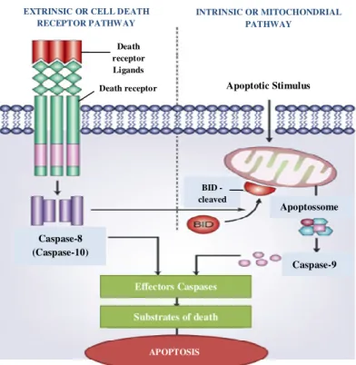
Figure 1 - Illustration of the two major apoptotic signalling pathways. Apoptosis can be
initiated by two alternative pathways: via death receptors on the cell surface (extrinsic pathway) or by mitochondria (intrinsic pathway). In both pathways, induction of apoptosis leads to the activation of an initiator caspase. The two pathways are correlated since caspase-8 (extrinsic pathway) activates the intrinsic pathway for cleavage of Bid, Bcl2 family member.
Adapted from 121. ... 23
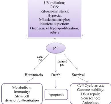
Figure 2 - Action of p53. A wide range of cellular stress stimuli induce p53, leading to co-ordinated changes in gene expression and various biological outcomes, depending on the cell type and the type, intensity and duration of the activating stress. Those events that induce p53 through the DNA-damage-response pathways are highlighted on the left-hand side in lilac. Regulation of some biological events may occur in a homoeostatic manner mediated by basal or low levels of p53. Adapted from122. ... 25
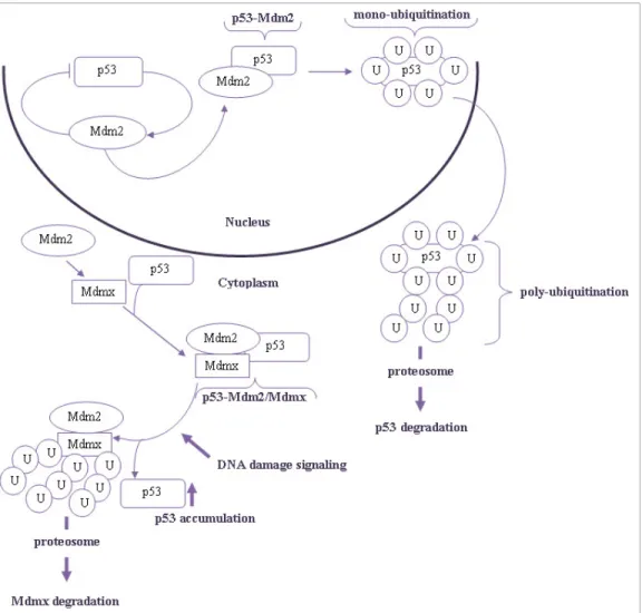
Figure 3 - Schematic diagram of p53 degradation regulated by MDM2 and MDMX. In unstressed conditions, p53 fails to accumulate due to constitutive degradation. Under genotoxic stress, cells activate an evolved response for MDMX degradation that stabilizes p53 and activity. U - ubiquitination. ... 28
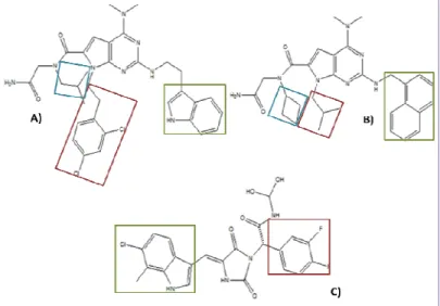
Figure 4 - 4.1) p53 structure (pink) with three hydrophobic residues Phe19 (Green), Trp23 (Yellow) and Leu26 (Orange). Image obtained using the UCSF Chimera software. 4.2) Chemical structures of: A) MI-77301, B) RG-7112, C) TDP665759, D) AMG-232 mimic the three hydrophobic residues Phe19 (Green), Trp23 (Red) and Leu26 (Blue). Image obtained using the ChemDraw software. ... 29
Figure 5 - Chemical struture of: A) and B) pyrrolopyrimidines; C) RO-5963 mimic the three hydrophobic residues Phe19 (Green), Trp23 (Red) and Leu26 (Blue). Image obtained using the ChemDraw software. ... 31
Figure 6 – a) Spirooxindole stucture; b) Pyrazoline structure. ... 35
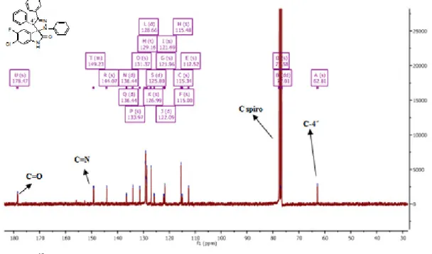
Figure 7 - 13C NMR spectra of compound 2g. ... 39
Figure 8 - 1H NMR spectra of compound 2g. ... 39
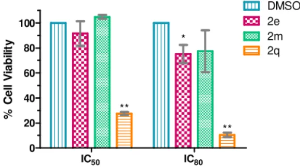
Figure 9 - Evaluation of cell viability in human normal colon fibroblasts (CCD-10co) following 72 h incubation ith compounds 2e, 2m and 2q at equitoxic (IC50 and IC80) concentrations using the MTS metabolism assay. *p<0.05; **p<0.01 0.01 vs respective DMSO control. Data are mean ± S.D. of three independent experiments... 43
Figure 10 - Assessment of death cell induction in human colorectal cancer cell line (HCT116 p53+/+) following 72 hours incubation with compounds 2e and 2m at equitoxic (IC50 and 2xIC50) concentration, or DMSO (vehicle control). Data are mean ± S.D. of three independent experiments. ... 44
15 Figure 11 - Evaluation of apoptosis in human colorectal cancer cell line (HCT116 p53(+/+))
following 72 h incubation with 2e and 2m at equitoxic concentrations (IC50 and 2xIC50), or
DMSO (vehicle control). **p<0.01; ***p<0.001vs DMSO. Data are mean ± S.D. of three independent experiments. ... 45
Figure 12 – Effect of compounds 2e and 2m in p53 and MDM2 protein levels, caspase-3
activation and PARP cleavage. Immunoblots analyzed in total cell extracts of HCT-116p53(+/+) cell line following 72 h incubation at equitoxic IC50 concentration, or DMSO
(vehicle control). Blots were normalized with ponceau staining as control. Cropped blot to exclude two other compounds used from other spirooxindole family.Data are mean ± S.E.M. of two independent experiments. ... 46
Figure 13 - Evaluation of cell cycle progression in human colorectal cancer cell line
(HCT116 p53+/+) following (a) 24 hours and (b) 48 hours incubation with compounds 2e and 2m at equitoxic (IC50) concentration, or DMSO (vehicle control). **p<0.01 vs respective
DMSO control. Data are mean ± S.D. of three independent experiments... 47
Figure 14 - Coefficient drug interaction (CDI) analysis of compound 2e with 5-Fluorouracil.
HCT.116 cells were exposed to compounds for 72h and cell viability was evaluated using the MTS metabolism assay. Threshold- CDI value threshold (CDI=1). **p<0.01; ***p<0.001. Data are mean ± S.E.M. of five independent experiments. ... 48
Figure 15 - The proteins of interest are fused to non-fluorescent fragments (F1 and F2) of a
fluorescent reporter protein. When the proteins of interest interact, the non-fluorescent halves get close enough to fold together and reconstruct the functional fluorophore. When inhibition of p53-MMDM2 interaction exist by an inhibitor molecule (I), occurs a reduced signal. ... 49
Figure 16 - Compound 2e and 2m decreases p53-MDM2 interaction by BiFC. HCT116 p53(– /–)cells were co-transfected for 24 hours. *p<0.05; **p<0.01. Data are mean ± S.D. of three
independent experiments. ... 49
Figure 17 - Evaluation of cell viability in primary mouse astroglial cell culture following 72 h
incubation with compound 2x at equitoxic (IC50 and IC98) concentrations using the MTS
metabolism assay. Data are mean ± S.D. of three independent experiments. ... 50
Figure 18 - Evaluation of apoptosis in GL261 cells, following 72 h incubation with
compound 2x at equitoxic (IC50 and twice of IC50) concentration, or DMSO (vehicle control).
Data are mean ± S.D. of two independent experiments. ... 51
Figure 19 - Evaluation of cell cycle progression in GL261 cells, following (a) 24 hours and
(b) 48 hours incubation with compound 2x at equitoxic (IC50) concentration, or DMSO
(vehicle control). Data are mean ± S.D. of three independent experiments. ... 52
Figure 20 - Evaluation of apoptosis in human colorectal cancer cell line (HCT-116 p53+/+)
16
concentrations, or DMSO (vehicle control). *p<0.05; **p<0.01; ***p<0.001 vs DMSO. Data are mean ± S.D. of three independent experiments. ... 56
Figure 21 - Effect of compounds 7f and 7z in p53 and MDM2 protein levels, caspase-3
activation and PARP cleavage. Immunoblots analyzed in total cell extracts of HCT-116 cell line following 72 h incubation at equitoxic IC50 concentration, or DMSO (vehicle control).
Blots were normalized with ponceau staining as control. Cropped blot to exclude two other compounds used from other spirooxindole family. *p<0.05 vs DMSO. Data are mean ± S.E.M. of two independent experiments. ... 57
Figure 22 - Evaluation of cell cycle progression in human colorectal cancer cell line
(HCT116 p53+/+) following (a) 24 hours and (b) 48 hours incubation with compounds 7f and 7z at equitoxic (IC50) concentration, or DMSO (vehicle control). *p<0.05; **p<0.01 vs respective DMSO (control). Data are mean ± S.D. of three independent experiments. ……..57
Figure 23 – Evaluation of cell viability in human normal colon fibroblasts (CCD-10co)
following 72 hours incubation with compounds 7f and 7z at equitoxic (IC50 and IC80)
concentrations using the MTS metabolism assay. Data are mean ± S.D. of two independent experiments. ... 598
Figure 24 - Evaluation of apoptosis in human breast cancer cell line (MDA-MB-231)
following 72 h incubation with compound 7h at equitoxic (IC50 and 2x IC50) concentrations,
or DMSO (vehicle control). *p<0.05; **p<0.01 vs respective DMSO (control) Data are mean ± S.D. of three independent experiments. ... 598
Figure 25 - Compound 7h induces activation of caspase-3 and -7 after 48 hours of exposition.
Caspase-3/7 activity was measured using the Caspase-Glo 3/7 assay (Promega) in total cell extracts of MDA-MB-231 cell line. **p<0.01 vs DMSO. Data are mean ± S.D. of two independent experiments. ... 60
17 TABLE INDEX
Table 1 – Main MDM´s antagonists57. ... 29
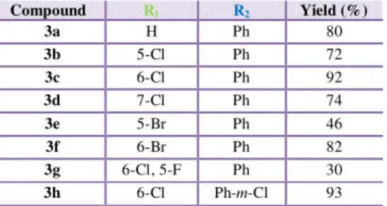
Table 2 - Synthesis of 2-indolinones 3a-3h. ... 37
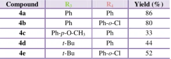
Table 3 - Synthesis of hydrazonyl chlorides 4. ... 38
Table 4 - Yields obtained in the synthesis of spiropyrazoline oxindoles 2a-2x. ... 38
Table 5 -In vitro antiproliferative activities of spiropyrazoline oxindoles 2a-x. ...40
Table 6 –Chemical structure of spiroisoxazoline oxindoles 1 and spiropyrazoline oxindoles 2. ...41
Table 7 - Stability of spiropyrazoline oxindole compounds (2e, 2m and 2x) in PBS. Data of three independent experiments. ... 52
Table 8 - Cell line general characterization. ... 81
Table 9 - Main feature of commercial chemotherapeutic drug used in the course of this project described as drug denomination, molecular weight, solvent, manufacturing company and molecular target. ... 86
18 SCHEME INDEX
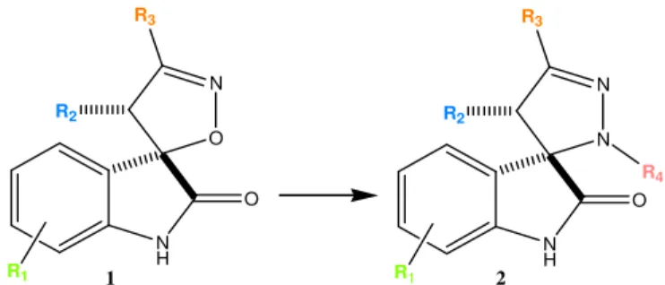
Scheme 1 - Optimization strategy: from spiroisoxazoline 1 to spiropyrazoline 2 oxindoles. .. 35
Scheme 2 - Synthesis of spiropyrazoline oxindoles 2a-x. (a) aromatic aldehydes, piperidine, EtOH in reflux; (b) Et3N/DIPEA, CH2Cl2, r.t. ... 36
Scheme 3 - Synthesis of hydrazonoyl 7 and hydrazonyl chlorides 4. ... 37
Scheme 4 - Chemical structure of spirotryazoline oxindoles 7f, 7h and 7z. ... 55
Scheme 5 - Schematic diagram of compound incubation. ... 83
Scheme 6 - Cytotoxic potential evaluation. The cells per well were seeded in 96-well plate at 37ºC, humidified atmosphere of 99% and 5% (v/v) CO2, and 24 hours later the cells were exposed to compounds A and B for more 72 hours in the same conditions. Then, each well was aspirated and 100 μL of a solution mixture of complete culture medium, MTS and PMS (100:19:1) were pipetted to each well. After 30 minutes of incubation the absorbance at 490nm was read in a microplate reader. ... 87
19
Chapter 1
20 1.1. CANCER: ORIGIN, INCIDENCE AND THERAPEUTIC OPTIONS
The maintenance of cellular homeostasis is essential for tissue integrity in multicellular organisms. In this regard, apoptosis, a form of regulated cell death, is an highly conserved mechanism that has evolved to maintain cell number and cellular positioning within tissues, therefore acting as a natural barrier to cancer development1,2.
Cancer is a multifaceted and multifactorial disease comprised of complex genetic and epigenetic aberrations that destabilize the normal balance of cellular life and death3. The main causes for cancer development are the nutrition habits, specific infections such as Helicobacter pylori, Hepatitis B and C viruses, and Human papillomaviruses, genetic determinants, and others factors4–6. According to the World Health Organization, cancer is a leading cause of death worldwide accounting for 8.2 millions of deaths each year. Female breast and colorectal cancers are among the five most diagnosed cancers and they are also the main contributors for mortality and loss of healthy life4,6.
In terms of the therapies available, they differ according to the type of cancer. Overall, surgery, chemotherapy, and radiation therapy are the most common treatments used against cancer. Chemotherapy usually refers to the use of medicines or drugs as the therapeutic agent and act throughout the body, meaning that it can kill cancer cells that have spread (metastasized) in parts of the body away from the original (primary) tumour7. Surgery and
radiation therapy remove, kill, or damage cancer cells in a certain area. These three types of therapy can and are currently used in combination for better efficiency of therapy8,9.
More recently, new forms of cancer treatment have emerged, including targeted therapy that uses drugs or other substances to more precisely attack cancer cells causing minor damage to normal cells, and immunotherapy that relies in the use of an individual’s own body immune system to tackle cancer. Immunotherapy may function in different ways: it can stimulate the immune system, or it can help the immune system to attack cancer cells. Finally, immunotherapy may also be used in combination with other types of treatment7.
Cancer research has been a topic of great interest for the past decades; however, investment in cancer control, prevention, diagnosis and treatment is still an urgent need5.
1.1.1. Colorectal Cancer
Colorectal cancer (CRC) is one of the most commonly diagnosed cancers and presents the highest cause of cancer deaths10,11. CRC develops in the colon or in the rectum, also known as the large intestine.
Most of the times, CRC begins as a noncancerous growth called polyp or adenoma that develops on the inner lining of the colon or rectum. Although all adenomas have the capacity to become cancerous, fewer than 10% are estimated to progress to invasive cancer. Cancer
21
that develops in glandular cells is called adenocarcinoma, which is approximately 96% of colorectal cancers12.
Surgery is one of the primary options for CRC treatment, often in combination with radiation therapy and chemotherapy. The most often used drugs for colorectal cancer include: 5-Fluorouracil (5-FU); Capecitabine, which is in pill form and once in the body is converted to 5-FU; Irinotecan; Oxaliplatin; and Trifluridine and tipiracil (Lonsurf), a combination drug in pill form. Sometimes, two or more of these drugs are combined to achieve more efficacy.
Targeted therapy drugs work differently from standard chemotherapy drugs. Sometimes they are effective when standard chemotherapeutics drugs are not, and they often have different and less severe side effects. Currently, the most commonly used target cells with EGFR changes and the blood vessel formation (VEGF) where the Cetuximab (Erbitux) and the Bevacizumab (Avastin) are used. These drugs can be used in combination with chemotherapeutic drugs or alone if chemotherapeutic drugs are no longer effective13.
1.1.2. Breast Cancer
Breast cancer is a heterogeneous disease with a wide spectrum of clinical, pathologic, and molecular features. Although it is much more frequent in women, men can also develop the disease14. The majority of breast cancers begin in the lobules and ducts where the mammary
glands are located15,16. Breast cancer can be defined as in situ or invasive depending on its extension.
In situ breast cancer
Ductal carcinoma in situ (DCIS) is when abnormal cells replace the normal epithelial
cells of the breast ducts. It is considered a non-invasive form of breast cancer and is the most common type of in situ breast cancer, representing about 83% of diagnosed cases. DCIS may or may not progress to invasive cancer and, in fact, some of these tumours grow so slowly that they may not affect the woman’s health even without treatment.
Lobular carcinoma in situ (LCIS), also known as lobular neoplasia, generally is not
considered a precursor of invasive cancer. Instead, it represents an indicator for increased risk for developing invasive cancer. LCIS accounts for about 13% of all female in situ breast cancers diagnosed.
Invasive breast cancer
The majority of breast cancer cases are invasive, or infiltrating, meaning that they have broken through the walls of the glands or ducts where they were originated, and have grown
22
into the surrounding breast tissue. Based on comprehensive gene expression profiling, breast tumours are classified into several subtypes: luminal and human epidermal growth factor receptor 2+ (HER2+), depending on if they express hormone receptors (HR+) and HER2, respectively15,16. Approximately 74% of all breast cancers are Luminal A (HR+/HER2-). These tumours tend to grow slowly and are less aggressive than other subtypes, probably because they are responsive to hormonal therapy16,17. Conversely, the triple negative tumours (HR-/HER2-) are the most aggressive, in part due to lack of specific therapies16,17.
The majority of breast cancers are treated surgically, which can be often combined with radiation therapy, chemotherapy, hormonal (endocrine) therapy, and/or targeted therapy16,18.
The benefit of different types of therapies depend on multiple factors, such as the size of the tumour, the number of lymph nodes involved, the presence of estrogen or progesterone receptors, and the amount of HER2 protein produced by cancer cells. For example, the triple negative and HER2+ breast cancers tend to be more sensitive to chemotherapy. Some examples of chemotherapeutics used are Paclitaxel, Carboplatin, 5-fluorouracil, Docetaxel among others16,18.
There is yet, targeted therapy which is divided in two principal targets: targeted therapy aimed at HER2-positive where Trastuzumab, Pertuzumabre are used and targeted therapy for hormone receptor-positive using Everolimus and Palbociclib16,18.
1.1.3. Glioblastoma
The brain is the most complex organ in vertebrates. It is composed primarily of two broad classes of cells, neurons and glial cells, which can develop into different types of tumours. The majority of brain tumours develop from glial cells that include astrocytes, oligodendrocytes, microglia and ependymal cells. Therefore, glioma is a generic term used to describe the different types of glial tumours: astrocytoma, oligodendroglioma, and glioblastoma. The most frequent and malignant of all is glioblastoma19.
All the types of treatment referred before can be used in brain and spinal cord tumours. Each treatment is based on the type of tumour and other factors, and often, more than one type of treatment is used. Some of the chemotherapeutic drugs used to treat brain tumours include cisplatin, irinotecan and others, such as temozolomide. For example, temozolomide acts by inducing cytotoxicity through pro-autophagic processes and also through the induction of apoptosis. Of note, autophagy is a type II programmed cell death that represents an alternative mechanism to overcome, at least in part, the resistance of many cancers to apoptosis-related therapies. On the other hand, targeted therapy targets vascular endothelial growth factor (VEGF) and (mammalian target of rapamycin) mTOR18,20,21.
23 1.2. CANCER BIOLOGY: PATHWAYS AND MECHANISMS
Tumourigenesis is a multistep process of acquiring successive genetic mutations that transform a normal cell into a cancer cell. These mutations give rise to hyperproliferative signals that sustain and enable the immortality of cells through an unlimited replicative potential, the invasion and metastization ability, the insensitivity to growth suppression signals, the resistance to cell death and the induction of tumour angiogenesis2,22. Deregulated
expression of transcription factors during tumourigenesis is essential for the promotion of proliferation and differentiation of a neoplastic population22.
1.2.1. Apoptosis
Cell death by apoptosis, is the principal strategy to control tumour development, in which cells receive specific stimuli to die. These stimuli are part of an important program of cell growth control, characterized by morphologic as well as biochemical changes. Apoptosis can occur by two major signalling pathways, the death receptors or extrinsic pathway, and the mitochondrial or intrinsic pathway. Overall, these two pathways are distinct, but ultimately converge to the same mechanism. The apoptotic process culminates in activation of a series of cysteine aspartyl-specific proteases, or caspases, that cleave key cellular proteins like polyADP-ribose polymerase (PARP) (Figure 1). Activation of caspases is considered the point of no-return, in which cell death becomes an irreversible process23,24.
Figure 1 - Illustration of the two major apoptotic signalling pathways. Apoptosis can be initiated by two
alternative pathways: via death receptors on the cell surface (extrinsic pathway) or by mitochondria (intrinsic pathway). In both pathways, induction of apoptosis leads to the activation of an initiator caspase. The two pathways are correlated since caspase-8 (extrinsic pathway) activates the intrinsic pathway for cleavage of Bid, Bcl2 family member. Adapted from 116.
EXTRINSIC OR CELL DEATH RECEPTOR PATHWAY INTRINSIC OR MITOCHONDRIAL PATHWAY Apoptotic Stimulus Apoptossome BID - cleaved Effectors Caspases Substrates of death Caspase-9 Caspase-8 (Caspase-10) APOPTOSIS Death receptor Ligands Death receptor
24 Extrinsic or Cell Death Receptor Pathway
The extrinsic or cell death receptor pathway is initiated by extracellular signals, resulting in binding of ligands to specific transmembrane receptors, collectively known as death receptors, belonging to the tumour necrosis factor (TNF) family. The death receptors are cell surface receptors that transmit apoptotic signals from the cell surface to activate intracellular signalling pathways25.
All death receptors work in a similar manner, via conformational changes that allow the assembly of a large multiprotein complex known as DISC (Death-Inducing Signalling Complex), ultimately leading to the activation of the caspase cascade. Pro-caspases are recruited to the DISC and subsequently are cleaved to result in active initiator caspases, including caspase-8 and -10, this initiator caspases have the ability to cleave and activate effectors caspases, such as caspase-3 and -7. Importantly, the extrinsic pathway is able to crosstalk with the intrinsic or mitochondrial pathway, thus amplifying the death signal25–27.
Intrinsic or Mitochondrial Pathway
The intrinsic apoptotic pathway is initiated by a non-receptor mediated stimuli that triggers intracellular signalling. Mitochondria activation in the intrinsic pathway is mediated by the Bid protein, a pro-apoptotic member of the Bcl-2 family. Bid is cleaved indirectly by p53 and directly by active caspase-8, and subsequently translocated to the mitochondria. The presence of cleaved Bid in the mitochondrial membrane regulates mitochondria permeabilization. When an apoptotic stimuli reach the mitochondria, a variation of the inner mitochondrial membrane potential occurs, as well as changes in the membrane permeability, leading to the passage of water and consequently organelle’s rupture. Consequently, mitochondria release pro-apoptotic factors into the cytoplasm, including cytochrome c, which together with the destabilization of the membrane potential leads to loss of cellular homeostasis, disruption of ATP synthesis and increased reactive oxygen species (ROS) production. The increase of ROS levels then leads to lipid, protein and nucleic acid oxidation by increasing the collapse of the inner mitochondrial membrane potential. Cytosolic cytochrome c also interacts with apoptotic protease activating factor 1 (Apaf-1) and pro-caspase 9, in the presence of ATP, to form a complex known as apoptosome. After that, caspase-9 is activated and subsequently activates effector caspases for the execution of apoptosis25–28.
1.3. p53 – THE TUMOUR SUPPRESSOR PROTEIN AND TUMOURIGENESIS
p53 tumour suppressor protein is regarded as a key player in tumour suppression, as it promotes growth arrest, apoptosis and cellular senescence, while also blocking angiogenesis29,30. Strikingly, half of all human tumours carry p53 inactivated by mutation 30–
25
inactivation of other signaling components, such as p53 endogenous negative regulators
murine double minute 2 (MDM2) and/or MDMX 29,32,33.
In some cases, frameshift or nonsense mutations result in the loss of p53 protein expression. However, missense mutations that lead to the substitution of a single amino acid in the p53 protein, can be stably expressed in tumours cells resulting in novel functions for p5334,35. These substitutions may occur throughout all the structural domains of p53 protein,
but they are more frequently clustered within the DNA binding region of p53. These mutations generally lead to a loss or diminution of the wild-type activity of p5334. Although
mutations have been detected in almost all of the p53 amino acids, a number of hotspot sites have been identified, especially at residues R175, G245, R248, R249 and R27333,36.
p53 has been called "guardian of the genome", due to its central role in coordinating the cellular response to a wide range of cellular stress37. One of the main functions of p53 is as transcription factor that both activates and represses an ample range of target genes37. The
regulation of p53 function has been described at multiple levels and consists in an array of post-translational modifications, that lead to protein accumulation and increase of its transcriptional activity, both during normal homeostasis as in stress-induced responses31,37 (see Figure 2). Even though, the key mechanism by which p53 is regulated is through control of its protein stability, mediated primarily by MDM2.
Figure 2 - Action of p53. A wide range of cellular stress stimuli induce p53, leading to co-ordinated changes
in gene expression and various biological outcomes, depending on the cell type and the type, intensity and duration of the activating stress. Those events that induce p53 through the DNA-damage-response pathways are highlighted on the left-hand side in lilac. Regulation of some biological events may occur in a homoeostatic manner mediated by basal or low levels of p53. Adapted from117.
26
As the p53 is frequently mutated or inactivated in cancer, this tumour suppressor has turned into a highly attractive therapeutic target for the treatment of the disease. Several studies have shown that p53 interaction with MDM2 and MDMX involves three main hydrophobic amino acids (Phe19, Trp23 and Leu26) in the p53 protein38–40. In case of cellular
stress, these interactions should be inhibited to activate the p53 response31. For that purpose, small molecules that mimic the three amino acid residues of p53-MDMs binding pocket have been widely developed and tested as potential antitumour agents38.
Further, as mutant p53 has been reported to confer drug resistance in different cancer therapies, its reactivation or neutralization can be beneficial in this context33.
1.4. NEGATIVE REGULATORS OF p53
Protein-protein interactions (PPIs) have an important role in cancer because they regulate various cellular processes, particularly in cell cycle or cell signalling pathaways41.
The small molecule inhibitors of protein-protein interactions have received considerable interest in the last years. In particular, the inhibitory interaction of p53 with regulatory proteins, MDM2 and MDMX, provided a focus of effort due to its importance in various types of cancer, and the prospect that inhibition of these interactions may provide new therapies42.
The MDM´s proteins are present at high levels and inhibit activity p53 in many human cancers. Thus, interrupting the p53-MDM interaction to reactivate p53 is a valuable therapeutic strategy for the treatment of tumours43.
Several studies currently focus on the design of small inhibitory molecules of both MDM´s (MDM2 and MDMX). Despite the efforts, there are no compounds in clinical stages for this purpose, since existing compounds for inhibiting MDM2 are inefficient in the inhibition of MDMX44. This problem derives, at least in part, from the "sub-pocket" Leu26 that represents the greatest challenge to design a dual inhibitor of MDM2/MDMX. The two proteins significantly differ in shape and in the properties of the residues surrounding the cavity39,44–46.
Therefore, the development of specific inhibitors for MDMX must take into account their unique structural properties that go beyond this "simple" difference. The well-defined binding furrows in MDM´s suggest the feasibility of designing small-molecule inhibitors to block their interaction with p5339.
1.5. p53-MDM2/MDMX PROTEIN-PROTEIN INTERACTION
The information of diverse proteins correlating their function in the tumour environment is emerging as an important aspect of studying protein function in cancer. The structural
27
changes of protein in the course of mutation with its altered cellular localization is a important point to understand better the progression of the disease22.
MDM2 belongs to the family of E3 ubiquitin ligase that contains a RING (really interesting new gene) domain and serves as the major ubiquitin ligase for p53 degradation32,39,47.
The MDM2 protein binds directly to the N-terminal domain of p53, thus inhibiting its transcriptional activity through multiple mechanisms including, direct occlusion of p53 transactivation domain, induction of p53 nuclear export and target of p53 for proteasomal degradation32,39. However, recent research has demonstrated that MDM2 alone is
insufficient to suppress p53 activity32.
Under normal conditions, MDM2, which is itself a product of a p53 target gene, inhibits the activity of p53 by physical interaction48. This inhibition is accomplished through a negative autoregulatory feedback loop in which p53 activates the transcription of its own inhibitor, as mentioned above. Therefore, in non-stressed cells, the low level of MDM2 mediate multiple mono-ubiquitination of p53, promoting its nuclear export29,37,49 (Figure 3).
When cells are challenged by a stress insult, p53 levels are rapidly up-regulated to enable repair. Meanwhile, the expression levels of MDM2 in cells under stress are also higher promoting the poly-ubiquitination and degradation of p53 after the repair has occurred. In the cytosol, mono-ubiquitinated p53 induces Bax oligomerization followed by mitochondrial translocation, eventually leading to the release of cytochrome c and other pro-apoptotic molecules into the cytosol. These mechanisms show that p53 ubiquitination by MDM2 is another layer of p53 activity regulation31,37,49, further demonstrating the importance of
p53-MDM2 binding as a therapeutic target in cancer.
MDM2 also interacts with its equivalent MDMX through the RING domain31.
In the same way that MDM2, MDMX is an important negative regulator of p53. MDMX is a homologue of MDM2, in particular in the domain that binds to p53. Nevertheless, both proteins present enough differences to turn Mdm2 inhibitors ineffective in inhibiting p53-MDMX interaction. It has been demonstrated that MDM2 regulates p53 mainly by protein degradation, whereas MDMX is a potent inhibitor of its transcriptional activity50. In fact,
unlike MDM2, MDMX does not have appreciable ubiquitin ligase activity44. Another key difference between Mdm2 and MDMX is that MDMX is primarily located in the cytoplasm, while MDM2 is located in the nucleus. MDMX nuclear translocation may occur to form the complex MDM2/MDMX and inhibit p5331. The formation of MDM2/MDMX heterodimers through the RING domains participate in p53 poly-ubiquitination and proteasomal degradation. In response to DNA damage signals, MDM2/MDMX heterodimers are poly-ubiquitinated, resulting in degradation of the complex and consequent p53 stabilization and activation45,49 (Figure 3).
28
In addition to having its own effects on p53, MDMX also plays an important role in stabilizing the MDM2 protein. Taking into account the role of MDMX in inhibiting p53, several molecules are being developed for MDMX inhibition.
In Table 1, are presented the main inhibitors developed for inhibition of the interactions between p53 and MDM´s proteins (Table 1).
Figure 3 -Schematic diagram of p53 degradation regulated by MDM2 and MDMX. In unstressed conditions, p53 fails to accumulate due to constitutive degradation. Under genotoxic stress, cells activate an evolved response for MDMX degradation that stabilizes p53 and activity. U - ubiquitination.
29 Table 1 – Main MDM´s antagonists51.
1.6. INHIBITORS OF p53-MDM2 INTERACTION
To date, there are over 20 different chemotypes that have been shown to antagonise the p53-MDM2 interaction, such as cis-imidazolines52 (eg, RG-7112), benzodiazepinediones53 (eg, TDP665759), spiropyrrolidine oxindoles54 (eg, MI-77301), and piperidinones55 (eg, AMG-232) (see Figure 4.2). Although these compounds share different scaffold structures, they all inhibit the p53-MDM2 interaction by mimicking the three hot spots residues of p5339 (figure 4.1).
1.6.1. Cis-Imidazolines (Nutlins)
The nutlins (cis-imidazolines) were identified as the first class of potent, non-peptide and specific small molecule MDM2 inhibitors, from a large chemical screen and chosen for their potency and selectivity for inhibition of the p53-MDM2 interaction35,39. Nutlin-3a, arrests proliferating cancer cells in the G1 and G2 cell cycle phases as well as induces apoptosis in
Strategy Compound
Interruption of p53 interaction with MDM2
Small molecules that bind the MDM2 N_ pocket: Nutlin-3a/RG7112, MI-219, MI-63, MI-319, MI-773, AM-8553, AMG-232, benzodiazepinediones, MK-8242 Small molecules that bind the p53 N-terminal domain: RITA
Interruption of p53 interaction with
MDMX Small molecules: WK 298, SJ-172550
Interruption of p53 interaction with both MDM2 and MDMX
Peptidic compounds: SAH-p53s, PMI peptide, pDI peptide
Small molecules: RO-5963, CGM097
Figure 4 - 4.1) p53 structure (pink) with three hydrophobic residues Phe19 (Green), Trp23 (Yellow) and Leu26
(Orange). Image obtained using the UCSF Chimera software. 4.2) Chemical structures of: A) MI-77301, B) RG-7112, C) TDP665759, D) AMG-232 mimic the three hydrophobic residues Phe19 (Green), Trp23 (Red) and Leu26 (Blue). Image obtained using the ChemDraw software.
30
wild-type p53 dependent manner, in a number of different cancer cell lines expressing wt-p53 including colorectal, lung, breast, prostate, melanoma, osteosarcoma and renal cancer35,56.
Further structure-based optimization of nutlin-3a led to compound RG-7112, the first MDM2 inhibitor advancing into clinical trials. In fact, RG-7112 presented enhanced binding affinity to MDM2 and more potency to inhibit wild-type p53 tumour growth35,39,57.
1.6.2. Benzodiazepinediones
The Benzodiazepinediones disrupt the p53-MDM2 interaction in a similar manner to the nutlins, by mimicking the action of the key amino acids involved in the binding of the p53 peptide to MDM235. One of these compounds is TDP66575953, which binds to the binding
cleft of MDM2, predominantly hydrophobic, and occupies the same pockets as the peptide side chains Phe19, Trp23, and Leu26 of p53. MDM2 interacts with the inhibitor through
nonspecific Van Der Waals contacts58.
1.6.3. Piperidinones
Compound AMG-232, was obtained by a structure-based design and wide optimization of a new class of MDM2 inhibitors containing a piperidinone scaffold. AMG-232 has entered in phase I clinical trials for human cancer treatment in 2012. In this compound the 3-chlorophenyl and 4-3-chlorophenyl groups are project into the Leu26 and Trp23 pockets,
respectively. The Phe19 pocket is occupied by the ethyl group. The sulfonyl tert-butyl group
resides in the small pocket around G58, and the carboxylic acid forms a salt bridge with the
His96 residue of MDM2, increasing the inhibitory effect of the compound on MDM2. Thus,
AMG-232 not only mimics the three key p53 residues but captures additional interactions to achieve a very high binding affinity with MDM257,59.
1.6.4. Spiropyrrolidine oxindoles
In the past years, several classes of small molecule inhibitors that disrupt p53-MDM2 binding, such as MI-219, have been revealed. Extensive optimization of spiropyrrolidine oxindoles family has yielded SAR405838 (MI-77301). This compound is in phase I of clinical trials for neoplasm malignant60. Before going to clinical trials, its ability to inhibit cancer cell proliferation and to inhibit tumour growth in xenograft models was reported. A good tissue bioavailability in mice, a desirable pharmacokinetic profile and an orally activity, were also reported for MI-7731054.
31 1.6.5. Dual Inhibitors
The need for a compound with a double ability for MDM2 and MDMX, led to the discovery of compound, RO-2443 that showed potent MDM2/MDMX inhibitory activity in
vitro, but poor water solubility. Optimization of RO-2443 yielded RO-5963, a close analogue
with slightly increased potency but substantially improved solubility. RO-5963 (figure 6) also showed p53-MDM2 inhibitory activity similar to that of nutlin-3a and p53-MDMX inhibitory activity was nearly equivalent to MDM2 activity but approximately 400-fold better than the MDMX potency of nutlin-3a39,44.
Studies have shown that RO-5963, inhibits cell growth, stabilizes p53 in a dose-dependent manner, and elevates protein levels of its transcription targets (p21 and MDM2). The RO compounds binding method is really interesting because, sub-pocket Leu26 is not occupied by these compounds. They have a very high affinity to both MDM2 and MDMX proteins, through their chloro-indole and di-fluoro-phenyl moieties, which fill Phe and Trp sub-pockets, respectively. These compounds also show, a high activity in cellular line cancer cells lines with wt-p5344.
A new class of molecules that mimic p53 α-helix, the pyrrolopyrimidines (Figure 5), and show effect on both MDM2 and MDMX proteins. These new molecules mimic α-helixes, secondary structures of most common proteins, which are involved in several protein-protein interactions (PPI). Pyrrolopyrimidines have several advantages over the majority of structures known as PPI inhibitors, such as improved aqueous solubility, excellent cell permeability and synergistic effect over MDM2 and MDMX61.
Figure 5 - Chemical struture of: A) and B) pyrrolopyrimidines; C) RO-5963 mimic the three hydrophobic residues Phe19 (Green), Trp23 (Red) and Leu26 (Blue). Image obtained using the ChemDraw software.
32
Chapter 2
33
The main objective of the studies presented in this thesis was the development and optimization of new anticancer agents that potentially target the molecular interaction between p53 tumour suppressor and its main negative regulators, MDM´s. Our strategy involved not only the chemical synthesis, but also the biological activity evaluation of the newly synthesized compounds, using several cellular models.
Primarily, we aimed at developing a novel spirooxindole scaffold containing a pyrazoline ring that functions as the rigid heterocyclic scaffold, from which the three lipophilic groups can be projected to mimic the p53 amino acids.
Secondly, we decided to study the biological effect of changing the CH-4´ (pyrazoline) by a N-4´ (tryazoline) on the five-member ring of the spirooxindole skeleton.
In parallel, we performed a broad range of molecular and biochemical studies to dissect the cellular mechanisms triggered by the most potent compounds. More specifically, our goals were to:
- Evaluate the antiproliferative capacity of the compounds in several cancer cell lines as well as in normal cells;
- Evaluate their ability to induce cell death, in particular apoptosis, and/or cell cycle arrest;
- Determine whether the compounds inhibit p53-MDM2 interaction;
- Evaluate the potential synergistic effect between the most potent molecule and a chemotherapeutic drug already in use;
34
Chapter 3
SPIROPYRAZOLINE OXINDOLES:
DESIGN AND EVALUATION
N H O R2 R1 + R3 N H N Cl R4 N H N N R1 R2 R4 R3 O
35
Spirooxindoles are reported as the core structure of a variety of medicinal agents with several applications, such as anticancer agents62. These molecules have a big potential because the oxindoles moiety can mimic the p53 Trp23 sub-pocket. Another advantage of
spirooxindoles, is that some derivatives were described to have specificity against cancer cell lines and not against normal epithelial cells35,57,63–66. This chemical structure is considered an important heterocyclic molecule, which belongs to a large family of bioactive compounds, such as compounds MI-77301 and RO-535365,67.
Pyrazoline is also a privileged unit in medicinal chemistry, and is described to have anti-viral, anti-tumour, antibacterial, anti-inflammatory, and anti-fungal activities68.
These two units are structural parts of our compounds, the spiropyrazoline oxindoles (Figure 6).
Figure 6 – a) Spirooxindole stucture; b) Pyrazoline structure.
In the last years, Santos’s group (iMed.ULisboa) has been investigating the potential of several families of spirooxindoles with 5-membered rings as potential anti-cancer agents. Previous work from the research group shown that spiroisoxazoline oxindoles 169 and spiropyrazoline oxindoles 270 have in vitro anticancer properties.
Spiroisoxazoline oxindoles 1 presented ability to disrupt the p53-MDM2 interaction, as determined by a Venus-based bimolecular fluorescence complementation assay (BiFC). However, the IC50 values were very high compared with other spirooxindoles described in the
literature (around 40µM for the colorectal cancer cell line HCT-116). Computational studies showed that the optimal Trp23 mimicry by the oxindole was lost due to the spatial orientation
of the groups: oxindole R2 and R3. Instead, these molecules, the oxindole is projected into
Phe19(p53) pocket, while aromatic substituent’s R2 and R3 are projected into Trp23(p53) and
Leu26(p53) pockets, respectively71. However, the inclusion of a fourth substituent to the main
Scheme 1 - Optimization strategy: from spiroisoxazoline 1 to spiropyrazoline 2 oxindoles.
N H O N R1 R2 R3 O N H N N R1 R2 R4 R3 O 1 2 N H R R R R O R NH N a) b)
36
scaffold and the substitution of the isoxazoline oxygen atom for a N-Ar group could allow a potential reorientation of the ligands in the MDM2 pocket (Scheme 1).
In this thesis, a family of spirooxindoles containing a five ring (pyrazoline) was synthesized and the antiproliferative activity evaluated. Previously, some spiropyrazoline oxindoles 2 were tested against the MCF7 and MDA-MB-231 breast cancer cell lines, and the most active compounds showed an IC50 value around 7µM, and were selective over
MDA-MB-231 tumour cells and non-cytotoxic against Hek-293T non-tumour cells. Therefore, in this work, we developed and optimized the previously developed spiropyrazoline oxindoles and assessed their biological effects.
0.1. SYNTHESIS OF THE TARGET COMPOUNDS
p53 interacts with MDM2 through the residues Phe19, Trp23 and Leu26 that occupy the
hydrophobic cleft of the MDM2 protein. These three residues represent the critical ones for binding between these two proteins. Having this into account 2-indolinones 3a-h and hydrazonyl chlorides 4a-e, containing substituent’s to mimic these important aminoacids, were synthesized (Tables 2 and 3). The R1 in indole part of the compounds mostly presents
halogens as the main choice. This choice was based on previous reports of potent MDM2 inhibitors (example MI-77301, SM13) that contain halogens in the indole moiety. The R2, R3
and R4 were synthesized taking into account the ability of an halogenated 5-membered
aromatic ring to insert into Phe19 pocket, and an often branched aliphatic moiety or an often
halogenated 5-membered aromatic ring to occupy the Leu26 pocket65,72.
Spiropyrazolines´ 2 have a basic molecular structure derived from the corresponding pyrazoline and are exemplified by a unique spiro junction at the C-5 position of a pyrazoline ring. The 1,3-dipolar cycloaddition is a very useful reaction for the synthesis of spirooxindole ring systems62,66,73–75.
The synthetic strategy employed to synthesize spiropyrazolines oxindoles 2a-x is depicted in scheme 2. The final spiropyrazoline oxindoles 2 were obtained by 1,3-dipolar cycloaddition between 2-indolinones 3a-h and nitrile imines 5 (generated in situ by dehydrohalogenation of hydrazonyl chlorides 4a-e) (Scheme 2).
N H O R2 R1 R3 N H N R4 Cl + N H O R1 N N R3 R4 R2 N N R4 R3 + _ N H R1 O a b 6 3 4 2 5
Scheme 2 -Synthesis of spiropyrazoline oxindoles 2a-x. (a) aromatic aldehydes, piperidine, EtOH in reflux; (b) Et3N/DIPEA, CH2Cl2, r.t.