Universidade de Trás-os-Montes e Alto Douro
Systemic factors and tumour progression
Tese de Dissertação de Mestrado em
Genética Molecular Comparativa e Tecnológica
Catarina Pinheiro
Orientação: Professor Doutor Sérgio Dias
Co-orientação: Professora Doutora Raquel Chaves
iii
Universidade de Trás-os-Montes e Alto Douro
Systemic factors and tumour progression
Tese de Dissertação de Mestrado em
Genética Molecular Comparativa e Tecnológica
Catarina Pinheiro
Orientação: Professor Doutor Sérgio Dias
Co-orientação: Professora Doutora Raquel Chaves
Composição do Júri:
Vila Real, 2019 Paula Filomena Martins Lopes
Maria dos Anjos Clemente Pires
v
Este trabalho foi expressamente elaborado com vista à obtenção de grau de Mestre em Genética Molecular Comparativa e Tecnológica.
vii
Acknowledgments
"If not now, when? If not me, who?”
Em primeiro lugar, quero agradecer ao Sérgio por me ter recebido no seu laboratório, por me ter integrado, e pela sua paciência e disponibilidade desde o primeiro dia. Muito obrigada!!
Aos meus colegas de laboratório por tornarem os dias mais fáceis, pela ajuda, disponibilidade e carinho, A TODOS sem excepção!
Em especial, à Ana Magalhães pelo todo (IMENSO) apoio e suporte (SOS) durante todo o tempo!
Ao António, porque tornou os dias sempre mais fáceis e ao LCosta Lab em geral pelo fantástico convívio, ajuda e partilha.
Ao IMM em geral, e todos aqueles com quem partilhei e aprendi, a todos, pela ajuda! À Professora Raquel Chaves, co-orientadora, pela disponibilidade e ajuda.
E porque eu acredito que somos um pouco de todos aqueles que se cruzam no nosso caminho, dedico este trabalho:
À minha mãe e ao meu pai, que se superaram todos os dias para que tudo acontecesse. Pelos sacrifícios, pela compreensão, pela liberdade e responsabilidade de sempre me deixarem decidir todos os meus passos e me apoiarem sempre independentemente da vontade deles.
Aos meus irmãos, porque sem eles não teria sido possível. Porque sem eles não seria 1/10 daquilo que sou, porque me ajudaram a crescer, porque me ensinaram os valores que para mim são essenciais, mas acima de tudo porque sempre foram exemplos para mim! Aos meus sobrinhos, Inês, Salvador, Santiago e Francisca, porque somos o reflexo daquilo que sentimos e amamos e seria impossível quantificar o quanto cada um deles altera a minha vida, e o quanto me fazem feliz.
Aos meus amigos simplesmente por estarem presentes mesmo aqueles que aparentemente ausentes ou distantes, porque sei sempre que estão lá! No entanto, quero individualizar aqueles que fiz perder imenso tempo para me acompanharem nesta aventura.
viii
À Lita porque fez metade daquilo que eu deveria ter feito, corridas sem fim, idas à Universidade, foi o meu segundo eu, sempre lá! Às vezes penso que exagero, mas sei que ela sabe, que eu não faria igual, faria mil vezes mais se fosse preciso! Obrigada!!
À Taninha porque me acompanhou sempre nas horas “extra” e nas corridas sem fim fora de horas. Pela paciência de ouvir as minhas frustrações científicas, e por ouvir mesmo quando não percebia nada.
À Cli, porque a amizade vai muito além das pessoas que por “imposições” da vida se cruzam no teu caminho e fazem parte do teu dia-a-dia. Mas podem passar dias, meses ou anos que há amizades que não mudam!
À Dé, João e Figueira porque obrigados a fazer parte da minha rotina, se tornaram parte importante do meu tempo e estiverem sempre presentes!
“Só vives uma vez, mas se souberes viver bem, uma vez é o suficiente.” Joe E. Lewis
ix
“Nothing in life is to be feared, it is only to be understood. Now is the time to understand more, so that we may fear less.”
xi
Resumo
Embora seja claro que a proliferação celular, por si só não causa cancro, a proliferação celular sustentada em um ambiente rico em células inflamatórias, factores de crescimento, estroma activado e agentes promotores de danos no DNA, certamente potencia, promove, o risco de desenvolvimento de neoplasias. Na verdade, há uma ligação estabelecida entre inflamação crónica e um risco mais elevado de desenvolvimento de cancro.
Um dos tipos celulares relevantes num contexto inflamatório são os macrófagos; estes possuem inúmeras funções relacionadas com a remodelação tecidual, inflamação, imunidade, e têm a capacidade de afectar o desenvolvimento e a progressão tumoral. No cancro, crê-se que os macrófagos possam desempenhar papéis aparentemente antagónicos. Assim, os macrófagos podem impedir o estabelecimento e a disseminação de células tumorais mas, por outro lado, podem executar funções para apoiar o crescimento e a disseminação do tumor. São células-chave na inflamação crónica e são células altamente versáteis, capazes de exibir fenótipos distintos e programas funcionais em diferentes tecidos. E tal heterogeneidade depende da sua capacidade de se submeter a diferentes formas de activação em resposta a sinais distintos, em particular, microambientes. Classicamente, os estados de polarização, são definidos com base no tipo de estimulação in
vitro, padrão de expressão da molécula de superfície, perfil secretor e propriedades
funcionais: M1 e M2 representam os extremos de um “continuum” de estados de activação. Os macrófagos M1 (classicamente activados) exibem funções inflamatórias, enquanto os macrófagos M2 (alternativamente activados) exibem funções anti-inflamatórias.
No caso de tumores, os macrófagos são recrutados a partir da circulação periférica por quimiocinas e geralmente são polarizados em direcção a um fenótipo M2.
Neste trabalho, testámos a hipótese de que interacções dos macrófagos com células endoteliais activas, possam influenciar a diferenciação M1/M2. As interacções de células endoteliais activas com células imunes são determinantes importantes da biologia do tumor, com as células inflamatórias a desempenhar papéis bem reconhecidos na progressão do tumor.
Com este trabalho, observamos um perfil semelhante de fenótipo M2 nas células CD11b+ aderentes após cocultura com células endoteliais activas. A nossa hipótese é que células endoteliais activas podem ser críticas para a polarização dos macrófagos num
xii
fenótipo M2, possivelmente pela activação da via de sinalização Notch, e pela expressão de Jagged1 e Notch1, respectivamente.
Palavras-chave: Macrófagos; Polarização de macrófagos; M1/M2; Células endoteliais
xiii
Abstract
Although it is clear that proliferation of cells alone does not cause cancer, sustained cell proliferation in an environment rich in inflammatory cells, growth factors, activated stroma, and DNA-damage-promoting agents, certainly potentiates and/or promotes the risk to develop neoplasia. In fact, there is an established link between chronic inflammation and a higher risk of developing cancer.
One of the relevant cell types in an inflammatory context is macrophages; these have innumerable functions related to tissue remodelling, inflammation, immunity, and have the ability to affect tumour development and progression.
In cancer, it is believed that macrophages may play apparently antagonistic roles. Thus, macrophages can prevent the establishment and spread of tumour cells but, on the other hand, may perform functions to support tumour growth and dissemination. They are key cells in chronic inflammation and are highly versatile cells capable of displaying distinct phenotypes and functional programs in different tissues. And such heterogeneity depends on their ability to undergo different forms of activation in response to distinct signals, in particular microenvironments. Classically, polarization states are defined based on the type of in vitro stimulation, surface molecule expression pattern, secretory profile, and functional properties: M1 and M2 represent the ends of a continuum of activation states. M1 (classically activated) macrophages exhibit inflammatory functions, while M2 (alternatively activated) macrophages exhibit anti-inflammatory functions.
In case of tumours, macrophages are recruited into the tumour from the peripheral circulation by chemokines and are usually polarized toward an M2 phenotype.
In this work, we tested the hypothesis that interaction of macrophages with active endothelial cells, can influence M1/M2 differentiation. The interactions of endothelial cells with immune cells are important determinants of tumour biology, with inflammatory cells playing well-recognized roles in cancer progression.
With this work, we observed a similar profile of M2 phenotype in adherent CD11b+ cells after coculture with active endothelial cells. Our working hypothesis is that active endothelial cells may be critical for macrophage polarization in an M2 phenotype, probably through Notch pathway activation, and Jagged1 and Notch1 expression, respectively.
Key Words: Macrophages; Macrophages polarization; M1/M2; Active endothelial cells;
xv
Index
Acknowledgments ... vii
Resumo ... xi
Abstract ... xiii
Figure index ... xvii
Table index ... xix
Abreviations and symbols ... xxi
1. Introduction ... 1
1.1. The links between inflammation and cancer ... 1
1.2. Myelopoiesis ... 4
1.3. Role of Macrophages in tumorigenesis ... 6
1.4. Macrophages activation ... 8
1.5. The Notch ligand Jagged2 in tumours ...11
1.6. Aims of the thesis ...14
2. Materials and methods ...15
2.1. Primary sample collection ...15
2.2. Single Cell Culture ...15
2.3. Endothelial cells activation ...15
2.4. Coculture assay ...15
2.5. Polarization ...16
2.6. qPCR ...16
2.7. Flow Cytometry and cell sorting ...17
2.8. Statistical analysis ...17
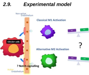
2.9. Experimental model ...18
3. Results ...19
3.1. Endothelial cells activation ...19
3.2. Macrophages polarization ...21
xvi
3.4. The Notch ligand Jagged2 ...26
3.5. The Notch ligand Jagged2 in tumours ...27
4. Discussion ...29
5. Conclusions and Future Perspectives ...33
xvii
Figure index
Figure 1: Schematic illustration of the canonical Notch signalling pathway. ...11
Figure 2: Experimental model ...18
Figure 3: Quantitative gene expression of E-selectin and VCAM-1. ...19
Figure 4: Ratio of absolute count of CD11b+ single cells. ...20
Figure 5: Standard polarization protocol and adherent CD11b+ cells comparative profile. ...22
Figure 6: Ratio M1:M2 by flow cytometry analysis. ...23
Figure 7: Quantitative gene expression of Jagged1 in ECs. ...24
Figure 8: Quantitative gene expression of Notch1 on adherent CD11b+ cells. ...24
Figure 9: Quantitative expression of Jag2 in adherent CD11b+ cells. ...26
xix
Table index
Table 1: Link between inflammation and cancer ... 3 Table 2: Primer List. ...17
xxi
Abreviations and symbols
APC – Allophycocyanin cDNA – Complementary DNA CRC – Colorectal Cancer CTC – Circulating Tumour Cells DC – Dendritic Cells
DLL – Delta-like ligand DNA – Deoxyribonucleic Acid EC – Endothelial Cell
ELAM – Endothelial Leucocyte Adhesion Molecule EMT – Epithelial-Mesenchymal Transition
FBS – Heat-inactivated fetal bovine serum FSC – Forward Scatter
HES – Hairy/Enhancer of Split HEY – Hes-related family
HSCs – Hematopoietic stem cells
HUVECs – Human umbilical cord vein endothelial cells ICAM-1 – Intercellular Adhesion Molecule 1
IFN-γ – Interferon gamma IL – Interleukin
iNOS – inducible nitric oxide synthase Jag1 – Jagged1
xxii LDL – Low-density lipoprotein
LPS – Lipopolysaccharides
M1 – Macrophages classica activation M2 – Macrophages alternative activation M-CSF – Macrophage colony stimulating factor MDSCs - Myeloid-derived suppressor cells mRNA – Messenger ribonucleic acid
NICD – Intracellular domain of the Notch receptor PCR - Polymerase Chain Reaction
PE - Phycoerythrin
PSGL-1 – P-selectin glycoprotein ligand-1
qPCR – Quantitative Polymerase Chain Reaction RNA – Ribonucleic Acid
SD – Standard Deviation SSC – Side Scatter
TAMs - Tumour-associated macrophages Th – T helper cells
TME – Tumour microenvironment TNF-α – Tumour Necrosis Factor-α
Introduction |
1
1. Introduction
1.1.
The links between inflammation and cancer
The connection between inflammation and cancer is not new. In 1863 Rudolf Virchow noted leucocytes in neoplastic tissues and suggested this connection. Virchow hypothesized that the origin of cancer was primarily at sites of chronic inflammation; it is interesting to note that in part based on his hypothesis - some classes of irritants, together with the tissue injury and ensuing inflammation they cause, enhance cell proliferation. Although it is clear that proliferation of cells alone does not cause cancer, sustained cell proliferation in an environment rich in inflammatory cells, growth factors, activated stroma, and DNA-damage-promoting agents, certainly potentiates and/or promotes neoplastic risk. Indeed, different lines of work led to a renaissance of the inflammation–cancer connection, and chronic inflammation has been associated with increased tumour risk (Reviewed in Coussens & Werb, 2002) (Biswas & Mantovani, 2010).
These links have important implications for cancer prevention and treatment. Later in 1986, Harold Dvorak noticed that cancers share several mechanisms commonly associated with the inflammatory (healing) process: angiogenesis and infiltration of inflammatory cells. In fact, the inflammatory cells and cytokines found in tumours are more likely to contribute to tumour growth, progression, and immunosuppression than they are to mount an effective host antitumor response (Balkwill & Mantovani, 2001).
Presently, the tumour microenvironment is considered an important therapeutic target, and is included in 5 of the 10 hallmarks of cancer (Hanahan & Weinberg, 2011).
Epidemiological studies have shown that chronic inflammation predisposes individuals to various types of cancer. It was estimated that underlying infections and inflammatory responses are linked to 15–20% of all deaths from cancer worldwide, a global total of 1.2 million cases per year. There are many triggers of chronic inflammation that increase the risk of developing cancer, including microbial infections (e.g. Helicobacter
pilori for gastric cancer and mucosal lymphoma), autoimmune diseases (e.g.
colitis-associated cancer), HPV infections can promote tumorigenesis in the cervix, chronic ulcerative colitis in Crohn’s disease, Hepatitis C infection in the liver predisposes to liver carcinoma, inflammatory conditions of uncertain origin (e.g. prostate cancer), and among many others examples (Quatromoni & Eruslanov, 2012) (Coussens & Werb, 2002) (Hagemann et al., 2006) (Alberto Mantovani, Allavena, Sica, & Balkwill, 2008) (Alberto Mantovani et al., 2008).
Introduction |
2
The links between inflammation and cancer have also been confirmed in a number of experimental models, e.g., in liver and colon cancers. Moreover, smouldering inflammation is a key characteristic of the microenvironment of most neoplastic tissues, including those not etiologically related to inflammatory processes (Prete et al., 2011) (Hagerling, Casbon, & Werb, 2014).
Chronic inflammation is an aberrantly prolonged form of a beneficial response to tissue injury and pathogenic agents. However, during cancer development, inflammation can be deregulated to promote malignant cell transformation. In other words, inflammation can increase the risk of cancer or promote tumour progression was already described, by providing effector molecules produced by infiltrating immune cells in the tumour microenvironment (TME). The TME consists of various, characteristic cellular and physical components. The TME includes many types of cells of the immune system. In addition, distinct physical features, such as hypoxia and an altered extracellular matrix critically contribute to the TME (Rokavec, Öner, & Hermeking, 2015).
In 2008, Mantovani and colleagues summarized several lines of evidence (Table 1) — based on a range of findings, from epidemiological studies of patients to molecular studies of genetically modified mice that have led to a general acceptance that inflammation and cancer are linked (Alberto Mantovani et al., 2008).
Introduction |
3 Table 1: Link between inflammation and cancer
Evidence described by Mantovani and colleagues
• Inflammatory diseases increase the risk of developing many types of cancer (including bladder, cervical, gastric, intestinal, oesophageal, ovarian, prostate and thyroid cancer). • Non-steroidal anti-inflammatory drugs reduce the risk of developing certain cancers (such as colon and breast cancer) and reduce the mortality caused by these cancers.
• Signalling pathways involved in inflammation operate downstream of oncogenic mutations (such as mutations in the genes encoding RAS, MYC and RET).
• Inflammatory cells, chemokines and cytokines are present in the microenvironment of all tumours in experimental animal models and humans from the earliest stages of development. • The targeting of inflammatory mediators (chemokines and cytokines, such as TNF-α and IL-1β), key transcription factors involved in inflammation (such as NF-κB and STAT3) or inflammatory cells decreases the incidence and spread of cancer.
• Adoptive transfer of inflammatory cells or overexpression of inflammatory cytokines promotes the development of tumours.
Introduction |
4
1.2.
Myelopoiesis
Myeloid cells are the most abundant hematopoietic cells in the human body. Myeloid cells arise from multipotent hematopoietic stem cells (HSCs) that develop into mature myeloid cells through sequential steps of differentiation. And since their first purification in 1988, numerous studies have refined the phenotype and functionality of HSCs and characterized their downstream progeny (Spangrude, Heimfeld, & Weissman, 1988) (Weiskopf et al., 2016).
Three groups of terminally differentiated myeloid cells — macrophages, dendritic cells (DC) and granulocytes are essential for the normal functions of the innate and adaptive immune systems. Classically these cells were known to protect organisms from pathogens, to eliminate dying cells, and mediate tissue remodelling. Although the contribution of myeloid cells to tumour pathogenesis has been recognized for over 100 years, only in the past two decades their crucial role in promoting tumour angiogenesis, cell invasion, and metastasis has been appreciated (Gabrilovich, Ostrand-Rosenberg, & Bronte, 2013) (Weiskopf et al., 2016).
More recently, conversion of myeloid cells into immunosuppressive cells in the tumour microenvironment, lead to the identification of so-called myeloid-derived suppressor cells (MDSCs) (Gabrilovich et al., 2013).
Myeloid cells are key players of the innate immune system, in healthy individuals, their functions include phagocytosis of foreign bacteria (macrophages), processing and presenting antigen (DC), first response to inflammation (neutrophils), and homeostasis and wound healing (platelets). Soluble factors, such as cytokines and chemokines, regulate the migration of myeloid cells from the bone marrow to the blood, and finally into tissues (Schupp
et al., 2017).
Thereby, the tumour exploits pathologically modulated myeloid cells to sustain constituent inflammatory mechanisms supporting the hallmarks of cancer: tumour proliferation, invasion and metastasis, immune escape, prevention of cell death, induction of neoangiogenesis, and the reorganization of cellular metabolism. Genetic or epigenetic traits such as promotion of inflammation and autophagy, and destabilization/mutation of DNA are serving as tumour promoters (Rokavec et al., 2015) (Schupp et al., 2017).
The importance of myeloid cells in cancer is not entirely surprising: tumours frequently employ pre-existing physiological programmes, particularly those involved in wound healing, that engage myeloid cells. The interplay between myeloid cells and adaptive
Introduction |
5
immunity is also emerging as an important regulator of cancer progression, with tumour-associated myeloid cells likely having an important role in cancer immune evasion (Engblom, Pfirschke, & Pittet, 2016).
Introduction |
6
1.3.
Role of Macrophages in tumorigenesis
The mononuclear phagocyte system plays a multi-faceted role in maintaining tissue homeostasis and responding to pathological processes such as autoimmune diseases, cancer, and aberrant wound healing (Olingy et al., 2017).
Back in 1939, Ebert and Florey reported the observation that monocytes migrated from blood vessels and developed into macrophages in tissues. Pro-inflammatory, metabolic and immune stimuli all promote the recruitment of monocytes to peripheral sites, where differentiation into macrophages and DC occurs, contributing to host defence, and tissue remodelling and repair (Ebert & Florey, 1939) (Gordon & Taylor, 2005).
Basically, myeloid precursors in the bone marrow give rise to monocytes that circulate in the peripheral blood, and which can be recruited to the tissues in order to become macrophages, a group of terminally differentiated myeloid cells (Helming, 2011).
Monocytes circulate in the bloodstream during steady state and are robustly recruited to sites of inflammation, where they exert functions that include clearance of cellular debris, promotion of angiogenesis, and restoration of tissue integrity (Olingy et al., 2017).
The morphology of mature monocytes in the peripheral circulation is heterogeneous, and these cells constitute 5–10% of peripheral-blood leukocytes in humans. They vary in size and have different degrees of granularity and varied nuclear morphology. Macrophages are also a heterogeneous population and a major component of the leukocyte infiltrate at sites of inflammation, infection, and tumour growth (Raggi et al., 2017) (Gabrilovich et al., 2013) (Guerriero, 2018) .
There is an extensive literature demonstrating that in both mouse and human macrophages are co-opted during malignancy to facilitate tumour growth. Their presence is associated with poor clinical outcome (Qian & Pollard, 2010), and their pivotal role in cancer was recently highlighted by the demonstration that TAMs (tumour-associated macrophages)
with a specific gene signature are associated with primary treatment failure in patients with Hodgkin’s lymphoma (Steidl et al., 2010). TAMs may be termed M2-like macrophages and are believed to mediate their effects via both non-immune and immune mechanisms (Gabrilovich et al., 2013).
Plasticity and diversity have long been known to be hallmarks of the monocyte-macrophage differentiation pathway. Indeed, adaptive responses to environmental signals are now recognized for both mature and immature elements in the myelomonocytic
Introduction |
7
differentiation pathway. In addition to acting as a first line of resistance against pathogens (the “unsung heroes of immunity”) and activating adaptive responses, myelomonocytic cells undergo reprogramming of their functional properties in response to signals derived from microbes, damaged tissues, and resting or activated lymphocytes (Alberto Mantovani et al., 2004) (Biswas & Mantovani, 2010).
Macrophages have numerous functions related to tissue remodelling, inflammation, immunity and thrombosis and have the capacity to affect tumour growth and progression. In cancer, macrophages can prevent the establishment and spread of tumour cells but, may also elicit functions to support tumour growth and dissemination. As discussed below, this paradoxical ambivalent relationship between macrophages and cancer reflects the functional plasticity of these cells, which can express distinct functional programs in response to different microenvironmental signals. Macrophages play an indispensable role in the immune system with decisive functions in both innate and acquired immunity. In innate immunity resident macrophages provide immediate defence against foreign pathogens and coordinate leukocyte infiltration. Macrophages contribute to the balance between antigen availability and clearance through phagocytosis and subsequent degradation of senescent or apoptotic cells, microbes and possibly neoplastic cells. Their role is essential for triggering, instructing and terminating the adaptive immune response. Macrophages collaborate with T and B cells, through both cell-to-cell interactions and fluid phase-mediated mechanisms, based on the release of cytokines, chemokines, enzymes, arachidonic acid metabolites, and reactive radicals. Macrophage activation can be either pro-inflammatory or anti-inflammatory, thus contributing to tissue cell destruction or to tissue regeneration and wound healing. These polar phenotypes are not expressed simultaneously, but regulated in such a manner that macrophages display a balanced, integrated pattern of functions (Guerriero, 2018) (Chen, Deng, Cui, Fang, & Zuo, 2018) (Gordon, 2018).
Introduction |
8
1.4.
Macrophages activation
The diverse functions of macrophages in tissue homeostasis appear to be related to their activation state or phenotype, which can shift rapidly in response to local environmental stimuli (Spiller et al., 2016).
Progress has been made in defining the surface phenotype, activating signals and molecular pathways associated with different forms of macrophage activation. Moreover, evidence has now accumulated showing that the orchestration of macrophage function has a key role in different pathological conditions (Biswas & Mantovani, 2010). Activation of macrophages has emerged as a key area of immunology, tissue homeostasis, disease pathogenesis, and resolving and nonresolving inflammation.
In 2000, Mills and colleagues proposed a new classification of macrophages as either M1 or M2. M1 (classically activated) macrophages exhibit inflammatory functions, while M2 (alternatively activated) macrophages exhibit anti-inflammatory functions. A useful oversimplification mirroring T helper type 1–T helper type 2 (TH1-TH2) polarization, two distinct states of polarized macrophages defined based on the type of in vitro stimulation, surface molecule expression pattern, secretory profile, and functional properties: M1 and M2 represent the extremes of a continuum of activation states (Biswas & Mantovani, 2010) (Raggi et al., 2017).
The factors regulating macrophages polarization are the focus of intense investigation and the nature of the activating stimulus and the combination of different stimuli can profoundly impact upon the type of response. A large body of evidence indicates that macrophages polarization state is not only determined by the type of activating stimulus but also depends on the local environment in which they differentiate (Raggi et al., 2017) (Gordon & Taylor, 2005) (Gabrilovich et al., 2013) (Stout & Suttles, 2004).
Macrophages are classically activated toward the M1 phenotype by microbial factors, like LPS, and Th1 proinflammatory cytokines, such as IFN-γ, TNF-α, and IL-1β, or a combination of the two. The M1 macrophage phenotype has been connected with the expression of IL-1, IL-12, IL-23, TNF-α, inducible nitric oxide synthase (iNOS), low amounts of IL-10 and high levels of the chemokine receptor CCR7 expression. A commonly accepted marker profile for M1-macrophages is also CD68+/CD80+ (Vrančić et al., 2012) (Hagemann
et al., 2006) (Raggi et al., 2017) (Biswas & Mantovani, 2010) (Almatroodi, McDonald, Darby,
& Pouniotis, 2016) (Gabrilovich et al., 2013) (Badylak, Valentin, Ravindra, McCabe, & Stewart-Akers, 2008).
Introduction |
9
In contrast, M2 subset comprises various forms of non-classically activated macrophages originating from exposure to different stimuli, such as the Th2 cytokines, IL-4, or IL-13 (M2a), immune complexes in combination with IL-1β or LPS (M2b), the anti-inflammatory cytokines, IL-10 and TGFβ, or glucocorticoids (M2c), IL-6, LIF, and MCF (M2d).
M2 macrophages are oriented to Th2-type immunoregulation and resolution of inflammation, exhibit tissue remodelling and repair functions, promote wound healing, angiogenesis, resistance to parasites, and tumour growth through the production of anti-inflammatory cytokines, extracelular matrix components, remodelling and proangiogenic factors (Martinez, Gordon, Locati, & Mantovani, 2006) (Alberto Mantovani et al., 2004).
M2 macrophages show more phagocytic activity, high expression of scavenger (CD163), mannose (CD206) and galactose receptors, production of ornithine and polyamines through the arginase pathway, and a phenotype of low expression of IL-12 and high expression of IL-10, the IL-1 decoy receptor and IL-1RA. M2 macrophages are also characterized as CD68+/CD163+ (Raggi et al., 2017) (Hagemann et al., 2006) (Biswas & Mantovani, 2010) (Almatroodi et al., 2016) (Gabrilovich et al., 2013) (Martinez et al., 2006) (Vrančić et al., 2012) (Badylak et al., 2008).
Martinez and colleagues, who identified the genes differentially expressed in M1 vs M2 macrophages, described CCR7 being expressed at a high M1:M2 ratio, while CXCR4 ratio was negative, indicating its expression was highest on M2 macrophages (Martinez et
al., 2006). In our work we decide to used these two membrane receptors to distinguish M1
and M2.
M1 and M2 macrophages have also distinct chemokine profiles, with M1 macrophages expressing TH1 cell–attracting chemokines such as CXCL9 and CXCL10 and M2 macrophages expressing the chemokines CCL17, CCL22 and CCL24. Chemokines can also affect macrophage polarization, with CCL2 and CXCL4 driving macrophages to an M2-like phenotype (Biswas & Mantovani, 2010).
As discussed by Mantovani and colleagues, the M1/M2 nomenclature is useful, but oversimplified because macrophages form a continuum of phenotypes. In our work we also didn’t distinguish M2 subtypes. Remains also controversial M1 and M2 terms, for the lack of tightly defined criteria to score phenotypes. Nevertheless, increasing efforts to define polarization are advancing (Alberto Mantovani, Sica, Allavena, Garlanda, & Locati, 2009) (Murray, 2017).
Introduction |
10
Monocyte/macrophage lineage exhibit extraordinary plasticity in response to endogenous as well as exogenous stimuli, which can allow overriding the initial M1/M2-polarization processes, for example M2-polarized macrophages can convert to an M1-activated status under certain conditions. Macrophage activation has been shown to be plastic, rapid, and fully reversible, suggesting that macrophage populations are dynamic and may first take part in inflammation and then participate in its resolution. Consequently, macrophages display progressive functional changes resulting from changes in the microenvironment (Benoit, Desnues, & Mege, 2008) (Murray, 2017).
In case of tumours, macrophages are recruited into the tumour from the peripheral circulation by chemokines and are usually polarized towards an M2 phenotype, which is characterized by an IL-10 high and IL-12 low expression pattern, and all phenotypic changes seen in macrophages are driven by tumour cell-macrophage interactions. There are many examples of macrophage-polarizing events during tumour progression, including the secretion of tumour-derived mediators and hypoxic tissue damage, as well as influences from other immune cells and stromal components. What is less known is how other cells from the tumour microenvironment may affect macrophage polarization (Hagemann et al., 2006).
Here we hypothesize that the interactions of macrophages with active endothelial cells may influence M1/M2 differentiation. More specifically, we hypothesize that endothelial Notch signalling is involved in such process. The Notch pathway has been shown to be involved in differentiation, but also in cell proliferation and survival in tumour cells, and is one of the most commonly activated signalling pathways in cancer. .
Introduction |
11
1.5.
The Notch ligand Jagged2 in tumours
The Notch signalling pathway is a highly conserved molecular cell signalling pathway that affects development in organisms ranging from sea urchins to humans and plays a crucial role in regulate proliferation, stem cell maintenance, cell fate specification, differentiation, and homeostasis of multicellular organism and implicates angiogenesis. It is also involved in cancer development and progression, including metastases formation (Perrimon, Pitsouli, & Shilo, 2012) (Venkatesh et al., 2018) (Lobov & Mikhailova, 2018).
Throughout the past several decades, the function of Notch proteins has been studied in diverse organisms, beginning in 1919 with its discovery and naming based on its effect on the partial loss of wings phenotype in Drosophila. Since then, numerous researchers have contributed to the discovery of the components and various regulators to the Notch signalling pathway involved in the loss of wings phenotype, Notch participates in a pathway that is normally activated by engagement of Notch receptors by Notch ligands expressed by adjacent cells (Artavanis-Tsakonas, 1988) (H. Wang, Zang, Liu, & Aster, 2015) (Hassan et
al., 2013) (Liu, Shen, Wen, Guo, & Zhang, 2016).
Notch ligands include the delta-like and jagged ligands (DLL1, DLL3, DLL4, JAG1, and JAG2). Interaction of these ligands with one of the Notch receptors (NOTCH1-4) results in γ-secretase–mediated cleavage and release of the intracellular domain of the Notch receptor (NICD). NICD functions as a nuclear transcription cofactor to activate Notch target genes, such as genes of the Hairy/Enhancer of Split (HES) and Hes-related (HEY) families of proteins (Figure 1) (Pietras, Stedingk, Lindgren, Påhlman, & Axelson, 2011).
Figure 1: Schematic illustration of the canonical Notch signalling pathway .
Introduction |
12
An important example to understand systemic cancer is the recruitment of cells from the bone-marrow to the cancer microenvironment, which has been shown to be important at different stages of the carcinogenic process.
The tumour microenvironment comprises different cell types, and some of them such as TAMs and MDSCs are recruited from the bone marrow into the tumour site.
As previously described by our lab, bone marrow–derived CD11b+Jag2+ cells, which infiltrate primary colorectal tumours are sufficient to induce epithelial to mesenchymal transition (EMT) in tumour cells, thereby triggering onset of metastasis. Furthermore, we argue that quantifying circulating CD11b+Jag2+ cells in patients may offer an indicator of colorectal cancer (CRC) progression to metastatic disease. In vitro coculture experiments showed that the bone marrow–derived CD11b+Jag2+ cells induced EMT through a Notch-dependent pathway.
Caiado and colleagues in our lab showed that peripheral blood levels of CD11b+Jag2+ cells in mouse models of CRC and in a cohort of untreated patients with CRC were indicative of metastatic disease (Caiado et al., 2013).
He and colleagues detected Jag2 protein expression in up to 95% of CRC cases and showed it was 3-fold upregulated in tumour cells compared to surrounding normal tissues, suggesting that Jag2 may have a role in the tumorigenicity of CRC. Their findings, together with our own results, implicate Jag2 in promoting aggressiveness of CRC, and lay the foundation for its future development as a therapeutic target for the treatment of CRC (He et
al., 2016).
Reedijk and colleagues also reported that Jag2 expression is increased in human CRC compared to that in the surrounding normal tissues, suggesting that dysregulation of Jag2 expression plays a role in CRC cell growth and progression to metastatic disease. These results suggest that Jag2 modulates the sensitivity of CRC cells to chemotherapeutic agents through p21. And Jag2 was identified as a novel target for therapeutic intervention of CRC (Reedijk et al., 2008).
Jag2 expression in primary tumour material was also correlated with gene signatures of vascular development and angiogenesis. In addition, coculture of vascular endothelial cells with hypoxic tumour cells revealed that Jag2 expression on tumour cells promoted endothelial cell tube formation. Together these results suggest that a hypoxia-regulated cross-talk between tumour and endothelial cells via Notch activation is involved during tumour angiogenesis (Pietras et al., 2011).
Introduction |
13
In breast cancer, increased Jag2 mRNA was found in the MDA-MB-435 derived Br4 variant cell line which specifically metastasizes to the brain, and knocking down of Jag2 significantly decreased migration and invasion abilities of the Br4 cells, suggesting that Jag2 plays an important role in tumour metastasis in breast cancer although the underlying mechanisms of the aberrant Jag2 expression is yet to be defined (Xing et al., 2011).
Up-regulation of Jag2 expression was also observed in others cancers, including pancreatic, bladder, and lung cancers, and was associated with the progression of these tumours (Vaish, Kim, & Shim, 2017).
Introduction |
14
1.6.
Aims of the thesis
Endothelial cells are crucial player in a variety of processes, such as inflammation, tumour progression, hematopoiesis and metastasis. Therefore, targeting specific pathways in endothelial cells often perturbs those events.
The results presented in this Thesis intended to scrutinize the role of active endothelial cells in macrophages polarization.
The objectives of this work can be summarized as follows:
I. To characterize the role of active endothelial cells in adhesion and polarization of macrophages.
II. To observe and characterize the involvement of Notch pathway signalling in macrophage polarization.
III. Scrutinize the role of Jag2 as a biomarker for metastatic disease.
Materials and methods |
15
2. Materials and methods
2.1.
Primary sample collection
Peripheral blood samples from healthy donors of both genders were kindly provided by Instituto Português do Sangue e Transplantação, IP, in a Compoflow system ®.
The blood was processed through Ficoll-Paque (Lymphoprep, Axis-Shield PoC AS) gradient and the red blood cells were lysed with RBC Lysis Buffer (Santa Cruz Biotechnology), CD11b+ cells were then labelling with anti-CD11b and sorted using cell sorter FACSAria IIu (BD Biosciences) according to the manufacturer’s protocol.
These cells are later used by the single cell culture, coculture and standard protocols for M1/M2 polarization.
2.2.
Single Cell Culture
Human umbilical cord vein endothelial cells (HUVECs) (Clonetics, Lonza) were cultured in EGM-2 (Lonza) and used until passage 6. Cells were used in passage 4 for all the experiments.
For single CD11b+ cells analysis, 4x10⁵ CD11b+ cells were plated alone and treated with same concentrations of TNF-α for 4/8 hours with culture for more 20 or 16 hours, respectively, or analysed immediately after the treatment with TNF-α.
2.3.
Endothelial cells activation
For the endothelial activation studies, cells were cultured in 6 well plates (Corning) at a density of 3x10⁵ cells/well in EGM-2 supplemented with 10% heat-inactivated fetal bovine (FBS) (Gibco). 24 hours after plating, cells were treated with 10 ng/mL TNF-α for 4 hours and then washed twice with culture medium and left untreated for 20 hours EGM-2 supplemented with 10% FBS .
2.4.
Coculture assay
For the coculture studies, ECs were plated in 6 well plates at a density 3x10⁵ cells/well in EGM-2 supplemented medium with 10% FBS for 24 hours and then were treated with 10 ng/mL TNF-α for 4h for activation. After this, cells were washed with cell culture medium and 4x10⁵ isolated CD11b+ cells were placed on top the ECs in RPMI 1640 supplemented medium (GIBCO) with 1% FBS. 20 hours later cells in coculture were washed
Materials and methods |
16
once in DMEM, collected by gently scraping cells into microtubes. Cells were then stained for flow cytometric analysis or labelling to sort for RNA extraction.
2.5.
Polarization
Macrophages were obtained by a standard polarization protocol, culturing CD11b+ (previously sorted) in RPMI 1640 supplemented with 20% FBS and 100ng/ml M-CSF (L-929) in 24 well plates at a density of 3x10⁵ cells/well.
7 days after non-adherent cells were discarded and polarization of the adherent cells (macrophages) was induced by culturing cells for an additional 18 hours in RPMI 1640 supplemented with 5% FBS and 100 ng/ml LPS plus 20 ng/ml IFN-γ (for M1 polarization) and 20 ng/ml IL-4 (for M2 polarization).
2.6.
qPCR
RNA extraction (TRIzol, BIO-RAD) and cDNA synthesis (Nzytech) were perfomed following the manufacture’s protocols
Concentrations of RNA were determined using Nanodrop ND-2000 (Thermo Scientific; Model: ND-2000).
Primers used for qPCR are listed in Table 2, and were designed using Ensembl BLAST/BLAT tool. For qPCR analysis, cDNA was amplified in an 8μl reaction with SYBR Green, PCR master mix (Applied Biosystems), and reactions were run in ViiA7 (Applied Biosystems). Reactions consisted in 2 min at 50 ºC, 10 min at 95 ºC followed by 40 cycles of 15 sec at 95 ºC and 1 min at 60 ºC.
Expression was normalized to 18S and expressed in arbitrary units. Results were expressed using the 2^(ΔΔCt) methods.
Materials and methods |
17 Table 2: Primer List.
Gene Forward Primer(5’ – 3’) Reverse Primer (5’ – 3’) h 18S GCCCTATCAACTTTCGATGGT CCGGAATCGAACCCTGATT
h Jagged1 ACCGCAACCGCATCGT GCCTCCACAAGCAACGTATAGG
h Jagged2 ACAGCGCCACTTGCAACA CAGGTGTAGTGGCCGAAAAAG
h NOTCH1 TGCCTCTTCGACGGCTTT GGGTACATGCTCCGCACAGT
h HES1 ACGACACCGGATAAACCAAA CGGAGGTGCTTCACTGTCAT
h CCR7 GGCTGGTCGTGTTGACCTAT AAGGGTCAGGAGGAAGAGGA
h CXCR4 CCGGGAACTACTCCGACATC GGCTTCCACCCCTATGCA
h CD163 TTTGTCAACTTGAGTCCCTTCAC TCCCGCTACACTTGTTTTCAC
h E-Selectin TTCCCTTGGTAGCTGGACTTTC TTTCCGAAGCCAGAGGAGAA
h VCAM-1 CCTGGGAAGATGGTCGTGAT TCTGGGGTGGTCTCGATTTT
2.7.
Flow Cytometry and cell sorting
Sorting of CD11b+ cells from peripheral blood or from cocultures was performed using an anti-CD11b antibody (APC, 101212, Biolegend) for staining and an Aria Cell Sorter equipped with FACS Diva 6.2 Software (BD Biosciences) for sorting. The sorted cells obtained with this method was > 95% pure according to flow cytometry.
For cell surface labelling, cells were washed with DMEM and incubated for 15 min at 4°C with the following antibodies: anti-CD11b+ (APC, 101212, Biolegend), anti-Jagged2+ (PE, 346904, Biolegend), anti-CD163 (PE, 12-1639-41, eBioscience) and anti-CD80 (PE, 560 925, BD Pharmingen).
Samples were acquired using BD Accuri C6 cell analyser and data analysed using FlowJo software 9.8.2 (Tree Star).
2.8.
Statistical analysis
Results are expressed as mean ± standard deviation (SD) of three independent experiments unless otherwise specified. Statistical analyses were performed using Student t test. All experiments were performed in triplicate and repeated independently at least 3 times. The p-value <0.05 was considered to be statistically significant. *P < 0.05, **P < 0.01, ***P < 0.001, t-test.
Materials and methods |
18
2.9.
Experimental model
Results|
19
3. Results
3.1.
Endothelial cells activation
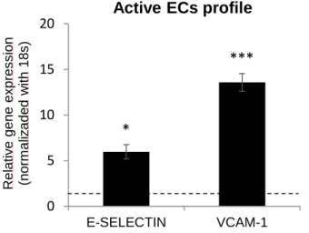
Čejková and colleagues defined the endothelial cell activation by the endothelial expression of cell-surface adhesion molecules, such as VCAM-1, ICAM-1, P-selectin and E-selectin (also known as the endothelial leukocyte adhesion molecule – ELAM). To activate endothelial cells, we added 10 ng/mL TNF-α for 4 hours, after which TNF was removed and cells were analysed after 20 hours in complete medium. As shown in Figure 3, we observed active ECs, characterized by the higher expression of VCAM-1 and E-selectin (Figure 3), 14-fold and 6-14-fold upregulated, respectively (Čejková, Králová-Lesná, & Poledne, 2016).
Figure 3: Quantitative gene expression of E -selectin and VCAM -1.
Relative mRNA levels of E -selectin and VCAM-1 in active ECs fold to control ECs (expressed as dotted line). Confluent ECs monolayer was previously treated 4 hours with TNF-α and left untreated for 20 hours. All experiments were performed in triplicate and repeated independently at least 3 times. The p-value <0.05 was considered to be statistically significant. * P < 0.05, **P < 0.01, ***P < 0.001, t-test.
As expected when the endothelium is activated, the adhesive ability of monocytes increased (Figure 4). 0 5 10 15 20 E-SELECTIN VCAM-1 Rel ati v e gene ex pres s ion (no rm al iz ad ed w ith 18 s )
Active ECs profile
***
Results|
20
Figure 4: Ratio of absolute count of CD11b+ single cells.
Ratio adherent:suspension cells , perfomed by BD Accuri c6. Confluent ECs monolayer previously treated 4 hours with TNF-α and coculture left untreated for 20 hours. All experiments were performed in triplicate and repeated independently a t least 3 times. The p-value <0.05 was considered to be statistically significant. * P < 0.05, **P < 0.01, ***P < 0.001, t-test. 0% 10% 20% 30% 40% 50% Control TNF-alpha % of absol ut e C ount of si ngl e cel ls
Ratio Adherent:Suspension cells
Results|
21
3.2.
Macrophages polarization
An important part of this work is to contribute towards a better understanding as to how macrophages differentiate into M1 or M2 in an inflammatory environment, to test this, we performed coculture with active ECs and CD11b+ cells.
M1 and M2 polarization states are defined by specific phenotypic and secretory patterns. Human macrophages were generated following a standard protocol, culturing CD11b+ cells with the maturation factor, M-CSF, and then polarized toward the M1 and M2 activation states by stimulation with LPS + IFN-γ and IL-4, respectively.
Stewart and colleagues described a problematic that we share: the binary classification of macrophage phenotypes, due to dual labelling of macrophages for both M1 and M2 markers. However, this challenge in classifying macrophages based on single markers is consistent with literature. For example, Qian and Pollard defined TNF-α as M1-specific, in agreement with Martinez and colleagues, who described TNF-α as upregulated in M1-macrophages and downregulated in M2-macrophages. However, Zeyda and colleagues showed TNF-α expression in both M1 and M2 polarized macrophages. Similarly, we originally defined CD163 as an M1 marker based on the work by Fuentes and colleagues and Qian and Pollard but ultimately reassigned it to an M2 pattern of expression on the basis of recently published studies and the work by Stewart and colleagues. Such discrepancies in “hallmark biomarkers” of macrophage differentiation or polarization make the interpretation of polarization markers challenging (Stewart, Yang, Makowski, & Troester, 2012). To overcome this problem we generated a positive control based on the expression profile of M1 and M2 cells, obtained using the Standard Polarization Protocol. We then compared this expression profile with the expression profile of our cells of interest, derived from culture and coculture experiments. This approach allowed us to quantify M1 versus M2 macrophages frequencies.
Thus, to quantify macrophages polarization in our experiments, we compared the expression of M1/M2 markers after the standard polarization protocol with the expression in cells after co-culture with ECs (Figure 5A) exposed or not to TNF-α (Figure 5B).
CCR7 was selected as a marker for macrophages with a M1 phenotype and CXCR4 and CD163 as markers for macrophages with a M2 phenotype.
We observe a similar profile of M2 markers in adherent CD11b+ cells when in coculture with active ECs. Our data suggests that active ECs may favour macrophages polarization into an M2 phenotype.
Results|
22
Figure 5: Standard polarization protocol and adherent CD11b+ cells comparative profile.
Adherent CD11b+ cells have a similar profile of M2 when in coculture with EC s previously treated with TNF-α for 4 hours. Expression of CCR7 as M1 marker, CXCR4 and CD163 as M2 marker (A) are analysed after standard polarization and adherent CD11b+ cells (B) after 20 hours coculture, with untreated ECs or ECs treated with TNF-α for 4 hours. All experiments were performed in triplicate and repeated independently a t least 3 times. The p-value <0.05 was considered to be statistically significant. * P < 0.05, **P < 0.01, ***P < 0.001, t-test.
Surface expression of prototypical M1 marker, CD80, and the M2 marker, CD163, was then analysed by flow cytometry to confirm polarization. As shown in Figure 6, the M1/M2 ratio is lower when the endothelial cells are active, which indicates that there is more polarization towards the M2 phenotype (Figure 6). This corroborated our previous PCR results.
Results|
23 Figure 6: Ratio M1:M2 by flow cytometry analysis.
Ratio of M1 (CD80) and M2 (CD163) in adherent CD11b+ cells. ECs treated 4 hours with TNF-α and 20 hours coculture with CD11b+ cells free of TNF -α. All experiments were performed in triplicate and repeated independently at least 3 times. The p -value <0.05 was considered to be statistically significant. *P < 0.05, **P < 0.01, ***P < 0.001, t-test.
3.3.
Notch signalling activation
Another important aspect of our studies was to analyse the expression of different factors (at the mRNA level) on adherent CD11b+ cells after coculture with active ECs, particularly the notch receptors and/or ligands and analyse the alterations in ECs after coculture and an eventual Notch signalling activation.
As previously described in our lab, in transgenic adenocarcinoma mouse prostate (TRAMP) mouse model, endothelial Jag1 positively regulate macrophages recruitment and polarization to M2, in vivo and in vitro. And we started by analyse the Notch pathway all ligands and receptors expression in ECs and adherent CD11b+ cells.
In our experiments, cultures of ECs alone treated with TNF-α result in a statistically significant induction of endothelial Jag1, and this was also observed with 4 hours treatment with TNF-α after coculture with CD11b+ cells (Figure 7). Next, we decided to quantify the expression of Notch ligand/receptors on adherent CD11b+ cells and scrutinize the involvement of the Notch pathway (Johnston, Dong, & Hughes, 2009) in the interaction between endothelial cells and macrophages polarization.
0
1
2
Adherent CD11b+ cells
Ratio M1:M2
Untreated EC Treated ECResults|
24
Figure 7: Quantitative gene expression of Jagged1 in ECs.
Only ECs are a confluent ECs monolayer treated 4 hours with TNF-α and 20 hours
culture free of TNF-α. Sorted ECs are the endothelial cells sorted after a coculture, a confluent ECs monolayer previously treated 4 hours with TNF-α and 20 hours coculture free of TNF-α with CD11b+ cells and sorted by Aria Iiu. All experiments were performed in triplicate and repeated independently a t least 3 times. The p-value <0.05 was considered to be statistically significant. * P < 0.05, **P < 0.01, ***P < 0.001, t-test.
The adhesion of CD11b+ cells and ECs make a bidirectional communication, and when we observe the Notch receptors in CD11b+ cells after coculture with active ECs, we noted a significant higher expression of Notch1 in adherent CD11b+ cells (Figure 8).
Figure 8: Quantitative gene expression of Notch1 on adherent CD11b+ cells .
mRNA levels of Notch1 expression in adherent CD11b+ cells , fold to Control.
Adherent CD11b+ cells analysed after 20 hours coculture free of TNF-α with endothelial cells previously treated 4 hours with TNF-α. All experiments were performed in triplicate
0 4 8 12
Only ECs Sorted ECs
m RNA lev el (F ol d to Cont rol )
JAGGED1 expression in EC
**
**
n.s.
Results|
25
and repeated independently a t least 3 times. The p-value <0.05 was considered to be statistically significant. * P < 0.05, **P < 0.01, ***P < 0.001, t-test.
These results lead us to think that there is a communication between these cells, via notch signalling pathway.
Results|
26
3.4.
The Notch ligand Jagged2
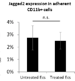
Apparently active ECs do not modify the Jag2 expression in adherent CD11b+ cells (Figure 9). But, in contrast we observed a higher expression of Jag2 when CD11b+ cells alone are treated with TNF-α for 8 hours (Figure 10).
Figure 9: Quantitative expression of Jag2 in adherent CD11b+ cells after coculture.
mRNA levels of Jag2 expression in adherent CD 11b+ cells after 20 hours coculture (untreated ECs and ECs previously treated 4 hours with TNF-α). All experiments were performed in triplicate and repeated independently a t least 3 times. The p-value <0.05 was considered to be statistically significant. * P < 0.05, **P < 0.01, ***P < 0.001, t-test.
Figure 10: Quantitative expression of Jag2 in adherent CD11b+ cells.
mRNA levels of Jag2 expression in adherent CD11b+ cells alone (treated 8 hours with TNF-α). All experiments were performed in triplicate and repeated independently a t least 3 times. The p-value <0.05 was considered to be statistically significant. * P < 0.05, **P < 0.01, ***P < 0.001, t-test.
Results|
27
3.5.
The Notch ligand Jagged2 in tumours
Part of the work presented in this Thesis was included in the “Oncodynamics Project” at IMM and Hospital Santa Maria, in close collaboration with L. Costa Lab – IMM, where we analysed the peripheral blood levels of CD11b+Jag2+ cells in metastatic patients.
In our cohort, 61 patients (breast=32, prostate=13, colorectal=13, melanoma=3 in a total of 98 analyses) we tried to correlate the levels of CD11b+Jag2+ cells with some variables like: sex, age, type of cancer or metastatic organ but we failed to find any statistical association.
But our aim was to try and analyse patient by patient and correlate the variation of CD11b+Jag2+ cells with circulating tumours cells (CTC), disease progression, treatment and disease free survival.
Analysing patient by patient can give us more information about how important are CD11b+Jag2+ cells in aggressiveness, treatment resistance, metastatic expansion and survival, but unfortunately this analysed is not be finished at time to write this work, for that reason, the results is not be presented here.
Discussion |
29
4. Discussion
The inflammatory response is one of the first defences the body has to fight against potential dangers, such as infections and injury, characterized by the rapid accumulation of immune cells and production of soluble mediators with the ultimate aim of protecting the organism from foreign invaders and initiating healing processes. During an inflammatory response, a variety of cytokines is produced by cells and trigger or enhance the specific inflammatory response (Ye, Wang, Jenson, & Yan, 2016).
The endothelium is an important homeostatic organ, which regulates the correct physiological state between the blood stream, including physical and chemical factors, and deeper layers of the vessel wall. In addition, the endothelium is often connected to pathophysiological processes that are essential for life. However, when the endothelium undergoes long-term stress, it may result in dysfunction and induces the development of associated diseases. If the endothelium is activated, the adhesive ability of monocytes is increased, as expected and observed in our results. Adhesion of immune competent cells is a key process for their migration to the inflammatory sites, without which the immune system cannot work (Čejková et al., 2016) (Helming, 2011).
The endothelial cell activation is typically induced by proinflammatory cytokines, such as TNF-α, and facilitates the recruitment and attachment of circulating leukocytes to the vessel wall. Monocyte recruitment from the blood stream is activated by a regulated multistep process and mediated by chemoattractants, cell adhesion molecules and their receptors. Extravasation may occur due to the nature of its completely physiological defence against infection, but also in pathological events. There are several stages involved in leucocyte recruitment into vascular tissue: (1) initial selectin-dependent tethering and rolling; (2) triggering of adhesion via chemokines and their receptors or through selectin binding to P-selectin glycoprotein ligand-1 (PSGL-1); (3) integrin-dependent adhesion and adhesion strengthening by integrin clustering; (4) and transmigration across the endothelium (Čejková
et al., 2016).
We observe the expression of VCAM-1 in active ECs, usually induced on endothelial cells during inflammatory diseases, influenced by several mediators, including reactive oxygen species, TNF-α, LDL (low-density lipoprotein) and oxidised LDL (Čejková et al., 2016). VCAM-1 encodes for an adhesion molecule known to play a critical role in inflammation by recruiting leukocytes to acute and chronic inflammatory sites.
Discussion |
30
After the endothelial cells activation, the next step of this work was to observe if this affected the macrophages polarization to M1/M2 phenotype. Cells belonging to the monocyte-macrophage lineage have long been recognized as heterogeneous, reflected the plasticity and versatility of these cells in response to exposure to microenvironmental signals (A. Mantovani, Sozzani, Locati, Allavena, & Sica, 2002) .
Mantovani and Sica have suggested that macrophages in tumours are more similar to alternatively activated M2 macrophages (rather than activated M1 macrophages), with gene profiling experiments on TAMs supporting these findings and confirmed in other works (A. Mantovani et al., 2002) (Guerriero, 2018).
Our in vitro findings suggested that adherent CD11b+ (macrophages) in coculture with active ECs have a similar phenotype to our M2 macrophages positive control, with this results we hypothesized that active ECs may be critical to polarize macrophages to M2. To analyse the macrophages polarization phenotype we used CCR7 as M1 marker, CD163 and CXCR4 as M2 markers in PCR analysis, and CD80 as M1 marker and CD163 as M2 marker in Flow Cytometry analysis.
The interactions of ECs with immune cells are important determinants of tumour biology, with inflammatory cells playing well-recognized roles in cancer progression. Despite the fact that macrophage–ECs communication is bidirectional, important questions remain about how ECs affect adjacent immune cells in context of tumour (Coussens & Werb, 2002). Our study investigated macrophages polarization, and gene expression in response to coculture with active ECs. Alterations induced by coculture was compared with alterations observed under normal conditions.
We had as a starting point some good results in our laboratory; Martins in her phD work, described in transgenic adenocarcinoma mouse prostate (TRAMP) mouse model, that endothelial Jag1 positively regulate macrophages recruitment and polarization to M2, in vivo and in vitro. And we analysed the Notch pathway expression in ECs and adherent CD11b+ cells.
Notch signalling is involved in the specification, proliferation, and migration of endothelial cells including the coordination of multiple aspects of endothelial behaviour during vessel patterning. Notch1 and Notch4, and their ligands DLL1, DLL4, and Jag1 are most prevalently expressed on endothelial cells (Takebe, Nguyen, & Yang, 2014).
Discussion |
31
In our experiments, active ECs have a statistically significant expression of Jag1, but more interesting have higher expression when active ECs are analysed after the coculture with CD11b+ cells (Johnston et al., 2009).
We also observe a Notch1 expression in adherent CD11b+ cells after coculture with active endothelial cell.
The adhesion of CD11b+ cells to ECs make a bidirectional communication, Notch pathway mediates juxtacrine cellular signalling wherein both the signal sending and receiving cells are affected through ligand-receptor crosstalk.
The Notch signalling activation was confirmed by the expression of HES1 in adherent CD11b+ cells after coculture with active ECs. It is known that in the absence of signalling, RBPJ inhibits the expression of HES1, and in coculture with non-treated ECs the Notch signalling is inactive, but we confirmed the Notch pathway involvement with expression of HES1 in adherent CD11b+ cells after coculture, when the ECs are active.
We know that active ECs polarized Macrophages to M2 phenotype, and Notch signalling is active, but, at this time, we can’t connected this results.
We doesn’t observed effect of active ECs in Jag2+ expression in the CD11b+ cells, as well as in the initial phase of Oncodynamics Project, although Jag2 overexpression as already described as a bad prognostic factor and metastatic indicator in CRC, we didn’t find any significant result.
Because Notch plays a critical role in many fundamental processes and in a wide range of tissues, it is not surprising that aberrant gain or loss of Notch signalling components has been directly linked to multiple human disorders. Deregulated Notch signalling is found in various diseases, such as T-cell leukaemia, breast cancer, prostate cancer, colorectal cancer and lung cancer as well as central nervous system malignancies (Kopan & Ilagan, 2009) (Yuan et al., 2015).
The role of the Notch signalling pathway was recognized decades ago when changes of Notch pathway members were found responsible for various human diseases. Since then, a list of aberrant Notch signalling has grown to include cancer, immune disorders and developmental syndromes (Kopan & Ilagan, 2009) (Liu et al., 2016).
Ample evidence has demonstrated that inhibition of Notch induces tumour growth arrest. Moreover, combined therapy of chemotherapy or radiotherapy with Notch inhibitors also results in a synergistic effect, suggesting Notch inhibitors may improve chemotherapy
Discussion |
32
response. Finally, Notch inhibitors combined with chemotherapy or radiotherapy hold great promise for cancer control (Yuan et al., 2015).
Notch signalling has already been demonstrated to play a critical role in the determination of M1 versus M2 polarization of macrophages. Compromised Notch signalling in macrophages lead to M2 polarization even in the presence of M1 inducers (Y. Wang et al., 2010).
Since macrophage polarization may occur in a variety of inflammatory processes, it is of the utmost importance to unravel the true consequences of macrophage polarization in cancer.