U
NIVERSIDADE DE
L
ISBOA
F
ACULDADE DE
C
IÊNCIAS
D
EPARTAMENTO DE
B
IOLOGIA
V
EGETAL
P
RODUCTION AND CHARACTERIZATION OF DIFFERENT TYPES OF
MALARIA PIGMENT
(
HEMOZOIN
)
AND THEIR POTENTIAL USE FOR
SCREENING FOR HEMOZOIN INHIBITING DRUGS
Ana Margarida Ferreira Góis
M
ESTRADO EM
M
ICROBIOLOGIA
A
PLICADA
U
NIVERSIDADE DE
L
ISBOA
F
ACULDADE DE
C
IÊNCIAS
D
EPARTAMENTO DE
B
IOLOGIA
V
EGETAL
P
RODUCTION AND CHARACTERIZATION OF DIFFERENT TYPES OF
MALARIA PIGMENT
(
HEMOZOIN
)
AND THEIR POTENTIAL USE FOR
SCREENING FOR HEMOZOIN INHIBITING DRUGS
Dissertação orientada por Prof. Dr. Thomas Hänscheid (FMUL)
e Prof.ª Dr.ª Margarida Gama Carvalho (FCUL)
Ana Margarida Ferreira Góis
M
ESTRADO EM
M
ICROBIOLOGIA
A
PLICADA
P
RODUCTION AND CHARACTERIZATION OF DIFFERENT TYPES OF
MALARIA PIGMENT
(
HEMOZOIN
)
AND THEIR POTENTIAL USE FOR
SCREENING FOR HEMOZOIN INHIBITING DRUGS
Ana Margarida Ferreira Góis
M
ASTER
T
HESIS
2012
This thesis was fully performed at the Institute of Microbiology in Faculty
of Medicine of the University of Lisbon under the direct supervision of
Prof. Dr. Thomas Hänscheid.
Prof. Dr. Margarida Gama Carvalho was the internal designated
supervisor in the scope of the Master in Applied Microbiology of the
Faculty of Sciences of the University of Lisbon.
i
Acknowledgements
I want to acknowledge Prof. Dr. Thomas Hänscheid for supervising me and for giving me the opportunity to be constantly learning. I want to thank the Institute of Microbiology in the Faculty of Medicine of the University of Lisbon, directed by Prof. Dr. José Melo Cristino, for allowing me to work in these facilities. I want to thank Prof. Dr. Margarida Gama Carvalho for her guidance and availability.
I want to thank the company Steris, namely Dr. Vincent Thomas, for the opportunity to collaborate with them in the study of the Ileal Fluid Dependent Organism (IFDO).
I would also like to thank Prof. Dr. Estrela Jorge and the technician Telmo Nunes from the Faculty of Sciences of the University of Lisbon for helping with the X-ray diffraction and scanning electron microscopy analyses. To Prof. Dr. Alexandra Paulo, Prof. Dr. Rui Moreira and Dr. João Lavrado from the Faculty of Pharmacy of the University of Lisbon, for the infrared spectroscopy analysis and important discussion about heme inhibition and drug discovery. To Prof. Dr. Cevayir Coban and Prof. Dr. Yoshikatsu Igari for sharing the procedure to assess remaining heme contamination in hemozoin samples by thin layer chromatography, and to Prof. Dr. Francisco Enguita for helping with the setup to perform the chromatography procedure.
Thank you, Rosangela, Maria, Cláudia and Márcia, for all the support, help and friendship. To everyone at Unidade de Microbiologia Molecular e Infecção, who were always kind and attentive whenever I asked for help.
I am grateful to my parents, my sister and my brother for always giving me confidence, motivation and courage, and for the permanent comfort. To my friends, who never stopped encouraging me. Thank you, Catarina, for your infinite understanding and help at all times that I would disturb your peace on the couch asking for opinions. Thank you so much, Zé, for being the best company to write, and for your sweet manner. For my grandparents, those who are close and those who are even closer.
ii
Abstract
Malarial pigment, hemozoin, appears to modulate the immune system, however it remains controversial if it has stimulatory or inhibitory effects. Biological properties may depend on the type of hemozoin, namely its origin and production method, morphology or size. Natural hemozoin can be obtained from P. falciparum cultures and identical β-hematin can be synthesized from heme. Characterization becomes crucial to confirm size, shape and possible contaminations.
Hemozoin production is also a unique antimalarial drug target. There are several heme inhibition assays to screen for drugs with the potential to inhibit hemozoin growth. Yet, these assays are often complex, use highly concentrated, toxic reagents, and not all confirm the end product’s nature.
This work aimed to produce and characterize natural hemozoin and synthetic hemozoin, as well as hemozoin-like crystals. Hemozoin-like crystals growth was also investigated to determine if drugs inhibit crystallization, and adapted in an assay format to explore their potential to screen for hemozoin-inhibiting drugs.
Hemozoin obtained from different origins was characterized by several methods, including scanning electron microscopy, X-ray diffraction and infrared spectroscopy. Hemozoin-like crystals were grown in broth medium with different drugs to investigate their ability to inhibit crystallization.
Despite an overall similarity in morphology and size, differences were detected and characterized between different types of hemozoin, which may be biologically relevant. Although very similar, X-ray diffraction and infrared spectroscopy showed that hemozoin-like crystals were not true hemozoin. However, inhibition results were consistent with previous heme inhibition studies, with chloroquine and amodiaquine presenting the highest potency, followed by other quinolines such as quinine and mefloquine.
In conclusion, hemozoin used in immunology should be thoroughly characterized by several complementary methods. Hemozoin-like crystals growth could be successfully used as a novel assay to screen compounds for their hemozoin inhibiting activity, and may be a helpful tool for antimalarial drug screening.
Keywords: hemozoin, hemozoin characterization, hemozoin-like crystals, antimalarial drugs,
iii
Resumo
A malária, uma das mais importantes doenças infecciosas da actualidade, permanece um problema de saúde pública a nível global, atingindo regiões tropicais e subtropicais em todo o mundo. Esta doença causa grande morbilidade e mortalidade e contribui para o atraso no desenvolvimento social e económico dos países afectados. A doença é causada por parasitas do género Plasmodium, transmitidos por fêmeas de algumas espécies de mosquito Anopheles. Após a fase sexual no mosquito e a fase hepática do ciclo de vida do parasita no Homem, é durante a fase sanguínea que se manifestam os sintomas da doença febril. Factores do hospedeiro e do parasita poderão contribuir para o desenvolvimento de malária severa, levando eventualmente à morte do doente, sendo que a espécie
Plasmodium falciparum é responsável pelo maior número de mortes associadas a malária.
Apesar dos esforços travados por organizações internacionais no sentido de combater este flagelo, os programas de controlo e erradicação da malária enfrentam vários obstáculos, como a resistência aos insecticidas usados para impregnar redes mosquiteiras, a inexistência de uma vacina eficaz e, principalmente, a grande disseminação de estirpes de parasitas resistentes aos fármacos anti-maláricos actualmente disponíveis. Ainda que sejam pontuais os relatos de resistência ao quinino, utilizado desde o século XVII, este provoca graves efeitos secundários. Independentemente do sucesso de fármacos sintetizados durante o século passado, compostos como a cloroquina, a mefloquina e os antifolatos são hoje inúteis em muitas regiões do mundo. Não obstante a notável eficácia das artemisininas, estes fármacos podem escassear e tornam-se bastante dispendiosos, devendo ser administrados apenas em combinação com outros, de forma a evitar a propagação de resistência. Assim, é urgente descobrir novos fármacos eficazes no combate à malária. O pigmento malárico, também designado hemozoína, é o produto da desintoxicação de moléculas de heme livre, formado por biocristalização no parasita durante a fase intraeritrocítica, após digestão do conteúdo celular do eritrócito infectado. Pensa-se que fármacos anti-maláricos pertencentes à classe das quinolinas interajam com as moléculas de heme, inibindo este processo de biocristalização e resultando na acumulação de heme livre tóxico responsável pela morte do parasita. Alguns autores defendem que as artemisininas actuam de forma semelhante, apesar de este ser assunto de debate. Esta via de desintoxicação de heme pelo parasita é bastante interessante no desenvolvimento de novos fármacos, já que é exclusivo do parasita e parece ser imutável, devido ao facto de não depender de enzimas codificadas pelo parasita passíveis de sofrer alterações por mutações que estivessem na origem de resistência.
Existem várias abordagens para investigação e descoberta de fármacos cujo alvo é a formação da hemozoína. Os ensaios in vitro de inibição da cristalização de heme tentam mimetizar a formação de hemozoína recorrendo à síntese de β-hematina, hemozoína
iv sintética considerada idêntica à hemozoína natural, e permitem identificar compostos com potencial para inibir o crescimento do cristal. Todavia, ensaios diferentes variam grandemente em termos dos resultados que produzem e dos próprios métodos e condições utilizados para a sua realização, e nem sempre são suficientemente reprodutíveis nem de simples execução.
O presente estudo teve como objectivo produzir e caracterizar hemozoína de diferentes origens, investigar a capacidade de vários fármacos para inibir o crescimento de cristais semelhantes a hemozoína, e o potencial destes cristais para identificar compostos antimaláricos inibidores da formação de hemozoína.
A hemozoína sintética foi obtida por catálise acídica, ao passo que a hemozoína nativa foi obtida quer por purificação, quer por simples extracção a partir de culturas de eritrócitos humanos infectados com P. falciparum. Os cristais semelhantes a hemozoína foram cultivados num meio bem definido suplementado com extracto de sangue. Os cristais produzidos foram caracterizados recorrendo a microscopia óptica, de polarização e microscopia electrónica de varrimento, bem como a difracção raio-X e espectroscopia de infravermelho, e investigados em relação à presença de contaminação por heme,
Mycoplasma, DNA ou proteína. Os cristais semelhantes a hemozoína foram ainda cultivados
em microplaca na presença ou ausência de vários antibióticos e fármacos antimaláricos. Numa primeira abordagem para a caracterização da hemozoína produzida, que visualmente se apresenta preta em suspensão ou pó seco, a microscopia óptica e de polarização permitiram observar algumas diferenças morfológicas, de cor e depolarização entre os cristais obtidos por diferentes métodos. A microscopia electrónica de varrimento revelou-se talvez o método mais adequado para avaliar a morfologia e homogeneidade, evidenciando a forma de paralelepípedo dos cristais de origem nativa e a forma acicular dos cristais de origem sintética e dos cristais semelhantes a hemozoína. Todavia, apenas recorrendo à difracção raio-X e à espectroscopia de infravermelho foi possível distinguir inequivocamente os cristais semelhantes a hemozoína dos cristais de verdadeira hemozoína.
Quando purificada, a hemozoína de origens natural e sintética apresenta ausência dos contaminantes investigados, ao passo que a hemozoína nativa extraída, não purificada, se encontra associada a proteínas e ácidos nucleicos. Ainda assim, e apesar de alguns estudos sugerirem um papel de estimulação do sistema imunitário para a hemozoína, a presença de proteínas e DNA pode ser interessante, caso se considere que a hemozoína poderá ser libertada dentro do vacúolo digestivo intacto ou associada a reminiscências desta ou de outras estruturas do parasita. A contaminação com uma quantidade apreciável de heme em cristais semelhantes a hemozoína contribuiu para estabelecer a diferença relativamente à verdadeira hemozoína. A quantificação realizada com o QuantiChrom™ Heme Assay Kit permitiu tirar partido da optimização para amostras de origem biológica e
v evita o uso de reagentes tóxicos. A obtenção de hemozoína de origem natural foi morosa e dispendiosa, ao passo que a produção de hemozoína sintética e cristais semelhantes a hemozoína foi mais simples e económica.
O IFDO (do inglês Ileal Fluid Dependent Organism) foi descrito por Burdon em 1989 após ter sido isolado do fluido de ileostomia de indivíduos com a doença de Crohn. Além de organismo auto-replicativo, pensou-se poder ser uma espécie de cristal formado por constituintes do meio. Semelhante a priões no respeitante à resistência a desinfecção e esterilização, a empresa Steris interessou-se pelo seu potencial como modelo para investigar este tipo de processos, e confirmada a sua natureza hémica, além da birrefringência e cor castanha, pensou-se que poderia ser hemozoína. Apesar da semelhança, uma caracterização mais completa em colaboração com a Steris revelou a diferença dos cristais, e estes foram então designados cristais semelhantes a hemozoína. A avaliação de compostos anti-maláricos e antibióticos como possíveis inibidores do crescimento dos cristais semelhantes a hemozoína permitiu criar um ensaio in vitro para identificar compostos cujo alvo seja a formação de hemozoína. Observando a microplaca após a incubação do cristal na presença de diferentes concentrações de fármaco, foi possível observar visualmente a presença ou ausência de um precipitado escuro no fundo do poço que indicam respectivamente o crescimento ou a inibição da formação dos cristais semelhantes a hemozoína.
O presente trabalho reforça a importância da caracterização da hemozoína obtida a partir de diferentes origens. Deve recorrer-se a vários métodos complementares para avaliação da morfologia, tamanho e identidade dos cristais, e testar a presença de moléculas contaminantes que possam apresentar propriedades químicas e biológicas em estudos imunológicos. Contribuir-se-á desta forma para esclarecer resultados ambíguos na investigação do papel da hemozoína na modulação do sistema imunitário do hospedeiro, bem como na avaliação da actividade de antimaláricos por inibição da formação do cristal. Os resultados obtidos sugerem ainda que o ensaio de inibição do crescimento dos cristais semelhantes a hemozoína pode ser usado a par dos ensaios existentes para testar compostos em relação à sua actividade de inibição da formação de hemozoína. O ensaio de inibição do crescimento dos cristais semelhantes a hemozoína é simples de realizar e requer infraestruturas laboratoriais básicas e reagentes relativamente económicos, podendo contribuir assim para a descoberta de novos antimaláricos e para os esforços no controlo da malária.
Palavras-chave: hemozoína, caracterização da hemozoína, cristais semelhantes a
vi
Index
1. Introduction 1
1.1. Malaria 1
1.2. Hemozoin, the malarial pigment 2
1.2.1. Plasmodium life cycle and hemozoin formation 2
1.2.2. Hemozoin characteristics and properties 3
1.3. Antimalarial drugs 4
1.4. Drug discovery 6
1.4.1. Drug screening methods 6
1.4.2. Heme inhibition assays for drug screening 7
1.5. Novel hemozoin-like crystal inhibition assay for drug screening 8
2. Objectives 9
3. Material and Methods 9
3.1. Culture media and solutions 9
3.1.1. Malaria Complete parasite Medium (MCM) 9
3.1.2. 10× AlbuMAX II® solution 9
3.1.3. 1× Phosphate-buffered saline (PBS) 9
3.1.4. Glycerolyte cryoprotective solution 9
3.1.5. Broth medium for HLC culture 9
3.1.6. Fresh blood extract 9
3.1.7. Wash buffer 10
3.1.8. Solution A 10
3.1.9. Solution B 10
3.2. Plasmodium falciparum cultures 10
3.2.1. Continuous Plasmodium falciparum cultures maintenance 10
3.2.2. Giemsa staining of blood smears 10
3.2.3. Frozen Plasmodium falciparum 3D7 strain stocks 11
3.3. Purification of Plasmodium falciparum hemozoin 11
3.4. Extraction of crude Plasmodium falciparum 11
3.5. Production of synthetic hemozoin 11
3.6. Production of hemozoin-like crystals 12
3.7. Light and depolarizing microscopy 12
3.8. Scanning electron microscopy 13
3.9. X-ray diffraction 13
vii
3.11. Heme contamination assessment 13
3.12. Mycoplasma contamination assessment 14
3.13. DNA contamination assessment 14
3.14. Protein contamination assessment 14
3.15. Hemozoin and hemozoin-like crystals quantitation (heme equivalents) 15
3.16. Novel Hemozoin-like crystal inhibition assay 15
4. Results 16
4.1. Characterization of hemozoin of different origins and hemozoin-like crystals 16
4.1.1. Light and depolarizing microscopy 17
4.1.2. Scanning electron microscopy 18
4.1.3. X-ray diffraction 18
4.1.4. Infrared spectroscopy 19
4.2. Hemozoin and hemozoin-like crystals quantitation 20
4.2.1. QuantiChrom™ Heme Assay Kit 20
4.2.2. Absorbance measurements 20
4.3. Purity and contamination assessment of hemozoin and hemozoin-like crystals 20
4.3.1. Heme contamination 20
4.3.2. Mycoplasma contamination 21
4.3.3. DNA contamination 22
4.3.4. Protein contamination 22
4.4. Novel hemozoin-like crystals inhibition assay 23
5. Discussion 25
5.1. Characterization of hemozoin and hemozoin-like crystals 25
5.1.1. Importance of characterizing hemozoin 25
5.1.2. Analysis of hemozoin from different origins and hemozoin-like crystals 26
5.1.3. Purity and contamination assessment 27
5.1.4. Quantitation and remaining heme contamination 28
5.2. Novel hemozoin-like crystals inhibition assay 29
5.2.1. Hemozoin-like crystals characterization and cultivation 29 5.2.2. Inhibition of hemozoin-like crystals growth by antimalarial compounds 30 5.2.3. Other assays and advantages of the novel hemozoin-like crystals
inhibition assay 31
6. Conclusions 33
1
1. Introduction 1.1. Malaria
Malaria is an ancient infectious disease, thought to have an effect on human history since its very beginning, in Africa, with recorded periodic fever episodes since 2700 BC in China, continuing through almost every society until the present eradication in North America and Northern and Western Europe [1] [2]. Protozoan parasites Plasmodium spp. cause the disease, and Plasmodium falciparum, Plasmodium vivax, Plasmodium ovale, Plasmodium
malariae and Plasmodium knowlesi are known to presently infect humans. It is an acute
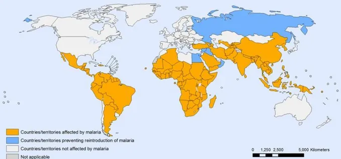
febrile illness, including episodes of rigors alternated with high fever and intense sweating and chills, vomiting, diarrhea and headache [3]. P. falciparum is the most deadly species [4] and can cause severe malaria, with loss of consciousness, cerebral malaria, severe malarial anemia, placental malaria or acute lung injury, whereas the other species cause milder disease, but severity and prognosis may result from diverse parasite and host factors [5]. Various Anopheles spp. female mosquitoes transmit Plasmodium parasites to humans, and their geographical distribution is associated with epidemiological patterns of the disease [6] [7], with tropical and subtropical regions worldwide being affected, as shown in Figure 1 [8].
Figure 1: Countries and territories affected by malaria in 2010. Tropical and subtropical regions are affected by malaria worldwide. [8]
Presently, the World Health Organization (WHO) reports a declining trend in malaria cases and mortality, with 81% of the total 216 million cases and 91% of the 655.000 estimated malaria deaths in 2010 occurring in Africa. Children less than five years of age are 86% of the deaths, and Sub-Saharan Africa accounts for the majority of morbidity and mortality. [4] Nonetheless, some authors believe in a higher global mortality burden, with circa 1.133.000 deaths in 2010 and a higher proportion of malaria deaths in individuals aged five years or
2 older (42%) [9]. In fact, in some African regions more adults are being infected and susceptibility to severe malaria is shifting towards older children [10].
Malaria contributes to prevent social and economic growth in already impoverished countries [11], and the United Nations set malaria combat as part of the Millennium Development Goals. Enormous efforts have been made towards malaria eradication, with mobilization of financial help and human resources allowing substantial advances in research and the implementation of vector control and drug therapy programmes. Nonetheless, an effective vaccine remains to be developed, and effectiveness of insecticide-treated bed nets is vulnerable to populations’ noncompliance and resistance to pyrethroids [12]. Drug therapy, a key part in malaria control, faces increased resistance worldwide, with no drug being universally effective, and so malaria continues to be a threatening global health problem [7].
1.2. Hemozoin, the malarial pigment
1.2.1. Plasmodium life cycle and hemozoin formation
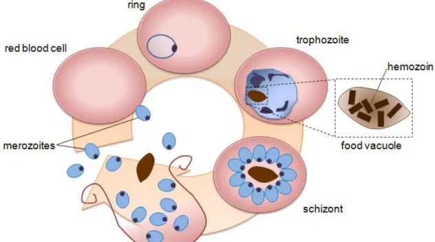
Plasmodium life cycle comprehends the hepatic and erythrocytic phases in the human host
and the sexual reproduction cycle in the mosquito vector. After the mosquito bite and the entrance of numerous sporozoites, multiplication takes place in the liver and merozoites are released into the blood stream, where they invade red blood cells. The blood stage of P.
falciparum life cycle is schematically represented in Figure 2. When inside the erythrocyte,
the parasite consumes host cellular resources so that it can grow, mature and become a schizont, the dividing form. [13] To obtain aminoacids, space and to maintain osmotic balance, the parasite digests up to 80% of the erythrocytic hemoglobin. [14] Inside the parasite acidic food vacuole, hemoglobin is oxidized to methemoglobin and then hydrolyzed into free heme and denatured globin by plasmepsins I, II and IV and histoaspartic protease. Denatured globin is hydrolyzed by falcipain and falcilysin into small peptides, which are only further degraded into amino acids by exopeptidases in the parasite cytoplasm. [15]
Free heme released as a byproduct of these reactions is toxic both to host and parasite. Reactive oxygen species generated by free heme may induce oxidative stress, leading to cell lysis and death, killing the parasite. In the human host, free heme may not only cause hemolysis, but also be involved in inflammation and organ toxicity. To avoid this, in mammals heme is detoxified into biliverdin by heme oxygenase and then into bilirubin by biliverdin reductase. Malaria parasite has different mechanisms to detoxify heme, of which the primary one is hemozoin formation in the food vacuole. Other mechanisms occur in the cytosol, such us detoxification by reduced glutathione, heme-binding proteins or degradation by hydrogen peroxide. [16]
As reviewed by Egan (2008) [17], a specific enzyme was thought to be responsible for polymerizing heme into the malarial pigment, hemozoin. However, it was then suggested that
3 hemozoin crystals were formed by autocatalysis, growing in a process of biomineralization. Despite some controversy, it is now believed that hemozoin crystals nucleate at the interface between the aqueous milieu and the surface of neutral lipid nanospheres in the digestive vacuole, rather than depending on the activity of a specific protein [18] [19].
Figure 2: Plasmodium falciparum blood stage cycle representation. Merozoite invades an erythrocyte and develops into an early trophozoite, the ring form. The early trophozoite grows and becomes a mature trophozoite, in which hemozoin formation starts. DNA synthesis begins, to prepare the dividing stage, the schizont. After erythrocyte rupture, merozoites are released in the blood stream together with hemozoin crystals. Parasites which differentiate into male and female gametocytes can be taken up by the mosquito vector and continue the cycle and transmission.
Hemozoin amount increases as the parasite grows. When the schizont is mature, the infected red blood cell goes through a controlled process of rupture and merozoites are released in the blood stream together with hemozoin crystals. Merozoites will invade other erythrocytes and some will differentiate into gametocytes, which may be taken by the mosquito during its blood meal, starting a new cycle. [20]
1.2.2. Hemozoin characteristics and properties
Hemozoin is a paramagnetic crystal of well-defined, flat faces [21], and with 0,5-1 µm in size [22]. The crystal is constituted by cyclic π-π dimers of ferriprotoporphyrin IX (heme) molecules, with coordination of the propionate group of one heme monomer to the Fe (III) centre of the other, and linked through hydrogen bonds between the propionate groups [23] [24]. Hemozoin from the malaria parasite is considered to be identical to synthetic β-hematin, as assessed by X-ray diffraction and infrared spectroscopy [25] [26]. Notwithstanding the brick-like morphology of P. falciparum hemozoin, synthetic β-hematin has a more needle-like
4 appearance when observed with scanning and transmission electron microscopy [27] [28] [29].
Being birefringent, hemozoin has the ability to depolarize light, and can be detected by optical methods such as darkfield and polarization microscopy, as well as flow cytometry. Hemozoin released in the blood is internalized by host phagocytes, playing an important role in the modulation of the immune system. Depending on the phase of the infection it may be beneficial or harmful to the host, with the pigment producing proinflammatory or immunosuppressive effects. [30] However, published studies on biological effects elicited by hemozoin are performed with different concentrations of hemozoin and the methods used to produce hemozoin are also different. Hence, as different methods yield different types of hemozoin, with distinct properties, they may as well produce different biological results [22]. Numerous studies have been published on hemozoin effect in immune responses and on adjuvant effects during vaccination. Characterization of hemozoin from different sources and the assessment of their differences become extremely important in order to be able to clarify the role of hemozoin in the modulation of immune responses.
1.3. Antimalarial drugs
Malaria therapy was for a long period of time limited to quinine, an alkaloid derived from the bark of the Cinchona tree, already used to treat the disease in the 17th century [2]. Nonetheless, it has serious adverse effects at therapeutic concentrations, which are referred to as cinchonism and include not only headache, nausea, vomiting and diarrhea, but also hearing and vision problems, hypoglycemia, hypotension and venous thrombosis as well as arrhythmia [31] [32] [33]. Last century, synthetic molecules similar to quinine were developed, such as chloroquine, amodiaquine, primaquine, halofantrine and mefloquine [34]. These were safer and easier to produce, and were thus preferentially used for malaria therapy, with chloroquine being the most used [31]. Their mechanism of action is thought to be the inhibition of malarial pigment (hemozoin) formation [35].
However, the parasite evolved to develop resistance to the commonly used drugs through a variety of mechanisms which emerged by the selective drug pressure, now thought to be originating in mutations or copy number changes of genes encoding parasite proteins that are either drug transporters or the drug target [36]. When a drug is administered and absorbed in the recommended dose and is accessible to the parasite, but still it is able to survive and multiply, that strain is considered resistant [34]. Quinine-resistant strains occur very sporadically since the first case in South America in the beginning of the 20th century, but chloroquine-resistant P. falciparum is now widespread all over endemic areas after being first reported to occur in late 1950’s in Southeast Asia and South America, and mefloquine resistance is frequent in Southeast Asia, where it appeared in late 1980’s [37].
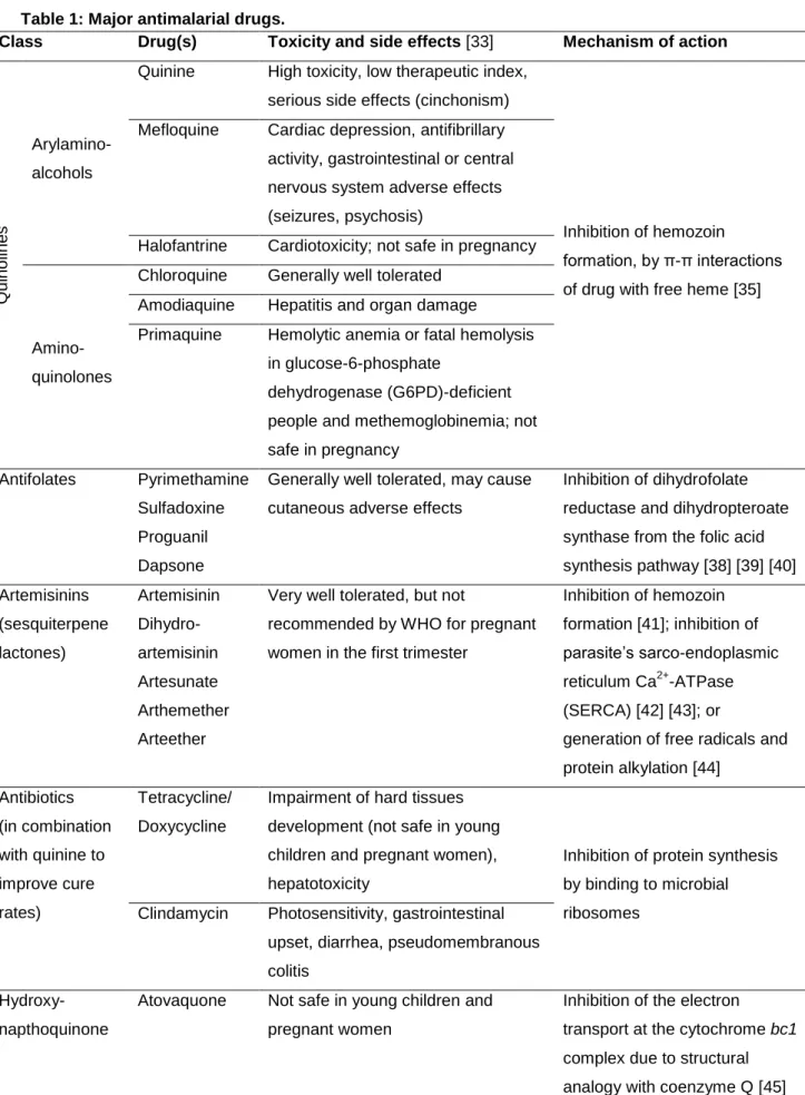
5 Meanwhile other drugs have been discovered that present antimalarial activity (Table 1). Table 1: Major antimalarial drugs.
Class Drug(s) Toxicity and side effects [33] Mechanism of action
Q ui no line s Arylamino- alcohols
Quinine High toxicity, low therapeutic index, serious side effects (cinchonism)
Inhibition of hemozoin
formation, by π-π interactions of drug with free heme [35] Mefloquine Cardiac depression, antifibrillary
activity, gastrointestinal or central nervous system adverse effects (seizures, psychosis)
Halofantrine Cardiotoxicity; not safe in pregnancy
Amino- quinolones
Chloroquine Generally well tolerated Amodiaquine Hepatitis and organ damage
Primaquine Hemolytic anemia or fatal hemolysis in glucose-6-phosphate
dehydrogenase (G6PD)-deficient people and methemoglobinemia; not safe in pregnancy
Antifolates Pyrimethamine Sulfadoxine Proguanil Dapsone
Generally well tolerated, may cause cutaneous adverse effects
Inhibition of dihydrofolate reductase and dihydropteroate synthase from the folic acid synthesis pathway [38] [39] [40] Artemisinins (sesquiterpene lactones) Artemisinin Dihydro- artemisinin Artesunate Arthemether Arteether
Very well tolerated, but not
recommended by WHO for pregnant women in the first trimester
Inhibition of hemozoin formation [41]; inhibition of parasite’s sarco-endoplasmic reticulum Ca2+-ATPase (SERCA) [42] [43]; or
generation of free radicals and protein alkylation [44] Antibiotics (in combination with quinine to improve cure rates) Tetracycline/ Doxycycline
Impairment of hard tissues development (not safe in young children and pregnant women), hepatotoxicity
Inhibition of protein synthesis by binding to microbial ribosomes
Clindamycin Photosensitivity, gastrointestinal upset, diarrhea, pseudomembranous colitis
Hydroxy- napthoquinone
Atovaquone Not safe in young children and pregnant women
Inhibition of the electron
transport at the cytochrome bc1 complex due to structural analogy with coenzyme Q [45] Based on data from publications on the toxicity, side effects [33] and mechanisms of action of the major antimalarial drugs [35] [38] [39] [40] [41] [42] [43] [44] [45].
6 Antifolates are synthetically developed antimalarials and include pyrimethamine, sulfadoxine, proguanil, dapsone and others [38] [39] [40]. Proguanil is commercialized as Malarone™ in a fixed-dose combination with atovaquone [34]. Nevertheless, resistant strains to antifolates spread even faster, with a prompt emergence of resistance to sulfadoxine-pyrimethamine in Southeast Asia and Africa in the 1960’s and the 1980’s.
The most efficient antimalarials are presently artemisinin and its derivatives, which are obtained from Artemisia annua herb. These are recommended by the WHO to treat malaria in combination with other antimalarial drugs, in an attempt to prevent the spread of resistant parasite strains [46]. Artemisinins mode of action is still a subject of much debate, as presented in Table 1, but some authors think it may also have a role in inhibiting hemozoin formation [41]. Importantly, combination therapy is expensive and artemisinin availability may be a limitation. Moreover, its potential toxicity in pregnant women makes it no option to this risk-group, and notwithstanding all the efforts to avoid it, there are reports of artemisinin resistance already in Cambodia [47].
So, despite the severity of its adverse effects, quinine is still the only choice when considering the prevalence of resistant-strains to alternative drug formulations in some areas or their toxicity in pregnant women in their first trimester. However, antibiotics are used in combination with quinine to improve cure rates, but these are toxic for children. [31]
Hence, widespread and multidrug resistance restricts the possibilities of an efficient therapy and holds off effective malaria control and elimination, making the need for new drugs even more urgent [48]. When aiming to discover new antimalarial drugs, hemozoin formation is still one of the most interesting targets, since it seems to be an immutable pathway [30]. Considering this, together with the fact that synthetic quinolines are easy to produce, very well tolerated drugs, the approach of synthesizing quinoline analogs is a very attractive one.
1.4. Development of novel antimalarial compounds
1.4.1. Drug discovery
Hemozoin formation remains a good antimalarial drug target for novel drug discovery. Inhibition of hemozoin formation hampers detoxification of free toxic heme originated by hemoglobin proteolysis in the parasite. Hemozoin formation does not seem to rely on the activity of a parasite-encoded molecule or enzyme and is an exclusive pathway of the parasite. Therefore, drugs targeting hemozoin formation are unlikely to create resistance and are highly selective. [30]
Antimalarial drug discovery is an urgent need, in face of the increasing drug resistance worldwide and the fact that malaria remains one of the more devastating infectious diseases, despite all the efforts to attain the opposite. Diverse strategies can be adopted to develop new compounds or use existing ones in a different formulation or with a novel purpose, in a
7 way that improves current therapy options. While improving existing antimalarial agents is possible by exploitation of analogs through chemical modifications, evaluating the activity of compounds from natural plants and extracts takes advantage of the knowledge of folk remedies, often used by natives in endemic areas for a long time as a solution to febrile illnesses. [49] And whilst new targets are being explored as to be the aim of new compound activity, perhaps an easier approach may be the identification of agents that are already used against other diseases, but prove to be of use in malaria treatment. To this end, high-throughput assays are performed that facilitate the task of screening previously developed compounds. [50]
Several alternatives exist as tools to screen for new antimalarial drugs, apart from their applicability to identify parasite resistance. In vitro sensitivity assays with monitoring of
Plasmodium cultures in the presence of the compound to be tested enables the evaluation of
its capacity to inhibit parasite growth. Parasite growth may be measured based on schizont maturation or parasitemia, using microscopy, automatic reading with flow cytometry and DNA stains, or incorporation of radiolabeled hypoxanthine or ethanolamine. Also, enzyme-linked immunosorbent assays exist that correlate accumulation of specific parasite proteins, such as lactate dehydrogenase and histidine-rich protein 2 (HRP-2), with parasite viability. [51]
1.4.2. Heme inhibition assays for drug screening
Heme inhibition assays allow the investigation of antimalarial drug candidates that target hemozoin formation. Several approaches may be taken to explore drugs which interact with heme molecules. Some focus on the interaction between candidate compounds and heme molecules, and others adapt synthetic β-hematin formation in assay formats to test the compounds’ ability to inhibit crystallization.
Multiwell plate assays provide information regarding the ability of compounds to bind to heme molecules, by analyzing inhibition of glutathione-dependent hemin degradation by those compounds [52] [53] [54]. Other types of analysis search for potential inhibitors of hemozoin formation by investigating drug-heme interactions by mass spectrometry [55] or UV-visible spectroscopy [56] [57].
Synthetic β-hematin formation can also be used as a process which mimics hemozoin biocrystallization, to perform in vitro assays where candidate compounds can be assessed for their ability to inhibit hemozoin growth [58]. Different β-hematin assays are performed according to different experiment formats, where reagents and growth conditions differ greatly [21]. The first assays to be described made use of P. falciparum derived hemozoin as an initiator for β-hematin formation and 14C-hemin as a way of quantifying the end product [59]. Meanwhile, the heme polymerization inhibitory activity (HPIA) spectrophotometric microassay was developed, which identifies ligands that bind axially with the protoporphyrin
8 iron [60]. Promoting coordination of pyridine to unreacted hematin, Ncokazi and Egan attained a colorimetric assay which enabled visual identification of inhibiting compounds by formation of an orange-pink color, and named it pyridine hemichrome inhibition of β-hematin (Phiβ) assay [61]. Parapini and colleagues further improved the HPIA assay into the β-hematin inhibitory activity (BHIA) assay, using the more stable hemin chloride instead of hematin and adjusting pH to 5, and concluded that these conditions allow for the identification of ligands undergoing π-π interactions with heme [62]. Several groups have made use of some kind of initiator or catalyst molecules to improve β-hematin inhibition assays, including detergents such as Tween20 [63] or NP-40 and neutral lipids [64], or lecithin [65].
Standardization of heme inhibition assays is difficult and results are not consistent between assays [59] [60]. The assays often involve complex procedures, make use of various conditions, reagents as well as different types of initiators [63] [61] [60] [62] [66] [64] [59], and some use rather toxic reagents [61] [64] [59]. Moreover, the end product of these heme inhibition assays is not always characterized, with little information about the assessment of the nature and quality of the final aggregates [67] [68] [69] [70].
1.5. Novel hemozoin-like crystal inhibition assay for drug screening
Ileal Fluid Dependent Organism (IFDO) was first described by Burdon in 1989, in consequence of the isolation of a “replicating agent” from the intestinal tract of patients with Crohn’s disease. After being “cultured” in specific broth and agar media, appearance of dark-brown “bacterial-like growth” was evident, but the hypothesis of IFDO being formed by crystallization from constituents of the medium instead of a biological process was already posed. [71] As IFDO is susceptible to inactivation treatments similar to prions, the company Steris became interested in exploring this agent as a model to test drugs and processes against prion propagation. Interestingly, detailed characterization of IFDO revealed it to be very similar to malarial pigment, hemozoin, as it is an aggregate of ferriprotoporphyrin IX (heme). At that point, Steris contacted the host laboratory and a collaboration project to study this crystal was initiated. IFDO particles were grown in a well defined medium and a comparative analysis was performed along with hemozoin obtained by different methods. A more extensive characterization of the crystal making use of X-ray diffraction and infrared spectroscopy led to the finding that, despite the similarity, IFDO is not true hemozoin, and so the crystals were termed hemozoin-like crystals (HLC). However, following Steris investigation on an assay to evaluate the therapeutic and decontamination efficiency against transmissible misfolded proteins, HLC were explored for the potential to be grown as a novel and simple inhibition assay for antimalarial drug screening.
9
2. Objectives
The aim of this work was to produce malarial pigment (hemozoin) from different origins, characterize it and establish its purity by several methods. The ability of antimalarial and antibiotic drugs to inhibit the crystallization of hemozoin-like crystals was investigated, together with the potential of these crystals to be used for screening for drugs inhibiting hemozoin formation.
3. Material and Methods
All reagents were purchased from Sigma-Aldrich® (Sintra, Portugal) unless stated otherwise.
3.1. Culture media and solutions
3.1.1. Malaria Complete parasite Medium (MCM): 500 mL of RPMI 1640 (1×, without L-glutamine, with NaHCO3) (Gibco™, Life Technologies, Madrid, Spain) supplemented with 50 mL of 10× AlbuMAX II® solution, 12 mL of HEPES Buffer Solution (1 M) (Gibco™, Life Technologies, Madrid, Spain), 5 mL of L-glutamine 200 mM and 500 µL of gentamicin 50 mg/mL (Gibco™, Life Technologies, Madrid, Spain).
3.1.2. 10× AlbuMAX II® solution: 25 g of AlbuMAX II® in a 500 mL aqueous solution of 5,2 g of RPMI 1640 (with L-glutamine, without NaHCO3) supplemented with 500 µL of gentamicin 50 mg/mL, 2,98g of HEPES, 1,67 g of sodium bicarbonate, 1 g of glucose and 0,1 g of hypoxanthine, adjusting pH to 7,2-7,4 and filtering 0,22 µm. All reagents from Life Technologies (Madrid, Spain) except HEPES which is from VWR (Carnaxide, Portugal). 3.1.3. 1× Phosphate-buffered saline (PBS): diluted 1:10 from 10× PBS pH 7,2 (Gibco™, Life Technologies, Madrid, Spain) in ultrapure water, obtained with Milli-Q Synthesis Q-Gard®1 water purification system (Millipore, Billerica MA, USA).
3.1.4. Glycerolyte cryoprotective solution: constituted by 57% of glycerol, 16 g/L of sodium lactate (VWR, Carnaxide, Portugal), 300 mg/L of potassium chloride and 25 mM of sodium phosphate pH 6,8.
3.1.5. Broth medium for HLC culture: prepared by autoclaving 35,5 g of Mycoplasma Broth Base (Oxoid, Basingstoke, England) in 950 mL of distilled water, obtained with Elix®10UV Progard®2 water purification system (Millipore, Billerica MA, USA) and 2 mL of Tween® 80; cooling at 50°C and adding 1,33 mL of horse serum (Gibco™, Life Technologies, Madrid, Spain), 20 mL of a 10% pancreatin solution in 1× PBS and 30 mL of a fresh blood extract. Medium is filtered at 0,22 µm and immediately used.
3.1.6. Fresh blood extract: prepared by washing red blood cells from healthy human donors, obtained from a “buffy coat” from Instituto Português do Sangue (IPS), three times with 1 vol. of 1× PBS; lysing RBC by addition of 1 vol. of sterile ultrapure water; submitting the
10 suspension to three successive freeze-thaw cycles and 5 min. of sonication using the 35 kHz ultrasonic bath Transsonic T570 (Elma®, Singen, Germany); centrifuging for 15 min. at 2400×g and discarding the pellet.
3.1.7. Wash buffer: constituted by 10 mM Tris hydrochloride (Merck, Lisbon, Portugal) pH 8.3, 50 mM potassium chloride and 1,5 mM magnesium chloride.
3.1.8. Solution A: constituted by 10 mM Tris hydrochloride pH 8,3, 100 mM potassium chloride and 2,5 mM magnesium chloride.
3.1.9. Solution B: constituted by 10 mM Tris hydrochloride pH 8,2, 2,5 mM magnesium chloride, 1% Tween® 20, 1% Triton® X-100 and 120 µg/mL proteinase K (Promega, Madison WI, USA).
3.2. Plasmodium falciparum cultures
3.2.1. Continuous Plasmodium falciparum cultures maintenance
Plasmodium falciparum 3D7 strain was obtained from Malaria Research and Reference
Reagent Resource Center (MR4; Manassas VA, USA) and continuous cultures of the parasite were maintained with human red blood cells (RBC), isolated from buffy coats. Healthy human blood from the buffy coat was washed three times with RPMI 1640 (1×, without L-glutamine, with NaHCO3) (Gibco™, Life Technologies, Madrid, Spain) and centrifuged 600×g for 5 minutes without brake to separate and isolate RBC from the leukocytes. Infected RBC were incubated at 37°C and 5% CO2, in MCM, which was changed every day.
Cultures were kept at <1% parasitemia and 5% hematocrit, unless when used to purify or extract hemozoin, for which it was necessary to have >10% parasitemia and 1% hematocrit, and 2-5% parasitemia and 5% hematocrit, respectively.
3.2.2. Giemsa staining of blood smears
To estimate the amount of infected RBC in culture, a smear was prepared daily with a small amount of blood from the culture, when the medium was changed. The smear was fixed in absolute methanol (Merck, Lisbon, Portugal) for 20 s, air-dried and stained with Giemsa’s azur eosin methylene blue solution (Merck, Lisbon, Portugal) diluted 1:100 in 1× PBS, for 20 minutes. Then, slides were rinsed with tap water and air-dried to be observed by bright field microscopy under oil immersion.
Parasitemia is determined as the average percentage of infected RBC on ten different visual fields when observing at 1000× magnification. When needed, the infected RBC were diluted by substituting the right volume with uninfected RBC to lower parasitemia and maintain the hematocrit.
11 3.2.3. Frozen P. falciparum 3D7 strain stocks
Frozen stocks of schizont-enriched P. falciparum 3D7 infected RBC from highly parasitized cultures can be maintained in vials at -80°C or in liquid nitrogen for long-term storage if transferred to a cryoprotective solution of glycerolyte. After obtaining the pellet of infected RBC from the culture by centrifuging for 10 min. at 600×g, 0,33 and then 1,33 volumes of glycerolyte were added drop by drop, gently mixing.
When needed, glycerolyte-frozen parasites were thawed to begin a new culture by gently washing the thawed infected RBC with 0,1 vol. of 12% and 10 vol. of 1,6% sodium chloride, centrifuging at 600×g and 20°C for 5 min., resuspending in 1× RPMI 1640 (without L-glutamine, with NaHCO3) (Gibco™, Life Technologies, Madrid, Spain), centrifuging again and finally resuspending in MCM. Uninfected RBC were added to obtain 5% hematocrit in the culture when it was necessary.
3.3. Purification of Plasmodium falciparum hemozoin
P. falciparum hemozoin (or native hemozoin, nHZ) was purified after saponin harvesting of
parasites from 1 L of P. falciparum (3D7 strain) cultures at 1% hematocrit, enriched in trophozoites at a parasitemia of at least 10%, as previously described by Coban et al. (2002) [72]. In short, parasites were extensively washed with 1× PBS, pellet was sonicated for 5 min., extensively washed with 2% sodium dodecyl sulfate (SDS) and then incubated overnight with 2 mg/mL proteinase K (Promega, Madison WI, USA). After being washed with 2% SDS again, the pellet was incubated for 3 h in 6 M urea and then washed with 2% SDS and ultrapure water. Purified nHZ was resuspended in 1 mL of ultrapure water, quantified as heme-equivalents using QuantiChromTM Heme Assay Kit DIHM-250 (BioAssay Systems, Hayward CA, USA), and stored at 4°C.
3.4. Extraction of crude Plasmodium falciparum hemozoin
P. falciparum hemozoin was extracted without further purification (cnHZ), as described by
Keller et al. (2004) [73], with some modifications. Briefly, infected RBC from 2 to 5% parasitemia P. falciparum (3D7 strain) cultures at 5% hematocrit were centrifuged at 800×g for 10 min. and the pellet was resuspended in 40 mL of 1× PBS with 1% of saponin for 10 minutes. After centrifuging at 16000×g for 15 min., the pellet was washed seven times in 1× PBS, resuspended in 1 mL of ultrapure water, quantified as previously described and stored at 4°C.
3.5. Production of synthetic hemozoin
Synthetic hemozoin (sHZ) was obtained by the method described by Slater et al. (1991) [25], with some modifications. Briefly, 475 mg of hemin chloride were dissolved in 100 mL of 0,1 N
12 sodium hydroxide and heme was precipitated by slowly adding 35 mL of glacial acetic acid (Merck, Lisbon, Portugal). Crystallization was promoted by overnight incubation of the mixture at 80°C. Non-crystalline heme was then removed by washing three times with 1 vol. of 100 mM sodium bicarbonate pH 9,1 during 3 h, centrifuging for 15 min. at top speed. The pellet was further washed three more times in 1 mL of ultrapure water and finally ressuspended, quantified as for nHZ, and stored at 4°C.
3.6. Production of hemozoin-like crystals
Original “IFDO” cultures were kindly provided by Dr D. Burdon to Steris and later on termed hemozoin-like crystals (HLC). Culture methods were adapted from the original protocol [71], in which a seeded medium is used to grow the particles (here designated Broth medium for HLC culture). Cultures were prepared by seeding 50 µL of a McFarland 6 HLC suspension in 10 mL of Broth medium for HLC culture, and incubating at 37°C and 5% CO2 for 5 to 7 days, in the CO2 incubator Heraeus® HERA® Cell (Fisher Scientific, Loures, Portugal).
A modified broth medium was also developed, replacing the fresh blood extract by 50 µM hemin chloride, added from a stock solution prepared in 0,4 N sodium hydroxide, and adjusting pH of the medium at 7,2 before filtering at 0,22 µm. An Agar medium for HLC culture was also prepared, with Mycoplasma Agar Base (Oxoid, Basingstoke, England). Besides these, other growth conditions were tested, including: adjusting pH of the medium to 5; incubation at ambient atmosphere instead of 5% CO2; using Brain Heart Infusion (Becton Dickinson, Madrid, Spain) broth or 1× PBS instead of Mycoplasma Broth Base; replacing horse serum by human serum (obtained from a healthy donor); excluding pancreatin from the medium; and seeding with sHZ instead of HLC.
Obtained HLC were washed three times with 1 vol. of 100 mM sodium bicarbonate pH 9,1 during 3 h, centrifuging for 15 min. at top speed, followed by three washes in ultrapure water. Then, HLC were finally resuspended in 1 mL of ultrapure water, quantified and stored at 4°C as performed for hemozoin. Alternatively, absolute ethanol (Merck, Lisbon, Portugal), absolute methanol (Merck, Lisbon, Portugal) and dimethyl sulfoxide (DMSO) were also used to wash HLC, in an attempt to reduce remaining heme contamination.
3.7. Light and depolarizing microscopy
Giemsa-stained smears of infected RBC from P. falciparum cultures (prepared as described above), and hemozoin and HLC preparations were observed with light and depolarizing microscopy, using the brightfield microscope Leica DM2500 (Leica Microsystems, Wetzlar. Germany).
13
3.8. Scanning electron microscopy
Ten microliters of hemozoin and HLC samples were allowed to dry overnight in air on top of a carbon tape on a metallic sample holder and were metalized for 30 min. using JEOL, JFC-1200 with a gold target. Scanning electron microscopy was performed with JEOL, JSM-2500 LV scanning electron microscope (JEOL, Tokyo, Japan), at Faculdade de Ciências da Universidade de Lisboa.
3.9. X-ray diffraction
Hemozoin and HLC were subjected to X-ray diffraction (XRD) analysis. Crystals were resuspended in 10 µL of absolute ethanol (Merck, Lisbon, Portugal) and allowed to air-dry on the silicon sample holders. XRD patterns were then acquired at Faculdade de Ciências da Universidade de Lisboa, with the automatic X-ray diffractometer Philips Analytical PW 3050/60 X’ Pert PRO (Ө /2Ө) (PANalytical, Almelo, The Netherlands), with an X’Celerator detector and automatic data acquisition with X’Pert Data Collector, version 2.0b. Cu Kα radiation (λ= 1,54060 Å) was used, operating with 30 mA and 40 kV. The diffractograms were recorded in the 2Ө range between 5° and 30° with a step size of 0.0170° (2Ө) and scan step time of 100 seconds.
3.10. Infrared spectroscopy
Synthetic hemozoin and HLC were dried overnight in a desiccator over phosphorus pentoxide and silica gel. Potassium bromide pellets of sHZ and HLC were prepared from the dried samples with a mortar and pestle and a compressor. Infrared spectra were obtained for each sample at Faculdade de Farmácia da Universidade de Lisboa, using IRAffinity-1 Fourier Transform Infrared Spectrophotometer and IR Solution Software (Shimadzu, Kyoto, Japan).
3.11. Heme contamination assessment
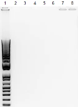
Hemozoin and HLC produced were assessed for remaining heme contamination using thin layer chromatography (TLC). A quantified sample was first diluted in absolute methanol (Merck, Lisbon, Portugal) to the higher concentration of hemin chloride to be eluted (0,2 mM). Then, 10 µL of the sample were eluted for 30 to 40 min. on a silica gel glass plate (Merck, Lisbon, Portugal) along with hemin solutions of known concentrations (0,2, 0,04 and 0,02 mM), inside a methanol-saturated tank. Images of the plates were acquired with Alpha Imager® HP System (ProteinSimple, Santa Clara CA, USA) and the result was analyzed by determination of integrated density using ImageJ software (National Institute of Mental Health, Bethesda MD, USA) and calculation of remaining heme contamination percentage in the sample.
14
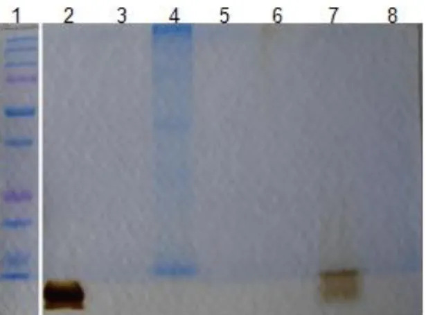
3.12. Mycoplasma contamination assessment
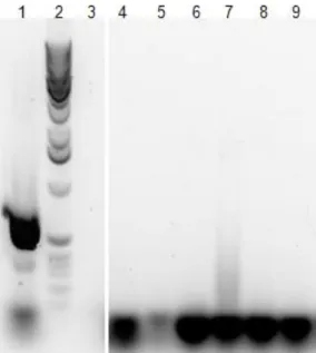
To assess for Mycoplasma contamination, Polymerase Chain Reaction (PCR) was performed as follows. One hundred microliters of sHZ, nHZ, cnHZ and HLC were diluted in 1 mL of 1× RPMI 1640 (Gibco™, Life Technologies, Madrid, Spain) and incubated in a 24-wells plate for 24h at 37°C and 5% CO2. After incubation, samples were collected into a microtube, centrifuged for 15 min. at 15700×g and 4°C, supernatant was discarded and pellet resuspended in Wash buffer. After centrifuging again, pellet was resuspended in 1:1 Solution A and Solution B, incubating at 60°C for 1 h followed by denaturing at 90°C for 10 minutes. PCR was performed using My Cycler™ thermal cycler (Bio-Rad, Amadora, Portugal), by subjecting the Reaction mix to the following program: 1 cycle at 95°C for 5 min. (step 1), 30 cycles for step 2 – 95°C for 0,5 min., 58°C for 1,5 min., 72°C for 1,5 min. – and 72°C for 10 min. (step 3). Reaction products were analyzed together with 1kb DNA Extension Ladder (Life Technologies™, Madrid, Spain) by electrophoresis at 60 V, for 1 h in 1× TAE Buffer, on a 1% agarose (NZYTech, Lisboa, Portugal) gel with 10000× GelRed® Nucleic acid gel stain (Biotium, Hayward CA, USA) diluted 1:20000. Positive results show a DNA amplicon of 717 bp.
Reaction mix: prepared to a volume of 25 µL in nuclease-free distilled water (Gibco™, Life Technologies, Madrid, Spain), with 1× NZYTaq Colourless Master Mix (NZYTech, Lisboa, Portugal), 0,1-1 µM upstream (Pr27 – 5’ TGC ACC ATC TGT CAC TCT GTT AAC CTC 3’) and downstream (Pr22 – 5’ ACT CCT ACG GGA GGC AGC AGT A 3’) primers (Fisher Scientific, Loures, Portugal), and <250 ng DNA template.
3.13. DNA contamination assessment
Hemozoin and HLC were assessed for DNA contamination by agarose gel electrophoresis. Ten microliters of each sample, pretreated or not with 10 µL of 1% saponin for 10 min., were loaded with 3,5 µL of Gel Loading Buffer on a 0,8% agarose gel with 10000× GelRed® Nucleic acid gel stain diluted 1:20000, and ran in 1× TAE Buffer for 30 min. at 100 mA.
3.14. Protein contamination assessment
Hemozoin and HLC were assessed for protein contamination by polyacrylamide gel electrophoresis under denaturing conditions. Twenty microliters of each sample were boiled for 5 min. at 100°C with 20 µL of 2× Loading buffer and then loaded onto a SDS-polyacrylamide gel (5% Acrylamide resolving gel/4% Acrylamide stacking gel) and ran in 1× Running buffer, for 40 min. at 180 V. The gel was stained in 1% Coomassie Brilliant Blue staining solution (Bio-Rad, Amadora, Portugal) for 20 min., destained overnight in Destain solution (constituted by 30% ethanol and 10% acetic acid in distilled water). After being
15 rinsed with abundant water, an image of the gel was acquired with Alpha Imager® HP System. Gels were dried on a filter paper with the DrygelSr Slab Gel Dryer SE1160 (Hoefer® Scientific Instruments, Holliston MA, USA), for at least 40 min. at 70°C.
2× Loading buffer: 10% glycerol, 5% β-mercaptoethanol, 3 % SDS, 62,5 mM Tris (Merck, Lisbon, Portugal) pH 8,8, and 0,01% bromophenol blue (Bio-Rad, Amadora, Portugal) in ultrapure water.
5% Acrylamide resolving gel: 5% acrylamide/0,1% bisacrylamide (Bio-Rad, Amadora, Portugal), 0,375 M Tris (Merck, Lisbon, Portugal) pH 8,8, 0,1% SDS, 0,05% ammonium persulfate and 0,1% TEMED in ultrapure water.
4% Acrylamide stacking gel: 4% acrylamide/0,1% bisacrylamide (Bio-Rad, Amadora, Portugal), 0,375 M Tris (Merck, Lisbon, Portugal) pH 8,8, 0,1% SDS, 0,05% ammonium persulfate and 0,1% TEMED in ultrapure water.
5× Running buffer: 15,15 g/L Tris, 72,05 g/L glycine (Merck, Lisbon, Portugal) and 0,5% SDS.
3.15. Hemozoin and Hemozoin-like crystals quantitation (heme equivalents)
The amount of hemozoin and HLC in the water suspensions was quantified as heme-equivalents using QuantiChromTM Heme Assay Kit DIHM-250 (BioAssay Systems, Hayward CA, USA), by colorimetric determination of total heme at 400 nm. Shortly, after solubilization in an aqueous solution of 20 mM sodium hydroxide for 1 h, samples were added to the Reagent solution and let react for 5 min., after which absorbance at 400 nm was read and the concentration in µM (heme equivalents) determined using the following expression:
Total heme concentration =
× 62,5 × Dilution factor,
which correlates absorbance at 400 nm with the concentration of heme present in solution.
3.16. Novel Hemozoin-like crystal inhibition assay
A McFarland 6 HLC suspension in water was sonicated for 5 min., using the 35 kHz ultrasonic bath Transsonic T570 (Elma®, Singen, Germany), and diluted 1:100 in Broth medium for HLC culture. One hundred and eighty microliters were distributed in wells of a 96-wells plate. A range of drug concentrations was tested in order to determine a minimum inhibitory concentration for HLC growth. Stock solutions of chloroquine diphosphate, amodiaquine dihydrochloride, quinine hydrochloride dihydrate, quinacrine dihydrochloride, clindamycin hydrochloride, gentamicin and ampicillin were prepared in sterile ultrapure water, and those of mefloquine hydrochloride (Roche, Amadora, Portugal), halofantrine hydrochloride and artemisinin were prepared in absolute methanol (Merck, Lisbon, Portugal). After being adjusted to pH 7 ± 0,2 and 0,22 µm-filtered, drug solutions diluted in Broth
16 medium for HLC culture and then diluted in the seeded wells to a final concentration of 50, 100, 250, 500, 750 and 1000 µM. Each drug was tested at least in duplicate, and plates were visually observed every day, for 5 days of incubation at 37°C and 5% CO2, to determine presence or absence of HLC growth – wells with less growth than unseeded control were considered as presenting absence of growth. Five microliters of all crystal-positive wells were seeded onto Tryptone Soya Agar (Oxoid, Basingstoke, England) and Mycoplasma Agar Base (Oxoid, Basingstoke, England) plates to confirm absence of bacteria. Five microliters from each well were also seeded onto fresh plates of Agar medium for HLC culture, to observe any HLC regrowth.
4. Results
4.1. Characterization of hemozoin of different origins and hemozoin-like crystals
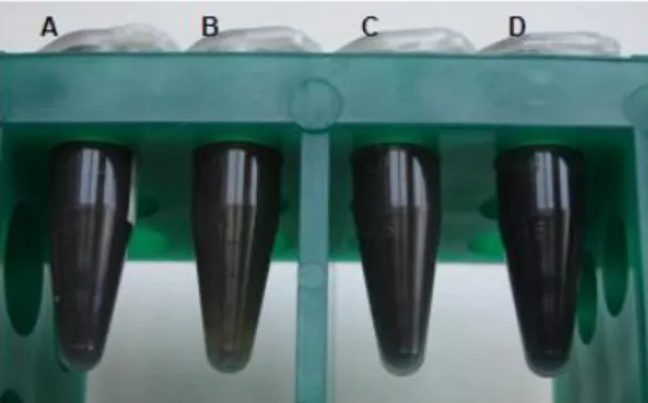
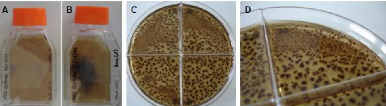
Hemozoin crystals are a dark-brown, black powder which when suspended in water give rise to a blackish suspension. Figure 3 shows the end-product of hemozoin synthesis and purification or extraction from P. falciparum (3D7 strain) cultures, as well as the product of hemozoin-like crystals (HLC) culture. Water suspensions of hemozoin obtained from the different origins and HLC were kept in microtubes at 4°C, after being quantified as for heme equivalents concentration. Visually, all these crystals are very similar, and when air-dried yield a black powder which presents no differences when observed with the naked eye.
Figure 3: Water suspensions of hemozoin and hemozoin-like crystals (HLC). Synthetic hemozoin (sHZ, A), native hemozoin (nHZ, B), crude native hemozoin (cnHZ, C) and HLC (D) suspensions in water were kept in microtubes at 4°C after quantification in terms of heme equivalents concentration.
During HLC incubation in broth medium at 37°C and 5% CO2, a dark-brown deposit appeared, as visible in Figure 4B. A small deposit also formed in the unseeded broth medium, but in a residual amount (Figure 4A), and not always being observed. Alternatively, an agar medium was used, in which seeded HLC originated dark-brown structures similar to bacterial or fungal “colonies”, which occurred beneath and on the surface of the agar, and were more numerous near the site of inoculation (Figure 4C and 4D).
17 Figure 4: Hemozoin-like crystals (HLC) cultures in broth and agar media. HLC unseeded (A) and seeded (B) broth in culture flasks and HLC seeded agar medium plates (C, D) were incubated for 5 days at 37°C and 5% CO2.
4.1.1. Light and depolarizing microscopy
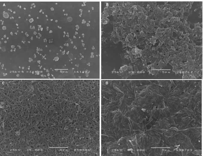
Hemozoin from the different sources and HLC have a crystalline appearance and depolarize light as observed with light and depolarizing microscopy at 1000× magnification, with the optical microscope Leica DM2500 (Figure 5). All preparations showed a more or less homogeneous crystal in size, but tend to aggregate in suspension, as shown for native hemozoin (nHZ) in Figure 5A and HLC in Figure 5D. Aggregates could be dispersed by incubating the samples in the ultrasonic bath (with a frequency of 35 kHz) for 5 min., as exemplified for synthetic hemozoin (sHZ) in Figure 5C, with the exception of HLC (Figure 5D). In the case of HLC, extensive sonication did not disperse crystal aggregates in suspension. The crude extract of native hemozoin (cnHZ) seems to consist of small aggregates of hemozoin surrounded by membranes (Figure 5B).
Figure 5: Microscopy observation of hemozoin and hemozoin-like crystals (HLC). Light (upper pannel) and depolarizing (lower pannel) microscopy observation of native hemozoin (nHZ, A), crude native hemozoin (cnHZ, B), synthetic hemozoin (sHZ, C) and HLC (D), at 1000× magnification, with the optical microscope Leica DM2500.
Some differences could be noticed in the way that hemozoin from different origins depolarized light, with needle-like particles appearing more bluish (Figure 5C, lower panel) and aggregates more brown-gold and green (Figure 5A, lower panel).
18
4.1.2. Scanning electron microscopy
Scanning electron microscopy allowed to analyze morphology and size of hemozoin crystals. As visible in Figure 6, sHZ, nHZ and HLC have a similar size, which ranges from 1,1 to 2,4 µm, HLC having the biggest crystals. Also, morphology of the crystals is very alike, showing protruding needle-like particles, as presented in Figure 6 (B, C and D). Native hemozoin appears to be more brick-like shaped (Figure 6B). At 3500× magnification, round structures are visible in images acquired from cnHZ samples (Figure 6A).
Figure 6: Scanning electron microscopy of hemozoin and hemozoin-like crystals (HLC). Crude native hemozoin (cnHZ A) (3500× magnification), and native hemozoin (nHZ B), synthetic hemozoin (sHZ C) and HLC (D) (5000× magnification) images obtained for air-dried samples with the scanning electron microscope JEOL, JSM-2500 LV.
4.1.3. X-ray diffraction
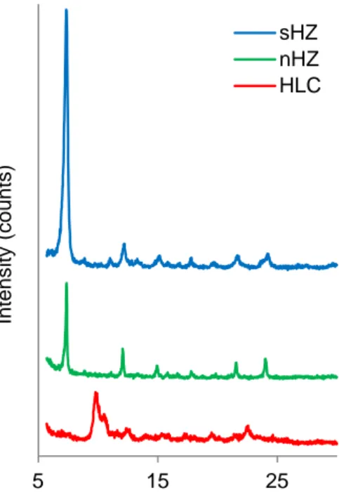
Hemozoin from synthetic (sHZ) and natural (nHZ) source was analyzed with the X-ray diffractometer Philips Analytical PW 3050/60 X’ Pert PRO and revealed to have identical X-ray diffraction peak patterns (Figure 7). HLC presented a different X-X-ray diffraction pattern, which is indicative of a difference relative to the other crystals.
19 Figure 7: X-ray diffraction patterns of hemozoin and hemozoin-like crystals (HLC). Synthetic hemozoin (sHZ, blue line), native hemozoin (nHZ, green line) and HLC (red line) air-dried samples were analyzed on silicon sample holders with the X-ray diffractometer Phillips Analytical PW 3050/60 X’ Per PRO using a Cu Kα radiation (λ= 1,54060 Å) source.
4.1.4. Infrared spectroscopy
When subjected to infrared spectroscopy analysis, sHZ produced a spectrum which presents the two hemozoin-characteristic peaks at 1663 and 1210 cm-1 (highlighted with arrows in Figure 8). HLC failed to show these peaks, thus confirming XRD data, which indicated the two crystals are in fact different.
Figure 8: Infrared spectra of synthetic hemozoin (sHZ) and hemozoin-like crystals (HLC). sHZ (blue line) and HLC (red line) samples were dried over phosphorous pentoxide and silica gel in a desiccator and analyzed in potassium bromide pellets with IRAffinity-1 Fourier Transform Infrared Spectrophotometer; arrows highlight the two hemozoin-characteristic peaks at 1663 and 1210 cm-1.
0 2700 5 15 25 Int en s ity ( c ou nts ) ° 2Ө sHZ nHZ HLC
20 Obtaining native hemozoin from parasitized cultures is considerably time consuming and costly and infrared spectroscopy analysis requires a relatively high amount of sample. Thus, considering that XRD results had already shown nHZ and sHZ to be identical, nHZ was not analyzed with infrared spectroscopy.
4.2. Hemozoin and hemozoin-like crystals quantitation
4.2.1. QuantiChrom™ Heme Assay Kit
After production and purification or extraction of hemozoin from different origins and HLC, the resultant suspensions in water were quantified as heme-equivalents using QuantiChromTM Heme Assay Kit, which correlates absorbance at 400 nm with the concentration of total heme in solution. Concentration of sHZ and HLC samples ranged between approximately 3 to 11 mM, depending on the initial total volume of reaction or culture. In spite of yielding larger pellets after extraction from the P. falciparum (3D7 strain) cultures, because cnHZ is not purified, it also contains membranes and proteins which contribute to pellet volume, without meaning it has a higher amount of hemozoin. Samples of cnHZ had concentrations of 0,2 mM. Purification from P. falciparum (3D7 strain) cultures produced water suspensions of nHZ at around 1 mM.
4.2.2. Absorbance measurements
Trying to follow HLC growth by absorbance measurement revealed to be a somewhat difficult task. When growing HLC in glass tubes, daily measuring A405 nm with WPA CO8000 Biowave Cell Density Meter (Biochrom, Cambridge, United Kingdom) reflected the dark brown deposit formation in the bottom of the tubes, augmenting from first day and stabilizing after 5 to 7 days of incubation. However, when cultures were carried out in polystyrene 96-well plates, neither A405 nm or A595 nm measuring with the microplate reader Infinite M200 (Tecan, Männedorf, Switzerland) were reliable HLC growth indicators, as they varied greatly between duplicates and in some cases unseeded wells had higher absorbance values than seeded ones, making it impossible to subtract a “blank” value from the growing HLC wells.
4.3. Purity and contamination assessment of hemozoin and hemozoin-like crystals
4.3.1. Heme contamination
Heme contamination in hemozoin and HLC samples was assessed by thin layer chromatography (TLC) and examples of the silica plates used are depicted in Figure 9. Hemozoin from natural source was not contaminated with remaining heme (Figure 9A and 9B). The synthetic crystal obtained from hemin chloride was contaminated with a residual amount of free heme, representing less than 1% of total heme content. However, HLC has