UNIVERSIDADE DE LISBOA
FACULDADE DE MOTRICIDADE HUMANA
Sexual dimorphism in heart rate recovery from peak exercise
Dissertação elaborada com vista à obtenção do Grau de Mestre em Exercício e Saúde
Orientador: Prof. Doutor Gonçalo Laima Vilhena de Mendonça
Júri: Presidente
Prof. Doutor Fernando Manuel da Cruz Duarte Pereira Vogais
Prof. Doutor Gonçalo Laima Vilhena de Mendonça Prof. Doutor Pedro Xavier Melo Fernandes Castanheira
Carolina Marques Teodósio 2016
2 “Working hard is important. But there is something that matters even more, believing in yourself."
3
4
Acknowledgements (in Portuguese)
Primeiro que tudo, um especial e grande agradecimento ao meu orientador e professor Gonçalo V. Mendonça, pelo convite para este estudo e principalmente por todo o apoio dado ao longo desta dissertação. Obrigado por todo o tempo despendido e todas as conversas partilhadas.
Segundo, quero agradecer à minha família por todo o apoio que me deu ao longo destes últimos anos. Tem sido uma longa jornada longe de casa, com muitos altos e baixos. Obrigada por todo o amor e coragem que me concederam.
Um especial agradecimento à minha querida mãe, por me apoiar em todos os momentos e me mostrar alegria nos momentos mais tristes.
Ao meu pai, por estar sempre presente quando eu mais preciso.
À minha irmã Helena, por todas as brigas e desentendimentos e à Nina, a nossa companheira para sempre.
Aos meus avós, por todo o carinho e amor.
Ao meu namorado Frederico, por me fazer rir e nunca me deixar desistir. Por toda a paciência, carinho e por todos os momentos passados juntos.
5 Aos meus queridos amigos e colegas de trabalho do Estádio Universitário de Lisboa, CEDAR, Ana, Andreia, Hugo e Miguel, por me ajudarem a crescer pessoal e profissionalmente ao longo destes últimos anos.
Por fim, um obrigado a todos os participantes deste estudo, pela sua dedicação e disponibilidade dispensada.
6
List of Acronyms
ANCOVA Analysis of covariance BMI Body mass index BP Blood pressure
CVD Cardiovascular disease DBP Diastolic blood pressure HR Heart rate
HRpeak Peak heart rate
HRR Heart rate recovery
HRR1min HRmáx – HR1 (Heart rate at the first minute of recovery) HRR2min HRmáx – HR2 (Heart rate at the second minute of recovery) MAP Mean arterial pressure
R2 Coefficient of determination RER Respiratory exchange ratio SBP Systolic blood pressure
SPSS Statistical package for the social sciences Ve Minute ventilation
VO2 Oxygen consumption
VO2peak Peak oxygen uptake
7
Units of measure
bpm beats per minute cm centimetres kg kilograms
kg/m2 kilograms per square meter L Litters
L.min-1 Litters per minute mmHg millimetres of mercury
ml.kg-1.min-1 millilitres per kilogram per minute rpm rotations per minute
8
List of Contents
Abstract (English) ... 10 Resumo ... 12 Epilogue ... 14 I. Introduction ... 15II. Aims and hypotheses ... 19
III. Methods ... 20 Participants ... 20 Measurements ... 20 Study Design ... 21 Statistical Analysis ... 23 IV. Results... 25 Rest ... 25 Exercise ... 26 Recovery ... 27 V. Discussion ... 31 Limitations ... 34 VI. Conclusions... 35 VII. References ... 36
9
Tables Index
Table 1. Descriptive characteristics of participants………..25 Table 2. Physiological and mechanical variables recorded at the ventilatory threshold (VT).26 Table 3. Physiological and mechanical variables obtained during peak exercise………27 Table 4. Multiple linear regression models for the 1st and 2nd minute heart rate recovery…..28
Figures Index
Figure. 1. Schematic representation of the study protocol...………...22 Figure 2. Heart rate obtained at the first and second minute of recovery post-peak exercise. ……….……….27 Figure. 3 Relationship between cardiodeceleration at 1 min of recovery (HRR1min) with peak
oxygen uptake (VO2peak). ……….29
Fig. 4 Relationship between cardiodeceleration at 2 min of recovery (HRR2min) with peak oxygen uptake (VO2peak) in men and women………29
10
Abstract (English)
Purpose: Delayed heart rate recovery (HRR) after peak exercise is associated with decreased vagal reactivation and represents a prognostic marker of cardiovascular disease. There is a lack of consensus on whether heart rate recovery (HRR) post-peak exercise follows a sexually dimorphic pattern. We hypothesized that two groups of men and women paired-matched for age and level of cardiorespiratory fitness (peak oxygen uptake - VO2peak percentile) would exhibit similar HRR from peak exercise intensities.
Methods: Forty healthy individuals (23 men and 17 women), aged 18 to 28 years, with above average cardiovascular fitness (VO2peak > 50th percentile), performed a peak cycle-ergometer tests with cardiorpulmonary measurements. HRR was obtained at the 1st (HRR1min) and 2nd min (HRR2min) of passive recovery. Student t tests were computed to explore possible differences between men and women for anthropometric and cardiopulmonary data obtained at rest, during exercise and recovery. Multiple linear regression analysis was used to determine whether the relationship between VO2peak and HRR differed between sexes. We used HRR1min and HRR2min as dependent variables and VO2peak, sex and the interaction between sex and VO2peak as independent variables.
Results: There were no between-group differences for the VO2peak percentile, RER or peak heart rate (p > 0.05). In contrast, men attained higher peak values for VO2 and work rate (p < 0.05). Both sexes had similar HRR post-peak exercise (p > 0.05). In multiple linear models, VO2peak explained 11.2% of HRR1min variance. As importantly, sex, VO2peak and their interaction were all significant predictors of HRR2min (explained variance: 29.2%) (p < 0.05). When the differences between sexes in VO2peak were controlled for, HRR remained similar between sexes both at 1 and 2 min of recovery.
11 Conclusion: This study shows that, for a given VO2peak percentile (VO2peak percentile > 50th percentile), HRR is similar between men and women. For this reason, we conclude vagal reactivation post-peak exercise does not follow a sexually dimorphic pattern.
Key Words: Heart rate recovery, aerobic capacity, peak oxygen uptake, cardiovascular fitness, passive recovery, peak exercise, autonomic function, vagal reactivation, VO2peak percentile, sexual dimorphism.
12
Resumo
Objetivos: O atraso na recuperação da frequência cardíaca (RFC) após o esforço associa-se a uma pobre reativação vagal e representa um marcador prognóstico de patologia cardiovascular. Atualmente, não existe consenso sobre se a RFC pós-exercício de pico segue um padrão de dimorfismo sexual. Colocou-se a hipótese de que dois grupos de homens e mulheres, emparelhados por idade e nível de aptidão cardiorrespiratória (percentil de VO2pico), apresentariam valores semelhantes de RFC.
Métodos: 40 participantes saudáveis (23 homens e 17 mulheres), com idades entre 18 e 28 anos, com aptidão cardiovascular acima da média (VO2pico > percentil 50), realizaram uma prova de esforço de pico em ciclo-ergómetro com medidas cardiorrespiratórias. A RFC foi obtida ao 1º e 2º minuto de recuperação passiva. Recorreu-se ao teste t Student para explorar possíveis diferenças entre homens e mulheres para dados antropométricos e cardiorrespiratórios obtidos em repouso, durante e depois do exercício de pico. Procedeu-se ainda à análise da regressão linear múltipla para determinar eventuais diferenças sexuais na relação entre VO2pico e a RFC. Definiram-se como variáveis dependentes a RFC ao primeiro e segundo minuto (RFC1min e RFC2min, respetivamente). Já o VO2pico, o sexo e a interação entre sexo e VO2pico foram definidos como variáveis independentes.
Resultados: Não houve diferenças entre os dois grupos para o percentil de VO2pico, quociente de trocas respiratórias de pico ou pico de frequência cardíaca (p> 0.05). No entanto, os homens obtiveram valores superiores do que as mulheres para o pico de VO2 e taxa de trabalho (p < 0.05). Ambos os sexos tiveram uma RFC semelhante após o esforço de pico (p > 0.05). Nos modelos lineares múltiplos, o VO2pico explicou 11.2% da variância da RFC. Já no que se refere à RFC2min, verificou-se que um modelo composto pelas variáveis sexo, VO2pico, e sua interação alcançou um poder explicativo equivalente a 29.2% da variância da RFC (p <
13 0.05). Quando as diferenças entre os sexos foram controladas com recurso à análise de covariância, a RFC subsistiu como semelhante entre sexos quer ao 1º como 2º min de recuperação.
Conclusão: Este estudo demonstra que, para um dado percentil de VO2pico (> percentil 50), não há dimorfismo sexual na frequência cardíaca de recuperação obtido ao 1º e 2º minuto de recuperação. Por este motivo, conclui-se que o perfil de reativação vagal pós-esforço de pico não se rege por um padrão de dimorfismo sexual.
Palavras – chave: Frequência cardíaca de recuperação; capacidade aeróbia; pico de consumo de oxigénio; capacidade cardiovascular; recuperação passiva; exercício de pico; função autónoma; reativação vagal; percentil de VO2pico; dimorfismo sexual.
14
Epilogue
This dissertation includes five sections (introduction, methods, results, discussion and conclusions). The introduction (section I) describes the overall findings of the available literature on the topic of sexual dimorphism in heart rate recovery. It also establishes a solid rationale for further exploring this issue. The definition of the aims and hypotheses are presented in the second section (Aims and hypotheses). The methods (section III) detail the methodological approach to this study. This section provides a description of the participants, as well as the experimental design (laboratory testing) used and statistical computations selected for data analyses. The results correspond to section IV. In this section findings are organized mainly in tables and figures. The discussion (section V) is focused on contrasting the present findings with those of previous investigations. It also identifies the major limitations to this study. Finally, the last section integrates the more important conclusions (section VI).
15
I. Introduction
Heart rate (HR) is partially controlled by the sympathetic and parasympathetic divisions of the autonomic nervous system at the level of the sinus node (Cataldo et al., 2013). At rest, HR is reduced and this reflects the dominant role of vagal activation over sympathetic at the sinus level (Sung, Choi & Park, 2006). Conversely, at the beginning of exercise, this balance is changed towards an increased sympathetic dominance. Then, after exercise, there is a shift in the sympathovagal balance towards vagal dominance once again. Nevertheless, full reestablishment of resting autonomic function may take a considerable amount of time (several minutes or even hours) and this varies substantially as a function of exercise intensity (Cataldo et al., 2013). Heart rate recovery (HRR) represents the return of heart rate from maximal/peak exercise-induced tachycardia to pre-exercise levels after cessation of exercise (Carnethon et al., 2005). Over the last years, some investigations have established that HRR is a physiologic parameter of great importance for prognosis of cardiovascular disease. There is compelling evidence that the chronotropic response during and post-exercise is a strong predictor of sudden and non-sudden cardiac death in apparently healthy adults (Jouven et al., 2005; Cole, Blackstone, Pashkow, Snader and Lauer, 1999). Additionally, HRR is also reduced within the context of specific pathological conditions such as metabolic syndrome and diabetes. Sung et al. demonstrated that patients with metabolic syndrome had lower HRR when compared to those without metabolic syndrome. According to these authors, this is likely explained by the failure of vagal reactivation in addition to sympathetic hyperactivity at post-exercise time point (Sung et al., 2006). In another experimental design, it was shown that men with diabetes exhibit attenuated HRR and that this is related to higher levels of cardiovascular disease and all-cause mortality (Cheng et al., 2003). Interestingly, this association persisted even after adjusting for the impact of age and cardiorespiratory fitness. Finally, in patients with hypertension, there is also evidence of delayed HRR after peak
16 exercise. In these patients decreased vagal reactivation post-exercise is associated with a non-dipping blood pressure (BP) profile (failure of nightime fall of blood pressure BP) (Polónia, Amaral, Bertoquini & Martins, 2006).
HRR can be obtained via exercise testing. Exercise represents an accessible and inexpensive tool that provides a variety of information on the state of autonomic nervous system and its responsiveness, both in apparently healthy individuals and in those diagnosed with cardiovascular disease (CVD) (Arena et al., 2007; Jouven et al., 2005). HRR value is a simple marker that is calculated following dynamic exercise and does not require a 24-hour Holter monitoring or specialized baroreflex-sensitivity testing (Cole et al., 1999).
From a physiological standpoint, HRR obtained at the first minute of recovery (HRR1min) from peak exercise is mainly dependent on vagal reactivation and second minute (HRR2min) is more closely related with a combination of vagal reactivation and sympathetic withdrawal (Shetler et al., 2001; Nishime, Cole, Blackstone, Pashkow & Lauer, 2000). Chronotropic deceleration during recovery allows discriminating between pathological and non-pathological conditions within general population. The cut-off value for abnormal HRR decreases as a function of exercise intensity (submaximal vs. peak), mode (treadmill vs. cycle-ergometer), type of recovery protocol (active vs. passive recovery), together with body position (upright vs. seated vs. supine) and timing of HRR measurement (1 vs. 2 min of recovery). For example, at the first minute of recovery after peak exercise, abnormal HRR is defined as a value < 12 bpm for peak treadmill exercise involving an active cool-down performed in the upright position (Cole et al., 1999; Nishime et al., 2000). In contrast, the cut off value for abnormal HRR at the second minute corresponds to a cardiodeceleration < 22 bpm or < 42 bpm for passive recovery performed post-treadmill exercise performed at peak and submaximal intensities, respectively (Cole, Foody, Blackstone & Lauer, 2000; Shetler et
17 the heart rate (bpm) from the peak heart rate (HRpeak) to the rate attained at one and two minute after the cessation of exercise (Cole et al., 1999).
Despite being influenced by multiple factors, such as heritability and genetic polymorphisms (Kohli et al., 2015; Ingelsson et al., 2007), HRR is particularly sensitive to the level of cardiorespiratory fitness of each individual, the intensity of exercise and chronobiological age (Darr, Basset, Morgan & Thomas, 1988; Carnethon et al., 2005). There are no differences in HRR for young and older men with similar cardiorespiratory fitness level. This means that trained individuals exhibit a HRR greater than those who fall within the boundaries of sedentarism (Darr et al., 1988). It has been previously shown that in persons with high levels of physical fitness (both men and women) there is a small incidence of HRR (Cole et al., 1999). Past research indicates that sex influences the relationship of HRR with other major cardiovascular risk factors. For instance, it has been shown that HRR is more strongly correlated with arterial stiffness, serum lipids, VO2peak, resting diastolic and systolic BP, metabolic syndrome, waist circumference and insulin sensibility in women compared to that seen in men (Arena et al., 2008; Nilsson, Hedberg, Jonason, Lonnberg & Ohrvik, 2007). In another study, Mora et al., 2005, showed that the combination of low HRR and poor exercise capacity was associated with substantially higher risk of cardiovascular death in asymptomatic women than men (Mora, Redberg, Sharrett and Blumenthal, 2005). These data suggest that the clinical significance of delayed HRR may follow a sexually dimorphic pattern. Despite this, there is no agreement on whether sex really affects HRR. Some investigations observed greater HRR recovery in women compared to men(Sung et al., 2006; Antelmi et al., 2008), while other described the exact opposite (Carnethon et al., 2005; Arena
et al., 2008; Kligfield et al., 2003; Kappus et al., 2015). This last set of authors recently
shown that sexual dimorphism in HRR is eliminated when controlling for sex differences in VO2peak as a covariate. It was concluded that sex does not impact HRR following peak
18 exercise in young healthy subjects, but instead, HRR is dependent on cardiovascular fitness (Kappus et al., 2015). They argued that discrepant findings on past research were likely caused by the inappropriate control for sex differences in VO2peak. Even though this last argument may well be true, it cannot be claimed that cardiovascular fitness or training status is similar among men and women exhibiting similar VO2peak values. There is general agreement that VO2peak follows a sexually dimorphic pattern because women have naturally lower aerobic capacity than men caused from differences in their body size and heart size (Plowman & Smith, 2003).Accordingly, to achieve similar VO2peak values as men at any age group, women have to be engaged in substantially higher training loads. Because HRR is strongly related to weekly training load and cardiorespiratory fitness (Buchheit & Gindre, 2006), sexual dimorphism in cardiodeceleration post-exercise cannot be completely excluded based on simple covariance analysis.
19
II. Aims and hypotheses
The main purpose of this study was to explore whether HRR post-maximal exercise follows a sexually dimorphic pattern. Additionally, we also aimed at determining the relationship between HRR and select cardiopulmonary parameters obtained during graded exercise testing both in men and women. We hypothesized that two groups of men and women paired-matched for age and level of cardiorespiratory fitness (peak oxygen uptake - VO2peak percentile) would exhibit similar HRR from peak exercise intensities.
20
III. Methods
Participants
Seventeen women and 23 men matched for age and VO2peak percentile participated in this study. Participants were recruited from the local university community via word-of-mouth. Participants were all non-obese and free of any known cardiovascular or metabolic disease, as assessed by medical history. All participants were non-smokers and normotensive (systolic and diastolic blood pressure - BP values repeatedly <135/85 mmHg) (Pickering et
al., 2005). None of the participants was using medical prescription or taking any medications.
We only included participants showing a VO2peak value above the 50th percentile for their age and sex group matched normative values (men: VO2peak > 43.9 mL.kg-1.min-1; women: VO2peak > 37.8 mL.kg-1.min-1) (ACSM, 2014). Past reports indicate that vagal activity, together with sympathetic baroreflex sensitivity, is enhanced by the influence of serum estrogen levels (Minson, Halliwill, Young & Joyner, 2000). Consequently, to control for possible effects of the menstrual cycle on vagal reactivation and sympathetic withdrawal during recovery from maximal exercise, women were tested during the early follicular phase of the menstrual cycle (days 2-6). None of the women were pregnant or using oral contraceptives at the time of the study. Additionally, they all had self-reported regular menstrual cycles of ~ 28 days. Written informed consent was obtained before study entry. The present experimental design was approved by the University’s internal review board and was carried out in accordance with the Declaration of Helsinki.
Measurements
Testing was performed during the morning period, between 7.00 and 11.00 h, in a private and quiet environment. After arriving to the laboratory, standing height and body mass
21 measurements were taken with participants wearing light-weight clothing and no shoes. Height was obtained using a stadiometer with measures obtained to the nearest 0.5 cm. Body mass was measured to the nearest 0.01 kg on a digital scale (BG 42, Breuer GmbH Söflinger, Germany). Body mass index (BMI) was calculated by dividing the participants’ mass in kilograms by the square of their height in meters. Subsequently, after 10 min of rest, testing was started with a 5-min resting period in the seated position (baseline). Then, after a brief warm-up period, each participant performed a maximal cycle-ergometer test consisting of a ramp protocol to volitional exhaustion. All cycle tests were conducted on the same friction-loaded cycle-ergometer (Monark Ergomedic 829E, Varberg, Sweden). Finally, at the end of maximal exercise, each participant remained quietly seated on the cycle-ergometer for an additional period of passive recovery. Expired gas measurements were made using a portable mixing chamber system (Metamax® I, Cortex, Leipzig, Germany), which was calibrated before each test with a known volume and with known gas concentrations. Heart rate data were obtained by means of a Polar RS 800 G3 heart rate monitor (Polar R-R recorder, Polar Electro, Kempele, Finland). Testing was carried out in a laboratory with an environmental temperature between 20 and 22 ºC and a relative humidity between 44 and 56%.
Study Design
Heart rate and brachial BP were obtained after 5 min of seated rest (Fig. 1). Resting values of systolic and diastolic BP were measured with an automated BP monitor, in duplicate (Tango SunTech, Medical Morrisville, NC). For analysis, the average of the two resting BP values was used. If the values were not within 5 mmHg, a third measurement was taken and the two closest values were averaged and used for analysis. Resting VO2 was defined as the participants’ mean of the last 5 min of the 10-min baseline period.
22
Fig. 1 Schematic representation of the study protocol. Resting blood pressure (BP) was taken after 5 min of physiological stabilization in the seated position. Then, each participant remained in the seated position for another 5 min for determination of baseline VO2. This was followed by a standardized 3-min warm-up (performed at 60 and 30 W for men and women, respectively) and an incremental ramp protocol using a friction-loaded cycle-ergometer. Oxygen uptake (VO2) and heart rate were continuously monitored during the exercise protocol and the work rate was increased 15 W per minute until the participant was unable to continue. Then, each participant remained quietly seated on the cycle-ergometer for an additional 5 min of passive recovery.
Graded exercise testing was performed using an incremental cycle-ergometer ramp protocol. Following a 3-min warm-up period (60 W for men and 30 W for women), the work rate was increased by 15 W per min until the participant was unable to continue. The participants cycled at a self-selected pedal rate between 50-60 rev.min-1 and the test was stopped when pedalling rate could no longer be maintained. The VO2 and heart rate data, measured throughout the incremental test, were displayed as 10-s averages. The highest VO2 attained at the end of the test was accepted as VO2peak if a plateau in the VO2 with an increase in exercise work rate was observed, or in accordance with the British Association of Sports
23 and Exercise Science criteria (respiratory exchange ratio > 1.06, heart rate which is > 95% of age predicted maximum and subjective of facial flushing, hyperpnoea and sweating) (Plowman & Smith, 2011) (Bird & Davidson, 1997). After exercise termination, each participant was required to remain quiet in a sitting position (passive recovery). Maximal heart rate was identified as the highest value recorded during each test. Finally, HRR was calculated as the difference between maximum heart rate obtained during the test and heart rate at 1 and 2 min of passive recovery (Cole et al., 1999; Pierpont & Voth, 2004; Shetler et
al., 2001). For each participant, the ventilatory threshold (VT) was determined from the time
course of the relationship between the ventilatory (Ve) equivalents for oxygen (O2) and carbon monoxide (CO2) (Ve/VO2 and Ve/VCO2). Accordingly, the VT was defined as the minimal work rate at which the Ve/VO2 exhibited a systematic increase without a concomitant increase in Ve/VCO2 (Wasserman et al., 1973). The VT was expressed both in terms of absolute (W) and relative intensities (fractional utilization of VO2peak).
Statistical Analysis
Before comparing both groups of participants, data were tested for normality and homoscedasticity with the Shapiro-Wilk and Levene’s tests, respectively. Comparisons of means between the women and men were performed using independent t tests. This was done with the purpose of exploring possible differences between men and women for anthropometric and cardiopulmonary data obtained at rest, during exercise and recovery. Multiple linear regression analysis was computed to determine whether the relationship between VO2peak and HRR differed between sexes. HRR1min and HRR2min were used as dependent variables and VO2peak, sex and the interaction between sex and VO2peak were used as independent variables. All independent variables were forced entered into the regression equation in one single block (enter method). Entry and removal values for the regression
24 analysis were performed at a significance level of 0.05. The coefficient of determination (R2) was used to assess the percent of variance explained by each model. Since HRR has been shown to vary as a function of VO2peak (Buchheit & Gindre, 2006), we also compared HRR1min and HRR2min between men and women controlling for VO2peak as a covariate (ANCOVA). Statistical analyses were performed using the Statistical Package for Social Science (SPSS 23.0 for Macintosh, SPSS Inc, Chicago, EUA). All data are reported as mean ± standard deviation. The statistical significance was p value < 0.05.
25
IV. Results
Rest
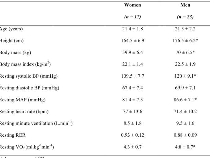
The anthropometric and cardiopulmonary characteristics of the participants obtained at resting conditions before maximal exercise are shown in Table 1. Even though both groups had similar age and BMI (p > 0.05), men were taller and heavier than women (p < 0.05). There were between-group differences in systolic BP, mean arterial pressure (MAP) and resting VO2 (p < 0.05). Men exhibited higher values than women for all these parameters. Conversely, diastolic BP, resting heart rate, minute ventilation (Ve) and the respiratory exchange ratio were similar between sexes at this specific time point (p > 0.05).
Table 1. Descriptive characteristics of participants.
Women Men
(n = 17) (n = 23)
Age (years) 21.4 ± 1.8 21.3 ± 2.2
Height (cm) 164.5 ± 6.9 176.5 ± 6.2*
Body mass (kg) 59.9 ± 6.4 70 ± 6.5*
Body mass index (kg/m2) 22.1 ± 1.4 22.5 ± 1.9
Resting systolic BP (mmHg) 109.5 ± 7.7 120 ± 9.1*
Resting diastolic BP (mmHg) 67.4 ± 7.4 69.9 ± 7.1
Resting MAP (mmHg) 81.4 ± 7.3 86.6 ± 7.1*
Resting heart rate (bpm) 77 ± 13.6 71.4 ± 10.2
Resting minute ventilation (L.min-1) 8.5 ± 1.8 9.5 ± 1.6
Resting RER 0.93 ± 0.12 0.88 ± 0.09
Resting VO2 (ml.kg-1min-1) 4.3 ± 0.7 4.8 ± 0.7*
Values are mean ± SD
* p < 0.05, significant differences between sexes.
Abbreviations: systolic BP, systolic blood pressure; diastolic BP, diastolic blood pressure; MAP, mean arterial pressure; RER, respiratory exchange ratio; VO2, oxygen uptake.
26
Exercise
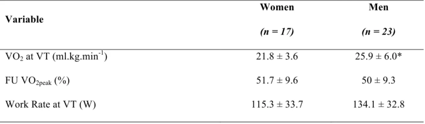
Table 2 shows the physiological and mechanical variables obtained during exercise at the VT. There were no differences between groups for absolute work rate or fractional utilization of VO2peak at the VT (p > 0.05). However, we found that men exhibited greater VO2 values than women at the VT (p < 0.05).
Table 2. Physiological and mechanical variables recorded at the ventilatory threshold (VT).
Variable Women Men (n = 17) (n = 23) VO2 at VT (ml.kg.min-1) 21.8 ± 3.6 25.9 ± 6.0* FU VO2peak (%) 51.7 ± 9.6 50 ± 9.3 Work Rate at VT (W) 115.3 ± 33.7 134.1 ± 32.8
Values are mean ± SD
* p < 0.05, significant differences between sexes.
Abbreviations: FU VO2peak, fractional utilization of peak oxygen uptake; VT, ventilatory threshold.
As can be seen in table 3, there were no between-group differences for the VO2peak percentile, respiratory exchange ratio or peak heart rate (p > 0.05). In contrast, men attained higher values than women for VO2peak (both relative and absolute values), work rate and Ve (p < 0.05).
27 Table 3. Physiological and mechanical variables obtained during peak exercise
Variable Women Men (n = 17) (n = 23) VO2peak (ml.kg-1.min-1) 42.4 ± 3.6 51.8 ± 5.6* VO2peak (ml.min-1) 2543.8 ± 384.2 3608.5 ± 368.3* VO2peak (percentile) 69.7 ± 14.7 78 ± 16.5
Peak Work rate (W) 216.2 ± 38.3 256.1 ± 33.4*
Peak Respiratory exchange ratio 1.16 ± 0.07 1.14 ± 0.08
Peak Ve (L.min-1) 88.7 ± 14.0 116.8 ± 22.1*
Peak heart rate (bpm) 181.6 ± 11.5 188.2 ± 8.7
Values are mean ± SD
* p <0.05, significant differences between sexes.
Abbreviations: VO2, oxygen uptake; Ve, minute ventilation.
Recovery
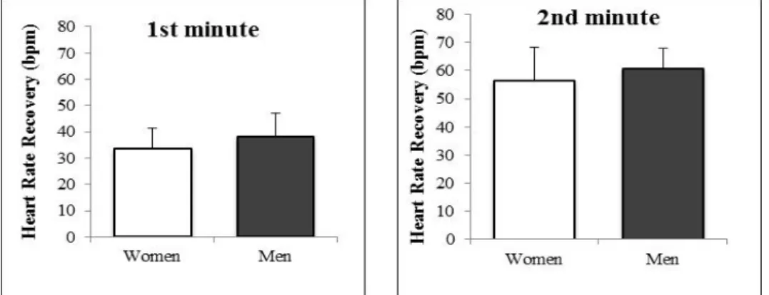
Figure 2 depicts HRR from men and women after peak exercise (1st and 2nd min of passive recovery). At the first and second minute of recovery, men exhibited an average heart rate of 150.2 ± 11.7 and 127.6 ± 10.2 bpm, respectively. Women had similar heart rate values as men at both time points of recovery (147.9 ± 14.4 and 125.2 ± 16.8 bpm, respectively). Thus, as shown in figure 2, both sexes had similar HRR post-peak exercise (p > 0.05).
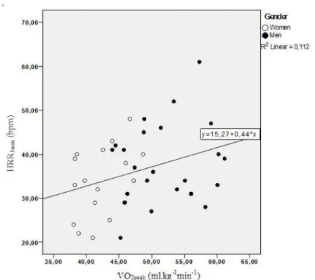
28 In multiple linear regression analysis, VO2peak (ml.kg-1.min-1), sex and the interaction between sex and VO2peak were found to be significant predictors of HRR2min (Table 4). Together, these three variables explained 29.2% of variance in HRR 2min (F = 4.9, p = 0.006). Conversely, for HRR1min, VO2peak was the only variable entering the model. VO2peak explained 11.2% of the variance in HRR1min (F = 4.8, p = 0.035) (Table 4). The graphical relationship between the dependent and independent variables is further depicted in figure 3 and 4.
Table 4. Multiple linear regression models for the 1st and 2nd minute heart rate recovery.
Values are mean ± SD
Abbreviations: HRR, heart rate recovery; VO2peak, peak oxygen uptake.
Beta p value R R2
Model – HRR1min
VO2peak (ml.kg-1min-1) 0.437 0.035 0.335 0.112
Model 1 equation: HRR1min = 15.270 + 0.437*VO2peak
Model – HRR2min
Sex (0 = women; 1 = men) 92.034 0.006
VO2peak (ml.kg-1min-1) 2.164 0.001 0.540 0.292
Sex x VO2peak -2.090 0.005
Model 2 equation for women: HRR2min = -35.264 + 2.164*VO2peak
29
Fig. 3 Relationship between cardio-deceleration at 1 min of recovery (HRR1min) with peak oxygen
uptake (VO2peak). The regression equation for prediction of HRR is also showed.
Fig. 4 Relationship between cardio-deceleration at 2 min of recovery (HRR2min) with peak oxygen
uptake (VO2peak) in men and women. The regression equations for prediction of HRR in each sex are also showed.
30 Finally, the analysis of covariance comparing HRR1min and HRR2min between men and women while controlling for VO2peak did not alter our findings of similar cardio-deceleration between sexes at post-exercise time points (HRR1min: F = 2.9, HRR2min: F = 2.1, p > 0.05).
V. Discussion
The results obtained in our study are in agreement with our hypothesis. Accordingly, our data indicate that cardio-deceleration at 1 and 2 min of recovery from peak exercise is similar between healthy young women and men with the same age and VO2peak percentile. For this reason, we conclude that, for a level of cardiorespiratory fitness above average (VO2peak > 50th percentile), HRR does not follow a sexually dimorphic pattern. These results were sustained even after controlling for sex differences in VO2peak. Thus, based on the ANCOVA results, it may be speculated that HRR would still be similar between men and women exhibiting comparable VO2peak values. Our findings corroborate those of previous research suggesting that HRR might be similar between men and women attaining the same VO2peak values (Kappus et al., 2015). Nevertheless, contrasting to the arguments used in that study (Kappus et al., 2015), we believe that the VO2peak should not be used for the purpose of pair-matching men and women in terms of cardiorespiratory fitness. As it is well known, women have naturally lower aerobic capacity than men and this is not secondary to different levels of physical activity. Conversely, it is a consequence of their smaller body size, heart volume and lower serum hemoglobin levels (Plowman & Smith, 2011). Based on this premise, women have to be exposed to greater training loads to increase their VO2peak to the levels typically seen men at any age group. As HRR has been shown to be chronically affected by the repeated exposure to training load, we contended that pair-matching the cardiovascular fitness of men and women using VO2peak for the purpose of exploring sexually dimorphism in HRR represented an important limitation of the existing literature. Thus, with the purpose of overcoming this limitation, we used VO2peak percentile as a surrogate measure of cardiovascular fitness among young healthy men and women (Darr et al., 1988). Furthermore, to avoid heterogeneity between groups, we only included participants exhibiting VO2peak
32 values > 50th percentile. We believe that the prevailing lack of consensus among available literature on the issue of sexual dimorphism in HRR is most likely a consequence of sampling limitations and study designs (i.e. comparisons between men and women not-paired for cardiovascular fitness or training load, or inexistence of age cut-off groups) (Arena et al., 2008; Cole et al., 1999; Cole, et al., 2000; Carnethon et al., 2005; Nishime et al., 2000; Shetler et al., 2001; Antelmi et al., 2008).
After using multiple linear regression statistics for confirmatory analysis, we found that VO2peak was the only predictor for changes in HRR (approximately 11% of variance). Based on these findings, we conclude that HRR1min is positively influenced by VO2peak (and possibly by training load as previously discussed). At the second minute of recovery, the interaction between sex and VO2peak, together with VO2peak and sex directly, explained approximately 29% of the variance in HRR. This implicates that, for individuals falling within the same values of VO2peak, sex partially determines cardio-deceleration at the second minute of recovery. HRR is mediated by both branches of the autonomic nervous system. The initial decrease in heart rate is mediated via prompt parasympathetic reactivation, with latter reductions due to continued parasympathetic reactivation and sympathetic withdrawal (Pierpont & Voth, 2004). Watson et al., 1980, noted that, after maximal exercise, the peak plasma level of norepinephrine in healthy individuals was attained at 108 s of recovery (Watson et al., 1980). Perini et al. further demonstrated that during 50 s of recovery from moderate- to high-intensity exercise, blood norepinephrine concentration did not start to decrease, maintaining a similar level as that attained during maximal exercise (Perini et al., 1989). Thereafter, norepinephrine concentration decreased exponentially. This suggests that, after high levels of exercise, the sympathetic drive may continue well into the first minute of recovery, masking the reactivation of parasympathetic system and contributing to a slower non-exponential decrease in heart rate. On the other hand, during the second minute of
33 recovery, the plasma norepinephrine clearance rate is higher and this unmasks the parasympathetic reactivation (Borresen & Lambert, 2008). Taking these mechanistic concepts into consideration, our findings suggest that sex may interact with cardiodeceleration at a recovery stage where chronotropism is under the influence of a progressive increase vagal tone and sympathetic deactivation.
As can be seen in figure 4, the slopes of the regression lines obtained for each sex at the 2nd minute of recovery are substantially different between men and women. Specifically, the slope of the regression line is steeper for women than men. This implicates that while women with above average cardiovascular fitness (VO2peak> 50th percentile) tend to improve their HRR for each unit of increase in relative VO2peak, this is not sustained for men of similar age. Different saturation (ceiling effect) of HRR improvement with training might be an explanation for such discrepancies in the slope of the HRR/VO2peak relationship between sexes. As demonstrated by Buchheit & Gindre (2006), persons who display the highest VO2peak values are generally those engaged in the highest training loads. When viewed from this perspective, our data suggest that the magnitude of HRR in women (HRR2min) may be influenced in a positive fashion by exercise interventions designed to improve their VO2peak beyond the 50th percentile. In contrast, this may not be the case for men. However, there exist individuals with advantageous predispositions and genetic parameters that determine high VO2peak values, even within the context of low-to-moderate training loads. Thus, as VO2peak may vary as a function of training load and genetic background, our data does not allow us to discriminate between the true nature of the interaction between HRR and VO2peak. Nevertheless, our models only explained 11.2% and 29.2% of the variance in HRR1min and HRR2min, respectively. Accordingly, it must be emphasized that there are other variables that also explain this variance. For instance, factors like heritability, age and body composition that have also been shown to affect HRR (Sing et al., 2008; Darr et al., 1988; Carnethon et
34
al., 2005; Inglesson et al., 2007; Lins, Valente, Filho & Barbosa e Silva, 2015). Heritability
estimates for HRR approach 34% and that HRR declines 2.5 bpm over a 7-years period (Carnethon et al., 2005). For body composition, there is evidence that higher BMI values are associated with slower HRR, independently of age (Sing et al., 2008; Lins et al., 2015).
Limitations
This study presents some important limitations. First, the sample size was relatively small (17 women and 23 men). Second, we only included participants with above average VO2peak values (> 50th percentile). Thus, it is not possible to assume whether these results are also extensive to subgroups of men and women exhibiting poorer levels of cardiovascular fitness (VO2peak < 50th percentile). Further research is warranted to explore whether these findings are extensive to between-sex comparisons in men and women showing VO2peak values < 50th percentile. Third, ovarian hormone levels were not measured. However, all women were tested during the early follicular phase of their menstrual cycle to minimize heterogeneity of serum estrogen concentrations. Yet, it could be argued that our findings might be restricted to sex comparisons made during the early follicular phase of the menstrual cycle. We believe that it is not the case because recent studies show that the vagal activity assessed in HRR is not affected by the menstrual cycle phase (Yazar & Yazici, 2016). Fourth, the recovery protocols have not yet been standardized in clinical practice. For this reason, the impact of a given exercise protocol and/or cool down period on HRR after exercise is uncertain at this stage (Antelmi et al., 2008). Lastly, we did not confirm the true existence of a ceiling/saturation effect in HRR of participants from each sex. Such confirmation would have required the reversible pharmacological inhibition of acetylcholinesterase activity and this was not possible.
35
VI. Conclusions
Briefly, this study demonstrates that, for a given VO2peak percentile (> 50th percentile), there is no sexual dimorphism in HRR obtained at 1 or 2 min of passive recovery. This corroborates our hypothesis. Even after controlling for the effects of VO2peak as a covariate, HRR (at 1 and 2 min of recovery) remained comparable between both sexes. We showed that sex, combined with VO2peak, explains approximately 29% of HRR2min. This emphasizes the notion that HRR depends on the interaction between sex and VO2peak. Importantly, this was only evident for the part of recovery depending both on vagal reactivation and sympathetic withdrawal (HRR2min). Conversely, for the first minute of recovery from peak exercise (more dependent on vagal tone reactivation), sex did not interact with cardiodeceleration. Under these circumstances, VO2peak was the only predictor HRR for both sexes. Thus, our findings support the concept that enhanced cardiorespiratory fitness contributes to faster HRR immediately post-exercise. This is in agreement with one previous report showing that HRR is faster in persons with higher aerobic capacity (Darr et al., 1988). However, since we obtained low levels of predictability (11.2% HRR1min and 29.2% for HRR2min), it is important to reinforce that there is a considerable amount of variance in post-exercise cardio-deceleration that cannot be explained by these two independent variables.
VII. References
1. Antelmi, I., Chuang, E., Grupi, C., Latorre, M., Mansur, A. (2007). Heart rate recovery after treadmill electrocardiographic exercise stress test and 24-hour heart rate variability in healthy individuals. Arq Bras Cardiol. 90(6): 380-385.
2. Arena, R., Myers, J., Williams, M., Gulati, M., Kligfried, P., Balady, G., Collins, E., Fletcher, G. (2007). Assessment of functional capacity in clinical and research settings: a scientific statement from the American Heart Associatio Committee on Exercise, Rehabilitation, and Prevention of the Council on Clinical Cardiology and the Council on
Cardiovascular Nursing. Circulation. 116: 329-343. DOI:
10.1161/CIRCULATIONAHA.106.184461
3. Arena, R., Arrowood, J., Fei, D., Shelar, S., Helm, S., Kraft, K. (2010) The influence of sex on the relationship between heart rate recovery and other cardiovascular risk factors in apparently healthy subjects. Scandinavian Journal of Medicine & Science in Sports. 20(2): 291-297. DOI:101111/i.1600-0838.2009.00883.x
4. Borresen, J., Lambert, M. (2008). Autonomic control of heart rate during and after exercise.
Sports Med. 38: 633-646.
5. Buchheit, M., Gindre, C., (2006). Cardiac parasympathetic regulation: respective associations with cardiorespiratory fitness and training load. Am J Physiol Heart Circ Physiol. 291(1): H451-H458. DOI:10.1152/ajpheart.00008.2006
37 6. Carnethon, M., Jacobs, D., Jr., Sidney, S., Sternfeld, B., Gidding, S., Shoushtari, C., Liu, K. (2005). A longitudinal study of physical activity and heart rate recovery: CARDIA, 1987-1993. Medicine & Science in Sports Exercise. 37(4): 606-612. Doi:10.1249/01.MSS.0000158190.56061.32.
7. Cataldo, A., Cerasola, D., Zangla, D., Russo, G., Sahin, F. N., & Traina, M. (2014). Assessment of autonomic function as marker of training status: the role of heart rate recovery after exercise. EJSS Journal 2(1): 89-97. doi.org/10.12863/ejssbx2x1-2014x3
8. Cheng, Y., Lauer, M., Earnest, C., Church, T., Kampert, J., Gibbons, L., Blair, S. (2003). Heart rate recovery following maximal exercise testing as a predictor of cardiovascular disease and all-cause mortality in men with diabetes. Diabetes Care. 26(7): 2052-2057
9. Cole, C. R., Blackstone, E. H., Pashkow, F. J., Snader, C. E., Lauer, M. S. (1999). Heart-Rate Recovery immediately after exercise as a predictor of mortality, The New England
Journal of Medicine. 341(18), 1351-1357.
10. Cole, C., Foody, J., Blackstone, E., Lauer, M. (2000). Heart rate recovery after submaximal exercise testing as a predictor of mortality in a cardiovascularly healthy cohort.
Ann Intern Med. 132(7): 552-555.
11. Darr, K., Bassett, D., Morgan, B., Thomas, D. (1988). Effects of age and training status on heart rate recovery after peak exercise. Am. J. Physiol. 254: H340-343.
Newton-38 Cheh, C., Drake, J., Musone, S., Heard-Costa, N., Benjamin, E. Levy, D., Atwood, L., Wang, T., Kathiresan, S. (2007). Heritability, linkage, and genetic associations of exercise treadmill
test responses. Circulation. 115(23): 2917-2924.
DOI:10.1161/CIRCULATIONNAHA.106.683821.
13. Jouven, X., Empana, P., Schwartz, P., Desnos, M., Courbon, D., Ducimetiere, P. (2005). Heart-rate profile during exercise as a predictor of sudden death. The New England Journal
of Medicine. 352(19): 1951-1958.
14. Kappus, R., Ranadive, S., Yan, H., Lane-Cordova, A., Cook, M., Sun, P., Harvey, I., Wilund, K., Woods, J., Fernhall, B., (2015). Sex differences in autonomic function following maximal exercise. Biology of Sex Differences. 6:28. DOI:10.1186/s13293-015-0046-6
15. Kligfield, P., McCormick, A., Chai, A., Jacobson, A., Feuerstadt, P., Hao, S. (2003). Effect of age and gender on heart rate recovery after submaximal exercise during cardiac rehabilitation in patients with angina pectoris, recent acute myocardial infarction, or coronary bypass surgery. American Journal of Cardiology. 92(5): 600-603. DOI:10.1016/S0002-9149(03)00733-1
16. Kohli, U., Diedrich, A., Kannankeril, P., Muszkat, M., Sofowora, G., Hahn, M., English, B., Blakely, R., Stein, C., Kurnik, D. (2015). Genetic variation in alpha2- adrenoreceptors and heart rate recovery after exercise. Physiol Genomics. 47: 400-406. Doi:10.1152/phsiolgenomics.00124.2014.
39 cardiac de recuperação após teste ergométrico e índice de massa corpórea. Portuguese
Journal of Cardiology. 34(1): 27-33.
18. Minson, C. T., Halliwill, J. R., Young, T. M., & Joyner, M. J. (2000). Influence of the Menstrual Cycle on Sympathetic Activity, Baroreflex Sensitivity, and Vascular Transduction in Young Women, Circulation, 101, 862–868.
19. Mora, S., Redberg, R., Sharrett, A., Blumenthal, R. (2005). Enhanced risk assessment in asymptomatic individuals with exercise testing and Framingham risk scores. Circulation. 112(11):1566-1572. DOI:10.1161/CIRCULATIONNAHA.105.542993.
20. Nilsson, G., Hedberg, P., Jonason, T., Lonnberg, I., Ohrvik, J. (2007). Heart rate recovery is more strongly associated with the metabolic syndrome, waist circumference, and insulin sensitivity in women than in men among the elderly in the general population. Am Heart J. 154(3): 460.e1-460.e7. doi: 10.1016/j.ahj.2007.06.025.
21. Nishime, E., Cole, C., Blackstone, E., Pashkow, F., Lauer, M. (2000). Heart rate recovery and treadmill exercise score as predictors of mortality in patients referred for exercise ECG.
JAMA. 284(11): 1392-1398.
22. Pescatello, L., Arena, R., Riebe, D., Thompson, P. (2014). ACSM'S guidelines for
exercise testing and prescritpion. Baltimore: Lippincott Williams & Wilkins. 9th edition.
23. Pickering, T. G., Hall, J. E., Appel, L. J., Falkner, B. E., Graves, J., Hill, M. N., Jones, D. W., Kurtz, T., Sheps, S. G., Roccella, E. J. (2005). Recommendations for Blood Pressure
40 Measurement in Humans and Experimental Animals Part 1 : Blood Pressure Measurement in Humans, A Statement for Professionals From the Subcommittee of Professional and Public Education of the American Heart Association Council on High Blood Pressure Research,
Hypertension, 45, 142-161. http://doi.org/10.1161/01.HYP.0000150859.47929.8e
24. Pierpont, G. L., & Voth, E. J. (2004). Assessing Autonomic Function by Analysis of Heart Rate Recovery from Exercise in Healthy Subjects, The American Journal of Cardiology, 94, 64-68. http://doi.org/10.1016/j.amjcard.2004.03.032
25. Perini, R., Orizio, C., Comandè, A., Castellano, M., Beschi, M., Veicsteinas, A. (1989). Plasma norepinephrine and heart rate dynamics during recovery from submaximal exercise in man. Eur J Appl Physiol. 58: 879-883.
26. Plowman, S., Smith, D. (2011). Exercise Physiology: for heath, fitness and performance. Lippincott Williams & Wilkins, a Wolters Kluwer business. 3th edition
27. Polonia, J., Amaral, C., Bertoquini, S., Martins, L. (2006). Attenuation of heart rate recovery after exercise in hypertensive patients with blunting of the nighttime blood pressure fall. International Journal of Cardiology. 106(2): 238-243. Doi:10.1016/j.ijcard.2005.02.006.
28. Sing, T., Rhodes, J., Gauvreau, K,. (2008). Determinants of heart rate recovery following exercise in children. Medicine & Science in Sports Exercise. 40(4): 601-605. DOI: 10.1249/MSS.0b013e3181621ec4
41 Myers, J. (2001). Heart Rate Recovery: Validation and Methodologic Issues, Journal of the
American College of Cardiology, 38(7), 1960-1967.
30. Sung, J., Choi, Y., Park, J., (2006). Metabolic syndrome is associated with delayed heart rate recovery after exercise. J Korean Med Sci. 21(4): 621-626.
31. Yazar, S., Yazici, M., (2016). Impact of Menstrual Cycle on Cardiac Autonomic Function Assessed by Heart Rate Variability and Heart Rate Recovery. Medical Principles and
Practice. 25(4): 374-377. DOI: 10.1159/00044
32. Wasserman, K., Whipp, J., Koyal S., Beaver, W. (1973). Anaerobic threshold and respiratory gas exchange during exercise. Journal of Applied Physiology. 35(2): 236-243
33. Watson, R., Hamilton, C., Jones, D., Reid, J., Stallard, T., Litller, W. (1979). Sequential changes in plasma noradrenaline during bycicle exercise. Clinical Science. 58: 37-43.
34. Winter, E., Jones, A., Davidson, R., Bromley, P., Mercer, T. (2007). Sport and Exercise
Physiology Testing Guidelines. The British Association of Sport and Exercise Sciences Guide.