Int J Anat Res 2015, 3(4):1593-96. ISSN 2321-4287 1593
Case Report
BILATERAL VARIATIONS IN BRANCHING OF AXILLARY ARTERY
Bikash Chandra Satapathy * ¹, Abu Ubaida Siddiqui ², Soumitra Trivedi ², Dhanesh
Kumar Sharma ³.
ABSTRACT
Address for Correspondence: Dr Bikash Chandra Satapat hy, Depart ment of Anat omy, All India Inst it ut e of M edical Sciences, Raipur-492099, India. E-M ail: bikash.sat apat [email protected]
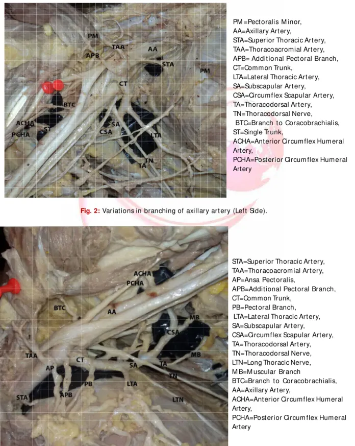
Bilat eral var iat ions in t he branching pat t ern of axillar y art ery w ere seen in a 78 year m ale cadaver dur ing rout ine dissect ion. On t he right side superior t horacic art ery w as a branch of second part of axillary art ery. Addit ional pect or al br anches w ere seen com ing out of t he second part on eit her side. Bilat erally from t he second part a com m on t runk originat ed w hich gave r ise t o lat eral t horacic and subscapular ar t ery. On t he left side an addit ional pect oral branch f rom t he com m on t runk w as present . From t he t hird part on eit her side a m uscular branch supplied t he coracobrachialis m uscle. On t he right side fr om t he t hird part of axillar y art ery a com m on t runk st art ed and divided int o ant erior and post erior circum flex hum er al art eries.
KEY W ORDS: Axi llary art ery, Com m on t r unk, Coracobrachialis, Clinical signif icance.
INTRODUCTION
Int ernat ional Journal of Anatomy and Research, Int J Anat Res 2015, Vol 3(4):1593-96. ISSN 2321- 4287 DOI: ht t p:/ / dx.doi.org/10.16965/ ijar.2015.294
Access this Article online
Quick Response code Web site:
Received: 09 Oct 2015 Accept ed: 04 Nov 2015 Peer Review : 09 Oct 2015 Published (O): 30 Nov 2015 Revised: None Published (P): 31 Dec 2015
Int ernat ional Journal of Anat omy and Research ISSN 2321-4287
ww w.ijmhr.org/ ijar.htm
DOI: 10.16965/ ijar.2015.294
¹ Senior Resident , ²Assistant Professor, ³Addit ional Professor
Depart ment of Anat omy, All India Inst it ute of M edical Sciences, Raipur, India.
Axi llar y ar t er y (AA) is t he cont inuat ion of sub clavian ar t er y an d ext ends f r om ou t er border of first rib t o low er border of subscapu-laris muscle. It is divided in t o t hree part s by pect oralis minor (PM ) muscle. The first part is bet w een out er border of first rib and medial border of pect oralis minor muscle. Superior tho-racic or supreme t hotho-racic artery (STA) is t he only branch from t he first part of axillary artery. The second part of axillary art ery is deep t o t he pect oralis m inor m uscle and commonly gives rise t o t horacoacromial artery (TAA) and lateral t horacic artery (LTA). The t hird part of axillary art ery ext ends from t he lat eral border of pect o-ralis m inor t o low er border of subscapularis muscle. Normally t here are t hree branches from t he t hird part of axillary artery, w hich are ante-rior circumflex humeral art ery (ACHA), post eante-rior
circumflex humeral artery (PCHA) and subscapu-lar art ery (SA) [1]. Axilsubscapu-lary art ery is know n t o have variat ions from t he above descript ion [2].
CASE REPORT
Int J Anat Res 2015, 3(4):1593-96. ISSN 2321-4287 1594
PM =Pect oralis M inor, AA=Axillary Art ery,
STA=Super ior Thoracic Art ery, TAA=Thoracoacrom ial Art ery, APB= Addit ional Pect oral Branch, CT=Comm on Trunk,
LTA=Lat eral Thoracic Art ery, SA=Subscapular Art ery,
CSA=Circum flex Scapular Art ery, TA=Thoracodorsal Art ery, TN=Thoracodorsal Nerve,
BTC=Br anch t o Coracobrachialis, ST=Single Trunk,
ACHA=Ant erior Circum flex Hum eral Artery,
PCHA=Post erior Circum flex Hum eral Artery
Bikash Chandra Sat apat hy et al. BILATERAL VARIATIONS IN BRANCHING OF AXILLARY ARTERY.
sides t he common t runk aft er giving rise t o lat eral t horacic art ery cont inued as subscapular art ery and divided int o a bigger branch called circumflex scapular art ery (CSA) and cont inued as a smaller t horacodorsal artery (TA) t hat accompanied t he t horacodorsal nerve (TN). On eit her side t here w as a branch from t he t hird part of axillary art ery supplying coracobrachialis muscle (BTC=branch t o coracobrachialis). On right side bot h ACHA and PCHA st art ed as a single t runk (ST) from t he t hird part of axillary art ery and t hen got divided. All ot her branches of axillary art ery w ere normal.
Fig. 1: Variat ions in branching of axi llary art ery (Right Side).
Fig. 2: Var iat ions in branching of axillary art ery (Lef t Side).
STA=Super ior Thoracic Art ery, TAA=Thoracoacrom ial Art ery, AP=Ansa Pect oralis,
APB=Addit ional Pect oral Branch, CT=Comm on Trunk,
PB=Pect oral Branch, LTA=Lat eral Thoracic Art ery, SA=Subscapular Art ery,
CSA=Circum flex Scapular Art ery, TA=Thoracodorsal Art ery, TN=Thoracodorsal Nerve, LTN=Long Thoracic Nerve, M B=M uscular Branch
BTC=Br anch t o Cor acobrachialis, AA=Axillary Art ery,
ACHA=Ant erior Circum flex Hum eral Artery,
Int J Anat Res 2015, 3(4):1593-96. ISSN 2321-4287 1595 DISCUSSION
Bikash Chandra Sat apat hy et al. BILATERAL VARIATIONS IN BRANCHING OF AXILLARY ARTERY.
Branching pat t ern of axillary is quit e variable and w ell document ed. Samt a et al [3] report ed t he incidence t o be as high as 28%. Verma et al [4] report ed a case in w hich t he left superior thoracic art ery was taking origin from the second part of axillary artery. In t he present case similar variat ion w as seen on t he right side.
Incidence of subscapular art ery t aking origin from second part of axillary art ery has been document ed t o vary from 4% by Samt a et al [3] t o 15% by Huelke [5]. Swamy et al [6] described a variant subscapular art ery t aking origin from second part of axillary art ery and giving rise t o posterior circumflex humeral and lat eral t horacic artery. Vasuki et al [7] encountered variat ions in t he t hird part of axillary art ery on t he right side. In t he present case a common t runk t ook origin from second part of axillary art ery and then gave rise to lateral t horacic artery and circumflex scapular art ery on bot h sides. The circumflex humeral art eries t ook origin from t he t hird part of axillary artery, but on left side anterior and post er ior circum flex hum er al art er ies w ere separat e branches, w hereas on t he right side t hey t ook origin as a common t runk and t hen divided int o ACHA and PCHA.
Chit ra and Anandhi [8] report ed absence of thoracoacromial t runk and presence of individual branches from t he second part such as delt oid, pect oral, acrom ial and clavicular on t he right side. In t he current case ext ra pect oral branches w ere t here. On left side t here w ere t w o ext ra pect or al br anches, one addit ional pect or al branch (APB) from second part of axillary art ery and one pect oral branch (PB) from t he common t runk from second part of axillary artery. On t he r igh t si d e t h e ad di t i on al p ect o r al b r an ch originat ed direct ly from second part of axillary artery.
Arey LB [9] gave some possible explanat ion for t he unusual blood vessels. Among w hich one w as “ oblit erat ion of norm ally ret ained blood vessel”, w hich could explain absence of t he subscapular art ery direct ly from axillary art ery on eit her side. The aut hor also suggest ed t hat incomplet e fusion and absorpt ion of usually dist inct part s could also lead t o unusual blood vessels. This process might have led t o fusion
of subscapular art ery w it h t he lat eral t horacic art ery t o form t he common t runk. The separat e pect oral branches might have result ed due t o t heir incom plet e fusion w it h t horacoacromial artery. The superior t horacic artery on right side w as taking origin from t he second part of axillary artery. This could be explained by incomplete fusion and absorpt ion of vessels.
Accor din g t o Ham ilt on an d M ossm an [ 10] art erial variat ions found in t he upper limb are because of defect in t he lim b bud’s vascular pl exus. These var iat i ons coul d have been p ossi bl e du e t o r et en t i o n , r egr essi o n o r reappearance of branches aft er arrest of normal vascular development .
CONCLUSION
In performing ant egrade cerebral perfusion in aort ic surgery for axillary art ery t hrom bosis variant anat om y of axillary art ery is crucial. Know ledge of branching pat t er n of axillar y art ery is essent ial for reconst ruct ing axillary art ery aft er t rauma, giving regional nerve block in t he axillary region. In performing surgeries in upper end of hum erus and axillary dissect ion during mastectomy for carcinoma breast and for ut ilising m edial arm skin flap, know ledge of dif ferent variat ions in branching of axillar y art ery is very valuable. So know ing t he varia-t ions in branching of axillary arvaria-t ery is beneficial for anaest het ist s, surgeons, ort hopedicians as w ell as int ervent ional radiologist s.
Conflicts of Interests: None
REFERENCES
[ 1] . Su san St an dr in g, edi t o r. Gray’s An at om y: The Anat om ical Basis of Clinical Pr act i ce. 40t h ed. London: Churchill Livingst one Elsevier; 2008. p. 817. [2]. Hollinshead WH. Anat om y for Surgeons in General surger y of t he upper lim b. The back and Lim bs. Volume 3. New York: Heber- Harper Book; 1958. 290-300 p.
[3] . Sam t a gaur, S K.Kat ariya, H Vaishnani, I N Wani, K V Bondr e GVS. A cadaver ic st udy of br anching pat t ern of t he axillary ar t ery. Int J Biol M ed Res. 2012 Feb 1;3(1):1388–91.
[4]. Verm a R, M ishra S, M ahajan A. Unilat eral variat ion i n t he br an chi n g p at t er n o f axi l lar y ar t er y: a cadaveric st udy. Int J Anat Res. 2014;2(1):292–4. [5] . Huelke DF. Var iat ion in t he origins of t he branches
Int J Anat Res 2015, 3(4):1593-96. ISSN 2321-4287 1596 Bikash Chandra Sat apat hy et al. BILATERAL VARIATIONS IN BRANCHING OF AXILLARY ARTERY.
[6]. Swam y R, Rao M , Kum ar N, Sirasanagandla S, Nelluri V. Unusual branching pat t er n of axi llar y ar t er y associat ed w it h t he high origin of ulnar art ery. Ann M ed Healt h Sci Res. 2013 Apr;3(2):265–7.
[ 7] . A.K.M anicka Vasuki, M .Nir m ala Devi, K.Kalyana Sundaram , Deborah Joy Hebzibah, T.K.Aleyem m a Fenn, M .Jam una.UNILATERAL VARIATION IN THE BRANCHING PATTERN OF RIGHT AXILLARY ARTERY: A CASE REPORT. Int J Anat Res 2015;3(3):1312-1315. DOI: 10.16965/ ijar.2015.218.
[8]. Chit ra PS, Anandhi V. A unique variat ion in branching pat t ern of axillary ar t ery. Int J Anat Var. 2013;6:1– 3.
[ 9] . Ar e y LB. De ve l o p m e n t al An at o m y. 6t h ed . Philadelphia: W B Saunders Com pany; 1957. 375-377 p.
[10]. Ham ilt on WJ, M ossm an HW. Cardiovascular syst em . Hum an Em bryology. 4t h ed. Balt im ore: W illiam s and Wilkins; 1972. p. 271–90.