Int J Anat Res 2015, 3(1):861-64. ISSN 2321-4287 861
Case Report
ANOM ALOUS LEFT VERTEBRAL ARTERY FROM ARCH OF AORTA-A
THOUGHT FOR SURGEONS?
Deepa Somanath *
1, Shivali Srivastava
2.
ABSTRACT
Address for Correspondence: Dr. Deepa Som anat h, Assist ant Professor, Dept . of Anat omy, Sri M anakula Vinayagar M edical College, M adagadipet , Puducherry- 605 107, India.
E-M ail: deepa.somanat [email protected]
* 1 Assistant Professor, Dept . of Anatomy, Sri M anakula Vinayagar M edical College, Puducherry, India. 2 Post Graduate St udent , Dept . of Anat omy, Sri M anakula Vinayagar M edical College, Puducherry,
India.
Art erial variat ions in t he branching pat t er n of arch of aort a ar e uncom m on and m ay occur as a result of developm ent al changes in t he fusion and absorpt ion of various phar yngeal ar ch ar t eries int o t he aor t ic sac. The present case repor t describes a finding in a m ale cadaver aged bet w een 60- 65 years about t he origin of left vert ebral art ery direct ly from the arch of aort a. Such variat ions should be kept in m ind during surgical procedures in t he super ior m ediast inum .
KEYW ORDS:Lef t ver t ebral ar t ery, Arch of aorta, Arch art ery, Anom aly.
INTRODUCTION
Int ernat ional Journal of Anatomy and Research, Int J Anat Res 2015, Vol 3(1):861-64. ISSN 2321- 4287 DOI: ht t p:/ / dx.doi.org/10.16965/ ijar.2015.102
The vertebral artery cont ributes t o t he blood supply of t he brain by helping in t he format ion of circle of W illis. It forms a major part of t he posterior cerebral circulat ion.
60- 85% of t he populat ion have t hree direct b r an ch es f ro m t h e ar ch of aor t a, n am el y brachiocephalic t runk, left common carot id and left subclavian artery. Somet imes, t he arch of aorta can give rise t o t he vertebral arteries of one or bot h sides, commonly t he left side. The vertebral artery on eit her side usually arises from t he f irst part of t he subclavian art er y, m edial t o t he scalenus ant er ior m uscle. It ascen d s v er t i cal l y t o ent er t h e f o r am en t ransversarium of sixt h cervical vertebra and
t raverses all cervical foramina t ransversaria up t o t he first cervical vertebra (at las). This part of t he artery from it s origin t o t he ent ry int o t he f or am en t ran sver sar i u m i s cal led t h e pr e vertebral segment . Then t he artery lies on t he posterior arch of t he at las in t he sub occipital t riangle, follow ing w hich it pierces t he at lant o-o cci p i t al m em b ran e an d d u r a m at er. Th e v er t ebr al ar t er y t h en en t ers t he fo ram en m agnum and unit es w it h t he corresponding artery of t he opposite side t o form t he basilar artery w hich in t urn terminates as t w o posterior cerebral arteries [1].
The present case report depict s t he anomalous origin of t he left vertebral artery from t he arch of aorta.
Access this Article online
Quick Response code Web site:
Received: 08 Jan 2015 Accept ed: 04 Feb 2015 Peer Review : 08 Jan 2015 Published (O):28 Feb 2015 Revised: None Published (P):31 M ar 2015
Int ernat ional Journal of Anat omy and Research ISSN 2321-4287
ww w.ijmhr.org/ ijar.htm
Int J Anat Res 2015, 3(1):861-64. ISSN 2321-4287 862
Deepa Somanat h , Shivali Srivast ava. ANOM ALOUS LEFT VERTEBRAL ARTERY FROM ARCH OF AORTA-A THOUGHT FOR SURGEONS?.
CASE REPORT
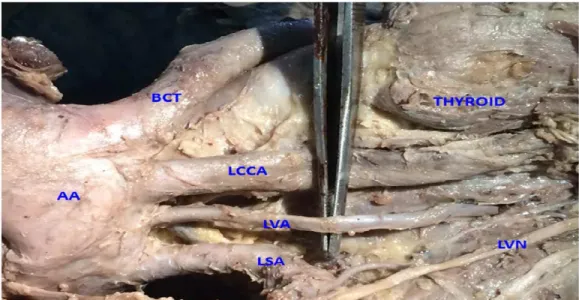
During routine dissect ion of a formalin fixed male cadaver aged 60- 65 years, for undergraduate st udent s of Sri M anakula Vinayagar M edical College and Hospit al, Pondicher r y, t he lef t vertebral artery was seen t o be originat ing from t he arch of aort a bet w een t he left com m on carotid and t he left subclavian arteries, w hereas, t he vert ebral art ery on t he right side had a normal origin from t he first part of t he right subclavian artery.
In t he superior mediast inum, t he left vertebral artery was seen ascending t o reach t he neck t o
Fig. 1: Show ing Anom alous Left vert ebral art ery.
AA-arch o f ao r t a,BCT-Br achi oceph al ic t r un k,LCCA-Lef t co m m on caro t id ar t er y,LVA-Lef t ver t eb ral ar t er y, LSA-lef t subclavian art ery,LVN-Left vagus nerve
enter scaleno vertebral t riangle. The artery t hen ran a ver t ical course t o ent er t he foram en t ransversarium of t he sixt h cervical vertebra. It t raversed t he cervical foramina t ransversaria up t o t he foramen t ransversarium of t he at las. The artery had a non t ort uous course and t he pre vertebral segment measured about 51 mm. The vertebral artery on t he right side originated from t he first part of right subclavian artery medial t o scalenus anterior muscle and follow ed a nor m al course of pre ver t ebr al segm ent entering t he foramen transversarium of t he sixt h cervical vertebra.
Fig. 2: Show ing Anom alous Left vert ebral art ery.
Int J Anat Res 2015, 3(1):861-64. ISSN 2321-4287 863 DISCUSSION
Deepa Somanat h , Shivali Srivast ava. ANOM ALOUS LEFT VERTEBRAL ARTERY FROM ARCH OF AORTA-A THOUGHT FOR SURGEONS?.
The vertebral artery is divided int o four part s; t he first part is derived from t he proximal part o f d or sal d i vi sio n o f t he seven t h cer v i cal intersegmental artery. The second part is derived from t he longit udinal com municat ions of t he post cost al anast om oses bet w een t he first t o sixt h cervical intersegmental arteries. The t hird part develops from t he spinal branch of t he first cervical intersegm ent al art ery and t he fourt h part develops from it s pre neural division [1, 2]. Albayram et al. have stated t hat an aberrant left vertebral artery from arch of aorta is due t o t he persistence of eight h intersegmental artery [3]. Aort ic arch anomalies are seen in chromosomal abnormalit ies such as 22q11 delet ion [4]. In a st udy by Shin et al. it was noted t hat t he left vert ebral artery arose from t he arch of aorta bet w een t he lef t com m on carot id and lef t subclavian art eries in 5.8% of cases [5] . In anot her st udy by Lemke et al. it was observed t h at 2.4- 5.8% cases h ad t h e ab ov e sai d variat ion[6].
Rekha et al. st at ed t he o ccu r ren ce o f lef t vertebral artery branching from the arch of aorta in 4.5% of cases in a sample size of 110 formalin fixed cadavers [7].
The incidence of t he left vertebral artery arising from t he arch of aorta was found t o be 3.33% in a st udy on 30 cadavers by Oza Sunil et al [8]. An unusual observat ion by Goray et al. depicted five direct branches arising from t he arch of aorta w hich included bot h vertebral arteries [9]. 1.6% of 62 cadavers w ere found to have an anomalous left vertebral artery from t he arch of aorta in a st udy by Nayak et al. [10]
Hassan et al. reported a case of a 40 year old fem ale w it h sym pt om s of cer vicom edullary st en o sis d ue t o co m p r essi o n o f t h e cervicomedullary junct ion by an anomalous left vertebral artery from t he arch of aorta [11]. An anomalous vertebral artery may or may not cause clinical sympt oms. How ever it is necessary t o b e vi gi lan t d u r in g p ro cedu r es o f t h e mediast inum and the neck to avoid misdiagnosis and iatrogenic complications. Since the vertebral artery forms an important part of t he posterior cerebral circulat ion, aberrat ions in it s origin,
course, diam et er and lengt h can af fect t he cerebral hemodynamics. This may cont ribute t o t he development of int racerebral malformations and dissect ion aneurysms [12]. As aforement io-ned, an anom alous vert ebral art ery w it h or w it hout aneurysm can cause compression of various st ruct ures on it s course.
Vertebral artery anomalies may also indicate t he presence of many ot her congenital malformat-ions of t he cardiovascular system, eg. Absence of inferior t hyroid artery, cardiac malformations, facial hemangiomas etc. [12]
Since the proximal part of direct branches of arch of aorta are more prone for at herosclerosis, any t hromboembolic disorder in t he vertebral artery can be hazardous as it cont ributes t he blood supply t o t he brain.
Know ledge of such variat ions is essent ial for surgeons and diagnost ic radiologist s in various procedures w hich require t he exposure of t he arch of aorta and its branches, such as aortic arch reconst ruct ion, graft technique for t ot al arch replacement , aneurysm repair and endarterec-t o m y. Oendarterec-t h er p r oced ur es w h i ch r eq u i re a t horough know ledge of vertebral artery and it s variat ions include angiography, low er cervical spine surgeries, t ranspedicular fixat ion, excision of cranio- cervical junct ion masses, endovascular procedures etc.
CONCLUSION
It is obvious from t he review of literat ure t hat left sided variat ions of t he vertebral artery are much more common than t hose of the right side. Certain chromosomal aberrat ions may give rise t o changes in t he developmental pat hway of t he art eries leading t o such anomalies. Variat ions such as the present case should be borne in mind dur ing t he above m ent ioned surgeries and procedures.
Acknow ledgem ent:
We w ould like t o t hank M aster Pranavan S, for helping us w it h t he phot ographs.
Conflicts of Interests: None
REFERENCES
Int J Anat Res 2015, 3(1):861-64. ISSN 2321-4287 864
Deepa Somanat h , Shivali Srivast ava. ANOM ALOUS LEFT VERTEBRAL ARTERY FROM ARCH OF AORTA-A THOUGHT FOR SURGEONS?.
Journal of Pharm aceut ical Sciences and Research 2013;5(10):196-198.
[ 2] . M o o r e KL. Th e Devel o p i n g Hu m an . Cl i n i cal l y Or i en t ed Em b r yo l o gy. 3r d Ed . W B Sau n d er s, Phi ladelphia. 1982;291–318.
[3] . Albayram S, Gailloud P, Wasser m an BA. Bilat eral Arch Or igin of t he Vert ebral Ar t eries. Am erican Journal of Neuro Radiology 2002;23:455–458. [ 4] . M o m m a K, M at su oka R, Takao A. Aor t i c ar ch
anom alies associat ed w it h chrom osom e 22q11 delet ion (CATCH 22). Pediat r Cardiol 1999;20(2):97-102.
[5] . Shin Y, Chung Y, Shin W, Im S, Hw ang S, Kim B. A m or phom et ric st udy on cadaveric aor t ic arch and i t s m aj o r b r an ch es i n 25 Ko r ean ad u l t s: t h e perspect ive of endovascular surger y. Jour nal of Korean Neuro Surgery Societ y 2008;44(2):78-83. [6] . Lem ke AJ, Benndorf G, Liebig T, Felix R. Anom alous
origin of t he right vert ebral art er y: review of t he lit erat ure and case report of r ight vert ebral art ery origin distal t o t he left subclavian art ery. Am erican Journal of Neuro Radiology 1999;20(7):1318-1321. [ 7] . Rekha P, Sent hi lkum ar S. A st udy on branching pat t ern of hum an aort ic arch and it s variat ions in sout h indian populat ion. Journal of M orphological Sciences 2013;30(1):11-15.
[8] . Oza S, Gujar S, M odasia U, Prajapat i S, Shekhavat J, Panda P et al. A st udy on t he variant origins of ver t ebral ar t ery. Int ernat ional Journal of M edical Science and Public Healt h 2014;3(3):349- 351. [9] . Goray VB, Joshi AR, Garg A, M erchant S, Yadav B,
M aheshw ari P. Aort ic arch variat ion: a unique case w it h anom alous or igin of bot h vert ebral art er ies as addit ional branches of t he aort ic arch distal t o lef t subclavian art ery. Am erican Jour nal of Neuro Radiology 2005;26(1):93-95.
[10]. Nayak SR, Pai M M , Prabhu LV, D’Costa S, Shett y P. Anat om ical organizat ion of aort ic arch variat ions in t he India: em bryological basis and review. J Vasc Bras 2006; 5(2):95-100.
[11]. Hassan HA, Ayad CE, Ibrahim TEM , Hassanand IA. An om al ous lef t ver t ebral ar t er y. Int er nat ion al Journal of Case Report s and Im ages 2014;5(3):247– 249.
[12]. M erw e B, Ackerm ann C, Scheepers S, M oosa S. Is anomalous origin of t he left vert ebral artery indeed a rare finding? S Afr J Rad2012;16(4):144-146.