From the Endocrine Unit of the Children’s Institute , Hospital das Clínicas, Faculty of Medicine, University of São Paulo.
ORIGINAL ARTICLES
PREMATURE THELARCHE: IDENTIFICATION OF
CLINICAL AND LABORATORY DATA FOR THE
DIAGNOSIS OF PRECOCIOUS PUBERTY
Thais Della Manna, Nuvarte Setian, Durval Damiani, Hilton Kuperman and Vaê Dichtchekenian
RHCFAP/3068 DELLA MANNA T et al. - Premature thelarche: identification of clinical and laboratory data for the diagnosis of precocious
puberty. Rev. Hosp. Clín. Fac. Med. S. Paulo 57(2):49-54, 2002.
PURPOSE: Two groups of girls with premature breast development were studied retrospectively. We tried to identify clinical, radiological, and hormonal parameters that could distinguish between a benign, nonprogressive premature thelarche and a true precocious puberty.
METHODS: The clinical outcome of 88 girls with breast enlargement before 6.1 years of age was analyzed. Taking into account the progression of their sexual maturation, we allocated the children into 2 groups: “Isolated Premature Thelarche” (n = 63) and “Precocious Puberty” (n = 25) groups. Chronological and bone ages, height and growth velocity centiles, computerized tomography of hypothalamus-pituitary area, pelvic ultrasonography, gonadotropin response to luteinizing hormone-releasing hormone stimulation as well as basal levels of luteinizing hormone, follicle-stimulating hormone, estradiol, and prolactin were studied in both groups. Statistical analysis were performed using the Student t test to compare the sample means. Fisher’s exact test and c2 test were used to analyze the nonparametric variables.
RESULTS: Isolated premature thelarche most frequently affected girls younger than 2 years who presented exaggerated follicle-stimulating hormone response to luteinizing hormone-releasing hormone stimulation test. The precocious puberty group had higher initial stature, accelerated growth rate and bone age, increased uterine and ovarian volumes, high spontaneous luteinizing hormone levels by immunofluorimetric assay, as well as a high luteinizing hormone response and peak luteinizing hormone/follicle-stimulating hormone ratio after luteinizing hormone-releasing hormone stimulation.
CONCLUSION: At initial presentation, girls who undergo true precocious puberty present advanced bone age, increased uterine and ovarian volumes in addition to breast enlargement, as well as an luteinizing hormone -predominant response after a luteinizing hormone-releasing hormone stimulation test.
DESCRIPTORS: Precocious puberty. Premature thelarche. Precocious puberty diagnosis. Pelvic ultrasonography. Luteinizing hormone-releasing hormone stimulation test.
Premature thelarche, the usually benign development of breasts in young girls, has been considered a variation of normal puberty1 due to its nonprogressive course. However, at the initial presentation, it is not always easy to distinguish it from a true pre-cocious puberty.
Since 1980, various longitudinal clinical trials and follow-up studies of premature thelarche have been
pub-lished. Theories about the cause have become polarized: one confirming the nonprogressive, benign process2-4 and the other suggesting that premature thelarche is a consequence of a defect in the hypothalamic-pituitary-ovarian
axis along with an exaggerated periph-eral response to sexual hormones5, 6.
#
stages. This period of higher FSH pro-duction could be responsible for the development of premature thelarche in some girls7,8.
The etiology of premature thelarche is still unknown; consequently, no spe-cific marker has been identified to dif-ferentiate such a process from the on-set of true precocious puberty.
With an intent to identify clinical, radiological, and laboratory features for forecasting the progression to pre-cocious puberty, we performed a retro-spective study of 88 girls with prema-ture breast development.
PATIENTS
Population
In a 17-year retrospective study, 88 girls who presented with premature breast enlargement were analyzed ac-cording to the following inclusion cri-teria: breast budding before 6.1 years of age based on Bierich’s standards of sexual precocity 9; breast development corresponding to Tanner10 stage B
2 or B3 at the first consultation, and a fol-low-up period of at least 6 months in order to classify their sexual matura-tion development as “regressive”, “sta-ble”, or “progressive”3. Girls who pre-sented a “regressive” or “stable” out-come were diagnosed as isolated pre-mature thelarche (IPT), and those with a “progressive” course were diagnosed as precocious puberty (PP).
The isolated premature thelarche group (n = 63) presented breast en-largement before 5.5 years of age; the mean age at the first appointment was 2.3 years, and the pubertal stages were B2P1 (n = 60) or B3P1 (n = 3); the clini-cal course was classified as “stable” in 28 girls and “regressive” in 35.
The precocious puberty group (n = 25) comprised various etiologies, such as hamartoma (n = 2), chronic encepha-lopathy (n = 2), hydrocephalus (n = 1)
and congenital toxoplasmosis (n = 2). Eighteen patients were idiopathic. The girls began breast development from the age of 1 month until 6.1 years; at the first consultation the mean age was 5.4 years, and the pubertal stages were B2P1 (n = 8), B2P2 (n = 6), B2P3 (n = 3), B3P1 (n = 1), B3P2 (n = 4), B3P3 (n = 3); all of them presented “progressive” signs of sexual maturation.
METHOD
The following variables were stud-ied retrospectively in both groups: chronological age (CA) at onset of the-larche; CA at the first appointment; height centiles calculated from height curves of the National Center for Health Statistics (NCHS)11 ; growth ve-locity centiles in the first year of fol-low up using the growth velocity curves of Tanner and Whitehouse12; initial bone age (BA) at the first ap-pointment was compared using the standards of Greulich and Pyle13 for hands and wrists; bone age after 1 or 2 years of follow up, analyzing the DBA/DCA ratio; computerized tomog-raphy or magnetic resonance of hypo-thalamus-pituitary area; pelvic ultra-sonography following the standards of Salardi et al.14 and Orsini et al.15 for uterine and ovarian volumes in chil-dren and adolescents, assuming an up-per limit for uterine volume of 4 mL and for ovarian volume of 2 mL as the normal upper limits for the prepuber-tal age group; the frequency of the microcystic condition of the ovaries; basal levels of LH and FSH were per-formed using commercially available radioimmunoassay (RIA) kits (RIA Gnost, Groupe Oris, France) and immunofluorimetric assay (IFMA) kits (Delfia - Pharmacia Diagnostic, Uppsala, Sweden). Gonadotropin re-sponse to LHRH (LHRH-Serono, 100 mg in bolus) was assessed by collect-ing blood samples for determination of
LH and FSH just before the adminis-tration of LHRH, and subsequently at 15, 30, 60, and 90 minutes. The basal levels of LH and FSH, the peak levels of LH and FSH, and the peak LH/FSH ratio were analyzed; estradiol and pro-lactin were performed by RIA using commercially available kits; prolactin measurements were also performed with fluorimetric kits. Informed writ-ten consent about the diagnostic tests used was obtained from the parents of all children. Our institutional ethics committee approved this retrospective analysis.
Statistical Analysis
Statistical analysis was performed by the c2 test and Fisher’s exact test for analysis the nonparametric variables, and the Student t test for
differentiat-ing between the means. Statistical sig-nificance was considered P < 0.05.
RESULTS
Breast enlargement began before 2 years of age in 90% of the IPT group, but in the PP group, the age distribu-tion was more scattered.
At the time of the first consultation, the IPT group had heights compatible with the normal population mean, with 79% of the cases below the 90th centile. However, in the PP group, a majority (60%) of heights were equal to or greater than the 90th centile (P < 0.001).
In the first year of follow up, the growth velocity was below the 90th centile in 78% of IPT patients, while in the PP group, 76% were girls ³ 90th centile (P < 0.001).
Computerized tomography or mag-netic resonance imaging of the hypo-thalamus-pituitary area was performed in 16 girls in IPT group and was nor-mal in 88% of them. In the PP group, 20 girls were analyzed with results as follows: 3 instances of anterior pituitary enlargement (15%), 2 of tuber cinereum hamartoma (10%), 2 of periventricular calcification (10%), 2 of ventricular asymmetry (10%), 1 of cortical atrophy (5%), and 1 of pituitary stalk enlarge-ment (5%). Nine (45%) were normal.
A uterine volume greater than 4 mL was found in only 1 out of 28 girls of the IPT group in which pelvic ultra-sonography was performed, but in 14 out of 15 girls analyzed in the PP group (P < 0.001). An ovarian volume
greater than 2 mL was observed in 1 out of 11 girls in the IPT group, and in all 3 girls analyzed in the PP group
(P < 0.001). An ovarian microcystic
appearance was equally frequent in both groups.
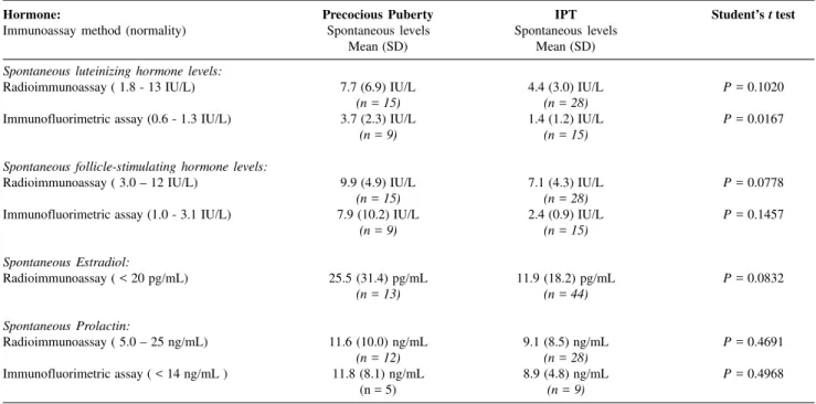
No statistical difference was found between the mean spontaneous LH lev-els: 7.7 and 4.4 IU/L, and FSH levlev-els: 9.9 and 7.1 IU/L in the PP group and
IPT group, respectively, when meas-ured with RIA at the time of the first appointment. However, on immuno-fluorimetric assay (IFMA), the mean spontaneous LH levels were 3.7 and 1.4 IU/L in the PP and IPT groups, re-spectively (P < 0.05). The mean
ba-sal FSH levels by IFMA were 7.9 and 2.4 IU/L for the PP and IPT groups, respectively (P > 0.05) (Table 1).
Upon LHRH stimulation, the mean peak LH response levels measured ei-ther with RIA (74.9 IU/L) or by IFMA (34.1 IU/L) were higher in the PP group (P < 0.01), but mean peak FSH
levels were higher in the IPT group (P
< 0.001), mainly when measured with
RIA (58.6 IU/L) (Table 2).
The peak LH/FSH response ratio was higher in the PP group (P < 0.05)
either by RIA or by IFMA (Table 2). The mean spontaneous estradiol levels measured with RIA were 25.5 and 11.9 pg/mL in the PP and IPT groups, respectively, and were not sta-tistically different (P = 0.83). Prolac-tin levels were not statistically differ-ent between groups, as measured by either method (RIA or IFMA).
DISCUSSION
In the IPT group, the clinical mani-festation of breast enlargement usually started before 2 years of age (90.5% of the cases) as has already been observed by others4,5,7,8. In the PP group, this age distribution was not seen.
In our patients, the bilateral breast budding was the rule in both groups without noticeable fluctuations in size in the IPT group, contrasting with re-ports by Stanhope et al.16.
The height centile of the IPT group at presentation was compatible with the norm, but in the PP group the children were predominantly taller for their chronological age. Likewise, the growth velocity centile was higher in the PP group (90th centile) during the first year of clinical follow up. Many authors con-sider that the accelerated growth veloc-ity could be considered as a sign of pre-cocious puberty17-19.
The PP group had advanced bone age at the first appointment (> 2 SD for the mean age of 5.4 years), but in the IPT group, the bone age was compat-ible with the girls’ chronological age
Table 1 - Comparison between spontaneous hormonal levels in precocious puberty and IPT groups.
Hormone: Precocious Puberty IPT Student’s t test
Immunoassay method (normality) Spontaneous levels Spontaneous levels
Mean (SD) Mean (SD)
Spontaneous luteinizing hormone levels:
Radioimmunoassay ( 1.8 - 13 IU/L) 7.7 (6.9) IU/L 4.4 (3.0) IU/L P = 0.1020
(n = 15) (n = 28)
Immunofluorimetric assay (0.6 - 1.3 IU/L) 3.7 (2.3) IU/L 1.4 (1.2) IU/L P = 0.0167
(n = 9) (n = 15)
Spontaneous follicle-stimulating hormone levels:
Radioimmunoassay ( 3.0 – 12 IU/L) 9.9 (4.9) IU/L 7.1 (4.3) IU/L P = 0.0778
(n = 15) (n = 28)
Immunofluorimetric assay (1.0 - 3.1 IU/L) 7.9 (10.2) IU/L 2.4 (0.9) IU/L P = 0.1457
(n = 9) (n = 15)
Spontaneous Estradiol:
Radioimmunoassay ( < 20 pg/mL) 25.5 (31.4) pg/mL 11.9 (18.2) pg/mL P = 0.0832
(n = 13) (n = 44)
Spontaneous Prolactin:
Radioimmunoassay ( 5.0 – 25 ng/mL) 11.6 (10.0) ng/mL 9.1 (8.5) ng/mL P = 0.4691
(n = 12) (n = 28)
Immunofluorimetric assay ( < 14 ng/mL ) 11.8 (8.1) ng/mL 8.9 (4.8) ng/mL P = 0.4968
#
(mean = 2.3 years). In our population, analysis of the advance in bone age during a 1-2 year observation period did not differentiate between the two groups.
Several authors14,20 using ultra-sonography have concluded that in pre-mature thelarche, ovarian and uterine volumes are normal and the ovarian microcysts are quite frequent. In the PP group of the present study, however, these volumes are higher, and the ap-pearance of larger follicular cysts is very frequent. In the present study, the increase in the uterine and ovarian vol-umes was only associated with the PP group; ovarian microcysts were ob-served in both groups and thus were not a useful means of distinguishing between them.
Random spontaneous LH levels measured by RIA have never enabled the diagnosis and monitoring of the on-set of puberty. This limitation has been explained by the intermittent and noc-turnal pattern of LH secretion reflecting the episodic activity of the LHRH neu-rons. In the late 1980s, with the intro-duction of immunometric assays, a
greater correlation index between such immunoassays and the bioactivity of LH was observed. Many authors21-23 found a significant difference between the random spontaneous LH levels of prepubertal and pubertal populations, thus indicating their value as a bio-chemical marker of puberty. No such difference was obtained for FSH levels. In the present study, at the first medical appointment, the random spontaneous LH and FSH levels meas-ured with RIA were compatible with the normal values for the method and did not differentiate between the IPT and PP groups. However, when meas-ured with IFMA, it was possible to dif-ferentiate the PP group, which had higher mean spontaneous LH levels. In our population, no difference occurred in the mean random spontaneous FSH levels between the IPT and PP groups whether measured by RIA or IFMA.
Pescovitz et al.18 used the LHRH stimulation test to differentiate IPT from the initial manifestation of central precocious puberty and observed that the IPT group had FSH-predominant responses to LHRH, but those with
complete sexual development had LH-predominant responses after stimula-tion; the intermediate groups had a great variation of plasma LH and FSH responses to LHRH. Oerter et al.24 re-vised and enlarged this study and con-cluded that the response to LHRH stimulation was the most useful vari-able in distinguishing between puber-tal and prepuberpuber-tal girls. Furthermore, a peak LH to peak FSH ratio above 0.66, measured with RIA, had 96% sensitivity and 100% specificity for di-agnosing pubertal gonadotropin secre-tion in girls. The response to LHRH stimulation is still the gold standard for diagnosing puberty; however, the ultra-sensitive immunometric methods have allowed a reduction in the number of blood samples to a collection between 20 to 40 minutes after a LHRH bolus (100 mg), and the peak LH/FSH ratio above 0.33 is indicative of puberty25, 26. In our population after LHRH stimulation, peak LH levels were sig-nificantly higher in the PP group, whether measured by RIA or by IFMA. On the other hand, the IPT group had peak FSH levels significantly higher as
Table 2 - Comparison between peak luteinizing hormone and follicle-stimulating hormone levels and peak Luteinizing hormone/follicle-stimulating hormone ratio upon luteinizing hormone-releasing hormone stimulation test in precocious puberty and IPT groups.
Luteinizing hormone- Precocious Puberty IPT Student t test
releasing hormone stimulation test: Mean hormone levels Mean hormone levels
immunoassay method (normality) (SD) (SD)
Peak luteinizing hormone levels:
Radioimmunoassay (> 15 IU/L ) 74.9 (68.1) IU/L 19.4 (11.3) IU/L P = 0.0071
(n = 15) (n = 29)
Immunofluorimetric assay (0.96 - 6.9 IU/L) 34.1 (20.7) IU/L 4.5 (3.0) IU/L P = 0.0042
(n = 9) (n = 15)
Peak follicle-stimulating hormone levels:
Radioimmunoassay (> 20 IU/L ) 31.3 (20.5) IU/L 58.6 (28.6) IU/L P = 0.0008
(n = 15) (n = 29)
Immunofluorimetric assay (6.7 - 24.5 IU/L) 16.3 (13.1) IU/L 24.4 (8.7) IU/L P = 0.1239
(n = 9) (n = 15)
Peak luteinizing hormone / follicle-stimulating hormone ratio: (prepubertal levels)
Radioimmunoassay (< 0.66) 2.44 (2.96) 0.33 (0.17) P = 0.0153
(n = 15) (n = 29)
Immunofluorimetric assay (< 0.3) 2.86 (2.05) 0.18 (0.09) P = 0.0043
determined by RIA, but not with IFMA. The peak LH/FSH ratio was signifi-cantly higher in the PP group, both with RIA and IFMA. Lee25 has suggested that peak LH levels higher than 15 IU/ L with RIA and higher than 6 IU/L with IFMA and a peak LH/FSH ratio higher than 0.66 (RIA) and higher than 0.3 (IFMA) may be used as diagnostic markers of pubertal activation in girls.
The spontaneous prolactin levels measured either by RIA or fluorimetric
assay were not useful in differentiating between the IPT and PP groups. The PP group had a mean estradiol level slightly higher than normal; however, this difference was not statistically sig-nificant in relation to that of the IPT group. This finding emphasizes the low sensitivity of most estradiol immuno-assays used recently, and perhaps sug-gests that the utilization of a biologi-cal assay such as hormonal vaginal or urinary cytology, indicating the level of
cell estrogenization, would be of greater diagnostic value5, 27.
Our data indicate that a girl could present true precocious puberty when, in addition to premature breast develop-ment, advanced bone age and increased uterine and ovarian volumes as deter-mined by pelvic ultrasonography, are observed. We conclude that an LH-pre-dominant response after a LHRH stimu-lation test appears to be an effective pa-rameter in differentiating IPT from PP.
RESUMO RHCFAP/3068
DELLA MANNA T e col. - Telarca precoce: identificação de dados clínicos e laboratoriais preditivos para o diagnóstico de puberdade precoce. Rev. Hosp. Clín. Fac. Med. S. Paulo 57(2):49-54, 2002.
OBJETIVO: A fim de distinguir o quadro de telarca precoce, benigno e auto-limitado, do início de um processo de puberdade precoce verdadeira estu-damos, retrospectivamente, dois grupos de meninas com desenvolvimento ma-mário prematuro, buscando identificar parâmetros clínicos, radiológicos e laboratoriais relacionados a cada quadro.
MÉTODOS: A evolução clínica de 88 meninas que apresentaram broto mamário antes dos 6,1 anos de idade foi analisada e classificada, segundo a progressão dos caracteres sexuais se-cundários, em um grupo portador de “Telarca Precoce Isolada” (n = 63) e um grupo portador de “Puberdade Pre-coce” (n = 25). Foram analisados
ida-de cronológica, estatura inicial e velo-cidade de crescimento em percentis, idade óssea, tomografia computado-rizada de hipotálamo-hipófise, ultra-sonografia pélvica, resposta gonado-trófica ao teste de estímulo pelo hormônio liberador do hormônio luteinizante, assim como níveis basais dos hormônios luteinizante, folículo-estimulante, estradiol e prolactina nos dois grupos. A análise estatística foi realizada pelo teste t de Student para comparação entre médias e pelos tes-tes do c2 e exato de Fisher para variá-veis não paramétricas.
RESULTADOS: A telarca precoce isolada afetou meninas menores de 2 anos, com resposta exagerada de hormônios luteinizante, folículo-esti-mulante no teste do hormônio liberador do hormônio luteinizante. O grupo pu-berdade precoce apresentou estatura inicial mais elevada, aceleração da ve-locidade de crescimento e da idade ós-sea, aumento dos volumes uterino e
ovariano, níveis de hormônios lutei-nizante basais elevados pelo ensaio imunofluorimétrico, com resposta exa-gerada de hormônios luteinizante e au-mento da relação de pico/hormônios luteinizante, folículo-estimulante no teste do hormônio liberador do hormô-nio luteinizante.
CONCLUSÃO: Frente a um quadro de desenvolvimento mamário prematuro, a presença de idade óssea avançada e au-mento dos volumes uterino e ovariano à ultra-sonografia pélvica sugerem puber-dade precoce verpuber-dadeira, que deverá ser confirmada por resposta predominante do hormônio luteinizante no teste de es-tímulo com hormônio liberador do hormônio luteinizante.
#"
REFERENCES
1. ROSENFIELD RL - Normal and almost normal precocious variations in pubertal development premature pubarche and premature thelarche revisited. Horm Res 1994; 41(suppl 2):7-13.
2. MILLS JL, STOLLEY PD, DAVIES J et al. - Premature thelarche. Natural history and etiologic investigation. Am J Dis Child 1981; 135:743-5.
3. CUELLO XA, VIVANCO XW & ABODOVSKY NG - Telarquia prematura. Rev Chil Pediatr 1985; 56:27-33.
4. VAN WINTER JT, NOLLER KL, ZIMMERMAN D et al. - Natural history of premature thelarche in Olmsted County, Minnesota, 1940 to 1984. J Pediatr 1990; 116:278-80.
5. ILICKI A, LEWIN RP, KAULI R et al. - Premature thelarche - Natural history and sex hormone secretion in 68 girls. Acta Paediatr Scand 1984; 73:756-62.
6. PASQUINO AM, TEBALDI L, CIOSCHI L et al. - Premature thelarche: a follow up study of 40 girls. Natural history and endocrine findings. Arch Dis Child 1985; 60:1180-92.
7. FAIMAN C & WINTER JSD - Sex differences in gonadotrophin concentrations in infancy. Nature 1971; 232:130.
8. JOB JC, GARNIER PE, CHAUSSAIN JL et al. - Effect of synthetic luteinizing hormone-releasing hormone (LH-RH) on the release of gonadotropins in hypophyso-gonadal disorders of children and adolescents. II. Precocious puberty and premature thelarche. Biomedicine 1973; 19:77-81.
9. BIERICH JR - Sexual precocity. Clin Endocrinol Metab 1975; 4 :107-42.
10. MARSHALL WA & TANNER JM - Variation in pattern of pubertal changes in girls. Arch Dis Child 1969; 44:291-303.
11. NATIONAL Center for Health Statistics - NCHS Growth Curves for Children 0-18 years.- United States, Vital and Health Statistics. Washington, DC, Health Resources Administration, US Government Printing Office, 1977: (Series 11, No 165). 12. TANNER JM & WHITEHOUSE RH - Clinical longitudinal standards
for height, weight, height velocity, weight velocity, and stages of puberty. Arch Dis Child 1976; 51:170.
13. GREULICH WW & PYLE SI - Radiographic atlas of skeletal development of the hand and wrist. 2nd ed. Stanford, California, Stanford University Press, 1955.
14. SALARDI S, ORSINI LF, CACCIARI E et al. - Pelvic ultrasonography in premenarcheal girls: relation to puberty and sex hormone concentrations. Arch Dis Child 1985; 60:120-5.
15. ORSINI LF, SALARDI S, PILU G et al. - Pelvic organs in premenarcheal girls: real-time ultrasonography. Radiology 1984; 153:113-6.
16. STANHOPE R, ADAMS J & BROOK CGD - Fluctuation of breast size in isolated premature thelarche [letter]. Acta Paediatr Scand 1985; 74:454-5.
17. STANHOPE R & BROOK CCD - Thelarche variant: a new syndrome of precocious sexual maturation? Acta Endocrinol (Copenh) 1990; 123:481-6.
18. PESCOVITZ OH, HENCH KD, BARNES KM et al. - Premature thelarche and central precocious puberty: the relationship between clinical presentation and the gonadotropin response to luteinizing hormone-releasing hormone. J Clin Endocrinol Metab 1988; 67:474-9.
19. TENORE A, FRANZESE A, QUATTRIN T et al. - Prognostic signs in the evolution of premature thelarche by discriminant analysis. J Endocrinol Invest 1991; 14:375-81.
20. FREEDMAN SM, KREITZER PM, ELKOWITZ SS et al. - Ovarian microcysts in girls with isolated premature thelarche. J Pediatr 1993; 122:246-9.
21. APTER D, CACCIATORE B, ALFTHAN H et al. - Serum luteinizing hormone concentration increases 100-fold in females from 7 years of age to adulthood, as measured by time-resolved immunofluorimetric assay. J Clin Endocrinol Metab 1989; 68:53-7.
22. GARIBALDI LR, PICCO P, MAGIER S et al. - Serum luteinizing hormone concentration, as measured by a sensitive immunoradiometric assay, in children with normal, precocious or delayed pubertal development. J Clin Endocrinol Metab 1991; 72:888-98.
23. NEELY EK, WILSON DM, LEE PA et al. - Spontaneous serum gonadotropin concentrations in the evaluation of precocious puberty. J Pediatr 1995; 127:47-52.
24. OERTER KE, URIARTE MM, ROSE SR et al. - Gonadotropin secretory dynamics during puberty in normal girls and boys. J Clin Endocrinol Metab 1990; 71:1251-8.
25. LEE PA - Laboratory monitoring of children with precocious puberty. Arch Pediatr Adolesc Med 1994; 148:369-376.
26. ECKERT KL, WILSON D, BACHRACH LK et al. - A single-sample, subcutaneous gonadotropin-releasing hormone test for central precocious puberty. Pediatrics, 1996; 97:517-9.
27. LENCIONE LJ – Urocitograma. 3rd ed. Buenos Aires, Panamericana, 1972.