O
RIGINALA
RTICLE Revista Brasileira de FisioterapiaMorphological effects of resumption of loading
after immobilization of skeletal muscles in
lengthened position in female rats
Efeitos morfológicos do retorno da sobrecarga após imobilização em
alongamento de músculo esquelético de ratas
Juliana C. Polizello¹, Leonardo C. Carvalho¹, Fernando C. Freitas¹, Natália Padula¹, Edson Z. Martinez², Ana C. Mattiello-Sverzut1
Abstract
Background: In rehabilitation, immobilization of skeletal muscles in the elongated position is performed as a countermeasure in order to reverse the effects of severe muscle shortening and postoperative events. The return to normal functional activities is believed to stimulate mechanotransducers capable of reorganizing the normal muscle cytoarchitecture, but few data describing the histopathological changes relating to these procedures are available in the literature. Objectives: To assess and quantify histological abnormalities induced by immobilization of the extensor digitorum longus (EDL) muscle in elongation and to compare them with free movement of the animal after this procedure. Methods: Eighteen female Wistar rats were used, divided into the following groups: Control; Immobilized in plantar flexion (EDL in an elongated position) for 14 days (GI); Immobilized for 14 days and released for 10 days (GIL). EDL fragments were frozen, sectioned and processed through immunohistochemical reactions for collagens I and III and histochemical methods for myofibrillar adenosine triphosphatase using hematoxylin-eosin. Results: GI animals presented slight increases in collagen I and fiber expression in a degenerative/necrotic process, and reductions in the proportion of FT2A fibers and in the diameters of all fiber types, compared with the controls. In GIL, the quantity of collagen I returned to control conditions; the proportion of FT2D decreased; the number of centralized nuclei increased; and the fiber diameter was smaller than in GI. However, FT2B and FT2D expression did not reach the reference values. Conclusions: The data presented show that the recovery of function over a 10-day period was partially efficient with regard to recuperation of the characteristics of the EDL muscle after the period of immobilization. If the data are extrapolated to physiotherapeutic clinical practice, use of procedures directed towards primary dysfunctions of the muscle may favor a morphofunctional response in the segment and its full recovery.
Key words: skeletal muscle; immobilization; morphology; collagen; muscle fibers.
Resumo
Contextualização:Na reabilitação, a imobilização em alongamento do músculo esquelético é realizada como contramedida para reverter efeitos de encurtamento muscular severo e em eventos pós-cirúrgicos. Acredita-se que o retorno às atividades funcionais normais estimule mecanotransdutores capazes de reorganizar a citoarquitetura normal muscular, porém a descrição das alterações histopatológicas relacionadas a esses procedimentos são escassas na literatura. Objetivos: Avaliar e quantificar anomalias histológicas induzidas pela imobilização em alongamento do músculo EDL (Extensor Digitorum Longus) e confrontá-las com a livre movimentação do animal após esse procedimento. Métodos:Foram utilizadas 18 ratas Wistar, distribuídas nos grupos: controle (GC); imobilizadas em flexão plantar (EDL em posição alongada) por 14 dias (GI); imobilizadas por 14 dias e liberadas por dez dias (GIL). Fragmentos do EDL foram congelados, seccionados e processados com reações imuno-histoquímica para colágenos I e III e histoquímica para Adenosina Trifosfatase Miofibrilar e Hematoxilina-Eosina. Resultados:Os animais do GI apresentaram discreto aumento da expressão de colágeno I e de fibras em processo degenerativo/necrótico, redução da proporção de fibras tipo (FT) 2A e do diâmetro menor de todos os tipos de fibras, quando comparados com os animais do GC. Para o GIL, observou-se retorno da quantidade de colágeno I às condições controle, além de redução na proporção de FT2D, aumento do número de núcleos centralizados e do diâmetro menor das fibras quando comparadas com o GI, porém a expressão de FT2B e FT2D não atingiu os valores de referência. Conclusões: Os dados apresentados mostram que a retomada da função durante dez dias foi parcialmente eficiente na recuperação das características do músculo EDL após o período de imobilização e que, se extrapolados os dados à clínica fisioterapêutica, a adoção de procedimentos orientados às disfunções primárias do músculo pode favorecer a resposta morfofuncional do segmento e o seu íntegro restabelecimento.
Palavras-chave: músculo esquelético; imobilização; morfologia; colágeno; fibras musculares.
Received: 23/03/2010 – Revised: 01/07/2010 – Accepted: 13/10/2010
1 Department of Biomechanics, Medicine and Rehabilitation of the Locomotor System, Faculdade de Medicina de Ribeirão (FMRP), Universidade de São Paulo (USP), Ribeirão Preto, SP, Brazil 2 Department of Social Medicine, FMRP, USP
Correspondence to: Ana Cláudia Mattiello-Sverzut, Departamento de Biomecânica, Medicina e Reabilitação do Aparelho Locomotor Faculdade de Medicina de Ribeirão Preto, USP, Av. Bandeirantes, 3.900, Monte Alegre, CEP 14048-900, Ribeirão Preto, SP, Brasil, e-mail: [email protected]
Introduction
In rehabilitation clinical practice, muscle stretching is maintained as a countermeasure to reverse the efects of severe muscle shortening, such as in the progressive elongation with plaster that is undertaken after administration of botulinum toxin1,2 and in cases of bone distraction to promote increases in limb length3,4. In addition, immobilization in the lengthened position is administered after surgery for muscle elongation5 or in cases in which tendon rupture occurs in the contralateral muscle group6,7.
Experiments on animals have demonstrated that immo-bilization in the lengthened position causes increased protein synthesis, reduction of the capacity to generate strength and in-creased quantities of connective tissue8,9. hey have also shown that this procedure causes less severe muscle atrophy, together with lower loss of elastic properties, than do procedures involv-ing immobilization in a shorteninvolv-ing position10. Moreover, it pro-motes an increase of nearly 17% in the number of sarcomeres in series11,12. Concerning the muscle composition of the difer-ent types of myosin heavy chain (MHC), studies have produced contradictory indings. In fast-contraction muscles, some authors have identiied an increase in the proportion of slow ibers, with a transition from the expression of fast isoforms of MHC towards slow ones11,13, while others have observed a transition from faster ibers to faster ones12,14. However, only a few isolated reports on the histopathological and morpho-metric abnormalities that are induced by sustained elongation exist in the literature, thereby making it diicult to understand the complex mechanism of organization of the costamere and mechanotransducer proteins in cells.
Release followed by a period of disuse induces injuries to muscle ibers15,16. he loading imposed on muscles after a pe-riod of inactivity can cause damage to the sarcolemma and to the protein mesh of the costamere17. Changes to these struc-tural proteins activate mechanoreceptors that, through a cas-cade of events, modify gene expression so as to favor protein synthesis18,19. his translates into increased collagen expression and synthesis20, satellite cell activation19 and increased produc-tion of contractile proteins21 and signaling proteins22,23. hese are the most important events relating to the resumption of loading activities after immobilization. hese events together favor recovery of muscle volume and organization of the struc-tural proteins, thereby reestablishing the biomechanical and functional characteristics. Recently, some studies have evalu-ated the changes imposed through resumption of loading after immobilization, but few of them have evaluated these changes in muscles that are subjected to continuous immobilization in elongation11. herefore, this study had the goals of evaluat-ing and quantifyevaluat-ing the histopathological changes induced by
release from a containment system after two weeks of immo-bilization of the extensor digitorum longus (EDL) muscle in an elongated position, in female rats.
Methods
Animals
Eighteen young adult female Wistar rats (Rattus norvegicus albinus) with a mean body mass of 200±30 g were kept in plas-tic cages in a controlled environment at a temperature of 24°C and 12-hour light and dark periods, with free access to standard food and water, at the vivarium of the Bioengineering Labora-tory of Ribeirão Preto Medical School (FMRP), Universidade de São Paulo (USP), Ribeirão Preto, SP, Brazil. his study was ap-proved by the Ethics Committee for Animal Research (protocol no. 06.1.692.53.8) of the Ribeirão Preto campus of USP.
he animals were randomly distributed into three groups of six animals: controls, which remained in plastic cages for a period of 14 days in order to be subjected to the same contain-ment period as experienced by the other groups; immobilized group (GI), which remained with the right hind limb immo-bilized in ankle plantar lexion for a period of 14 consecutive days; immobilized and released group (GIL), which had their right hind limb immobilized in plantar lexion for a period of 14 days and were then released into the cages for a period of ten consecutive days.
Subsequently, they were sacriiced followed by extraction of the EDL muscle. he central portion of the dissected muscle was obtained, immersed in powder and subjected to fast freez-ing in liquid nitrogen. he fragments were stored in a freezer at -70°C until the time of sample processing.
Immobilization techniques
The right hind limb was immobilized after administra-tion of an intramuscular anesthetic, using a mixture of ket-amine hydrochloride (80 mg/kg) and xylazine hydrochloride (15 mg/kg), at a dose of 0.05 mg/100 g of the animal’s body weight. A plaster device was then created to include the pel-vis, hip and knee in total extension and the ankle in plantar flexion, in order to promote stretching of the EDL muscle ( for further details, consult Mattiello-Sverzut et al.16).
Immunohistochemical reaction for collagen
he technique used in the Neuropathology Laboratory of the Department of Pathology, FMRP-USP, consisted of administra-tion of anti-collagen type I monoclonal antibodies (clone COL-1,
Sigma) and anti-collagen type III monoclonal antibodies (clone FH-7A, Sigma) in order to characterize collagen types I and III, at dilutions of 1:3000 and 1:2000, respectively. he diferent anti-bodies were ixed on diferent slides on which muscle fragments of thickness 5 μm were mounted. he slides were then ixed in iced acetone for 20 minutes, washed in phosphate-bufered sa-line (PBS) and incubated with anti-collagen type I monoclonal antibody and anti-collagen type III monoclonal antibody at 4°C overnight. On the next day, the slides were washed and incubated using the NovoLinkTM Max Polymer kit (Novocastra), which was composed of a post-primary polymer and a conjugated polymer. Firstly, the post-primary polymer was administered, and the mus-cle slices were left to incubate for 10 minutes at room tempera-ture. Washing and incubation with conjugated polymer were then performed for another 10 minutes at room temperature. After this procedure, the samples were washed again in order to incubate them with diaminobenzidine (DAB) for 15 minutes. Subsequently, the samples were washed with distillated washed, counterstained with hematoxylin for one minute, dehydrated, diaphanized and, inally, mounted in Permount (Fisher). his protocol resulted in myonuclei and membrane proteins that were stained blue and connective tissue ibers that were stained brown. he diferent col-lagen types of the EDL muscle were assessed semi-quantitatively, following the protocol described by Kurose et al.24, which consisted of immunostaining of collagen types I and III performed by three independent examiners. For the analysis, the following reactivity classiication was used: (-) negative; (±) slightly positive; (+) weakly positive; (++) moderately positive; (+++) strongly positive.
Morphological analysis
he slides were analyzed quantitatively under an optical microscope (Leica DM 2500) by two examiners. Generic mor-phological characteristics were evaluated and the anatomo-pathological changes to the muscle tissues of each animal were accounted for through the hematoxylin-eosin reaction.
Morphometric analysis
Using the QualiView software (Atonus), three random ields of the muscle fragment of each animal were photomicro-graphed, processed using the mATPase reaction25 in pre-incu-bation pH 4.6 and compared with muscle sections processed at pH 4.3 and 9.4. he diferent types of ibers present in the EDL muscle were counted on each image, and the smallest diam-eter of each iber was measured. Fibers that were incomplete on the images were excluded from the analysis. he measure-ment of the smallest diameter was selected to represent the cross-sectional area, in order to eliminate possible distortions in iber area caused by oblique sectioning of the sample.
Statistical analysis
he statistical analysis on the smallest diameter and pro-portions of EDL muscle ibers in each animal was performed between the groups using the linear mixed-efect model, with a signiicance level of 5% (α=5%) and a conidence interval of 95% (CI=95%), using the PROC MIXED routine of the SAS soft-ware, version 9.2.
Results
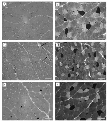
he fragments of the EDL muscles of the control group animals presented polyhedral ibers, peripheral nuclei and variations in iber size (Figure 1A). he immobilization caused a slight increase in the number of ibers undergoing degenera-tive/necrotic processes (Figure 1C). he GIL animals (Figure 1D and 1E) presented increased quantities of centralized nuclei compared with the controls (Figure 1A). Table 1 presents the mean and standard deviation values of the anatomopathologi-cal indings from the animals of the diferent groups analyzed. It
Figure 1. Photomicrographs of the EDL muscle. HE (A, C and E); mATPase pH 4.6 (B, D and F).
Control group (A and B), immobilized group (C and D), immobilized and released group (E and F) (bar: 50 μm). Note the following: (A) variation in fiber size and (B) predominance of fast-twitch type fibers and rarefaction of type 1 fibers (dark fibers); (C) fibers undergoing degenerative/necrotic process (dark arrow) and (D) presence of fibers with intermediated color (light arrow); (E) nuclear centralization (arrow head); and (F) predominance of fast-twitch fibers and rarefaction of type 1 fibers (dark fibers).
A B
C D
E F
can be seen that, in absolute values, the nuclear centralization in the GIL animals and the degenerative/necrotic processes in the GI animals were the features with greatest modiication in relation to the control group indings.
he assessment on the intramuscular connective tissue of the EDL muscle showed that there was a slight increase in the quantity of collagen type I in the GI animals and a slight reduc-tion in the quantity of collagen type III in the GIL animals, as shown in Table 2.
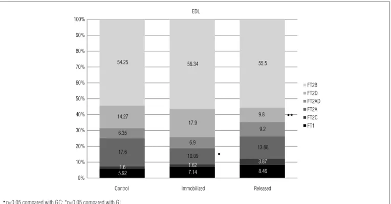
he analysis of the proportions of the diferent types of i-bers in the EDL muscle indicated that immobilization caused a reduction only in the number of FT2A ibers (GI vs. controls, p<0.001), while release into the cage after the immobilization period reduced the number of FT2D ibers (GIL vs. controls, p<0.01; GIL vs. GI, p<0.0001), in relation to the control and GI animals (Figure 2). he data on the smallest iber diameters showed that immobilization caused a reduction in the smallest size of all iber types in relation to the respective control values (GI vs. controls GC, p≤0.03) (Table 2). On the other hand, release of the animals into the cage caused an increase in the diameter values of FT1, FT2A, FT2AD, FT2D and FT2B, compared with those of the GI animals (GIL vs. GI, p<0.01) (Table 3).
Discussion
In rehabilitation clinical practice, there are still some ques-tions with regard to the readaptation of some variables of muscle tissue that is subjected to procedures involving disuse and subsequent release for activities of daily living. Eicient indication of speciic therapeutic approaches or decisions not to indicate certain procedures can be discussed in relation to several factors. Among these are the type of disuse, the period of movement restraint, the contractile characteristics of the muscle (or muscle group) damaged and the morphological ind-ings. In this respect, the present experimental study conducted
Figure 2. Percentages of different types of fibers in the EDL muscle in the groups analyzed.
5.921.6 7.14 8.46
1.62 3.87
17.6
10.09
13.88 6.35
6.9
9.2 14.27
17.9
9.8
54.25 56.34 55.5
0% 10% 20% 30% 40% 50% 60% 70% 80% 90% 100%
Control Immobilized Released
EDL
FT2B FT2D FT2AD FT2A FT2C FT1
t*
t
● p<0.05 compared with GC; *p<0.05 compared with GI.
Table 1. Histopathological changes to fibers [mean (± DP)] in the EDL muscle in the different groups analyzed.
GC=control group; GI=immobilized group; GIL=immobilized and released group.
GC GI GIL
Nuclear centralization 1.3 (±1) 2.8 (±2.3) 6.3 (±7) Lobulated fibers 0.5 (±1.2) 0.2 (±0.4) 0.5 (±0.5) Degeneration/necrosis 3 (±1.7) 4.2 (±1.9) 1.3 (±0.8) Regeneration 0.2 (±0.4) 0.2 (±0.4) 0 Splitting 1.2 (±1.6) 0.3 (±0.8) 1 (±0.9)
Table 2. Semi-quantitative assessment of interstitial collagen in the EDL muscle.
GC=control group; GI=immobilized group; GIL=immobilized and released group; - nega-tive; ± slightly positive; + weakly positive; ++ moderately positive; +++ strongly positive.
GC GI GIL
Collagen I ±/+ + ±/+
Collagen III ++ ++ +/++
on female Wistar rats found that 14 days of immobilization of the EDL muscle in elongation and subsequent release of the animals into the cage for another 10 days gave rise to signii-cant changes to the expression of interstitial collagen and the proportions and tropism of muscle ibers.
Effects of immobilization in elongation
Speciically, the procedure of immobilization of the EDL mus-cle, in the elongated position for 14 days, caused atrophy of the diferent types of muscle ibers, reduction only in the proportion of FT2A and a slight increase in the quantity of collagen I. Immobili-zation in an elongated position is considered to stimulate longitu-dinal tension, but it does not cause overload on skeletal muscle. In the other hand, it may induce modiications to factors relating to myogenic regulation. A study conducted by Gomes et al.27 found that daily sessions of stretching of the soleus of normal rats that were maintained for 30 minutes a day for 7 days only modiied the expression of mRNA to atrogin-1, and did not change the expres-sion of mRNA to MyoD and myostatin. According to Kim et al.28, there is a relationship between the expression of atrogin and ubiq-uitin-ligase, which plays an important role in the process of protein degradation and, consequently, in the regulation of muscle mass. In parallel, studies conducted by Williams et al.29 and Ahtikoski et al.30 found that immobilization in an elongated position for a week did not cause any change to the amount of connective tissue. In the second of these studies, it was found that keeping a muscle in elongation prevented reduction of the mRNA levels for collagen I. In addition, the mRNA concentrations for type I and III collagens showed reductions on the third day of immobilization of the tibialis anterior, but returned to the control values after the seventh day31. On the other hand, Jósza et al.11 found that the quantity of con-nective perimysial tissue in the soleus and gastrocnemius muscles of animals subjected to immobilization in dorsilexion increased during the irst week post-procedure, but progressed to ive times this amount after the third week. he data highlighted above sug-gest that the duration of joint restriction is an important variable relating to occurrences of changes to the collagen and conjunctive tissue, like those identiied in the present study. In the extracellular matrix, collagen type I determines tensile strength and stifness and therefore, from a biomechanical viewpoint, reduction of the level of this protein can impair the capacity to support loading in the elastic phase32,33, which occurs before the rupture, thus making the tissue more vulnerable to longitudinal stress.
Immobilization of the fast-twitch type ibers in elongation can cause an increase in the fraction of fast ibers, thereby leading to a transition from fast ibers to ibers that are even faster12. Since the EDL is mainly composed of fast-twitch ibers, it could be seen that after the period of immobilization of this muscle, there was a signiicant reduction in FT2A, although
without statistical increases in the other iber types. herefore, it is believed that the duration of the immobilization used in the present study was insuicient to conirm the direction of the transition that was induced by the intervention to main-tain stretching, as highlighted by previous authors.
he average value of the smallest diameter of the animals’ ibers immobilized in elongation was smaller than that of the control animals, as also observed by Chopard, Pons and Marini34, Stelzer and Widrick35 and Gehrke et al.36 On the other hand, Yang et al.37 immobilized the EDL muscle in an elongated position for six days and found that there were increases in muscle mass and in local expression of IGF-1. A study devel-oped by De Deyne et al.3 showed that the changes to IGF-1 may contribute little towards the transformations undergone by the EDL immobilized in an elongated position. Hence, it is possible that the duration of immobilization was an important factor, or that this protein has other roles, i.e. not only for promotion of muscle growth. Although the methodology used in the pres-ent study did not allow investigation of ultrastructural altera-tions and proteins speciic to the costamere, the presence of ibers undergoing a process of degeneration/necrosis and others with nuclear centralization makes it possible to suggest that damage to the sarcolemma and subsequent architectural modiication to the proteins of the costamere occurred. his is of fundamental importance with regard to the stability of the sarcolemma and nucleus positioning38, especially for the EDL muscle, as identiied by O’Neill et al.39. Destructuring of the sar-colemma and the proteins associated with it may, in turn, have evoked a cascade relating to lysosomal proteolysis via cathep-sin and ATP-dependent ubiquitin proteasome40,41. In addition, titin, a structural protein of importance for maintaining the structure of the sarcomeres, may have been damaged by the in-tensity of the elongation developed during immobilization. he Table 3. Mean smallest diameter (μm) with respective confidence interval for the different fiber types in the EDL muscle.
GC=control group; GI=immobilized group; GIL=immobilized and released group;●p<0.05
compared with GC; * p<0.05 compared with GI.
GC GI GIL
FT1 24.16
(22.25 - 26.07)
20.80●
(18.94 - 22.66)
24.31* (22.41 - 26.20)
FT2C 23.89
(21.07 - 26.71)
20.19●
(17.45 - 22.92)
22.77 (20.58 - 24.97)
FT2A 23.72
(22.04 - 25.40)
20.87●
(19.10 - 22.65)
23.22* (21.41 - 25.02)
FT2AD 24.59
(22.70 - 26.47)
21.49●
(19.57 - 23.39)
23.64* (21.75 - 25.53)
FT2D 29.95
(28.24 - 31.66)
23.37●
(21.70 - 25.05)
28.46●*
(26.57 - 30.35)
FT2B 32.65
(31.05 - 34.23)
23.12●
(21.54 - 24.71)
31.48●*
(29.88 - 33.08)
elongation may have exceeded the functional amplitude of the strain of the PEVK domain (proline-glutamine-valine-lysine-rich domain), thus causing disorganization of the contractile myoibrils and, consequently, signaling iber degeneration42,43. In parallel with the disarrangement of the sarcolemma, tissue repair and regeneration was highlighted by nuclear centraliza-tion and the slight increase in type I collagen. Summarizing, it should be borne in mind that immobilization promotes a reduction in protein synthesis and an acceleration in protein degradation. However, the levels of atrophy and modiication to collagen synthesis seem to be diferent for each muscle, de-pending on the type of iber composing the muscle30 and the position in which the muscle remained immobilized35.
Effects of the lengthening position on
post-immobilization release
Free movement among the animals was able to restore the diameter ratio to the proportions seen in the control group, for almost all types of ibers except FT2B and FT2D. Since the EDL muscle is mainly composed of fast-twitch ibers, which are the irst to be recruited, the return to activity may have caused greater damage to these ibers during the initial phase of re-lease44, thereby delaying their recovery process and volume gain. Studies conducted by our research group have shown that after immobilization of the plantar muscle (which is also composed predominantly of fast-twitch ibers) in a shortened positions for ten days, its morphological characteristics are recovered after ten days of rehabilitation, either by stretch-ing or by eccentric trainstretch-ing (Cornachione et al.45, unpublished results). Furthermore, we observed a slight reduction in the expression of collagen III.
his type of collagen is responsible for compliance of the extracellular matrix, thereby allowing greater mobility between the muscle ibers. As observed by other authors on the seventh day post-immobilization in an elongated position30,31, the pres-ent study also showed that the collagen type III contpres-ent did not difer from what was observed in the control group on the fourteenth day after immobilization. Although there are no reports in the literature on mRNA and/or pre-collagen type III expression after restraint removal using the methodology of the present study, it can be inferred that the release from restraint favored reorganization of these macromolecular structures, as suggested by Coutinho, DeLuca and Salvini46. Furthermore, in muscles with a predominance of glycolytic ibers, exercise
loading increases the expression levels of mRNA and of metal-loproteinase 2 (MMP-2) itself47. his, in turn, besides acting to degrade the connective tissue of the extracellular matrix, in-creases the activation of satellite cells48, thereby assisting in the process of tissue regeneration49. his is shown by the increased presence of centralized nuclei, as found in the present study. In parallel, the increase in the smallest diameter of most of the ibers can be explained in terms of the activation of a cascade of events involving growth factors and muscle regeneration, such as IGF-1 and MGF27,50.
Considering the variables analyzed and the methodology used, it can be concluded that immobilization in the length-ened position for 14 consecutive days caused histopathological changes in the EDL muscle that were consistent with disuse, featuring atrophy of all kinds of ibers and increased levels of collagen I. he return of loading for ten days, with the release of the animal, generated mechanical stimuli that were capable of promoting activation of mechanisms that were responsible for retrieving the patterns of the EDL muscle, except for the tropism and the proportion of fast-twitch ibers. From the re-sults obtained in this study on female rats, it is hypothesized that some care should be taken in clinical rehabilitation when subjecting segments to loading after brief periods of immobili-zation in the lengthened position.
It is important to highlight the fragility of the cellular and interstitial components to longitudinal stress, as well as the likely functional muscle weakness characterized by atrophy and reduced proportions of fast ibers. herefore, it is sug-gested that the recovery of function should be gradual based on the use of exercises that can increase the activity of oxida-tive and glycolytic ibers (FT2A), which sufered a reduction in number and volume. One parallel action would be to use elec-trical stimulation of mean frequency51 in the muscle group that is kept stretched through immobilization, which would help in the speciic recovery of these ibers and, thus, in restoring muscle strength and function.
Acknowledgement
Financial support was received from the Coordenação de Aperfeiçoamento de Pessoal de Nível Superior (CAPES), the Fundação de Amparo à Pesquisa do Estado de São Paulo (FAPESP), the Education, Research and Attendance Support Foundation of HCFMRP-USP and the Pro-rectory of Research of USP.
References
1. Farmer SE, James M. Contractures in orthopaedic and neurological conditions: a review of causes and treatment. Disabil Rehabil. 2001;23(13):549-58.
2. Kay RM, Rethlefsen SA, Fern-Buneo A, Wren TA, Skaggs DL. Botulinum toxin as an adjunct to serial casting treatment in children with cerebral palsy. J Bone Joint Surg Am. 2004;86-A(11):2377-84.
3. De Deyne PG, Kinsey S, Yoshino S, Jensen-Vick K. The adaptation of soleus end edl in a rat model of distraction osteogenesis: IGF-1 and fibrosis. J Orthop Res. 2002;20(6):1225-31.
4. Thorey F, Bruenger J, Windhagen H, Witte F. Muscle response to leg lengthening during distraction osteogenesis. J Orthop Res. 2009;27(4):483-8.
5. Fucs PM, Svartman C, Santili C, De Assumpção RM, de Almeida Leite LF, Quialheiro LS, et al. Results in the treatment of paralytic calcaneus-valgus feet with the Westin technique. Int Orthop. 2007;31(4):555-60.
6. Leppilahti J, Orava S. Total Achilles tendon rupture. A review. Sports Med. 1998;25(2):79-100.
7. Metz R, Kerkhoffs GM, Verleisdonk EJ, van der Heijden GJ. Acute Achilles tendon rupture: minimally invasive surgery versus non operative treatment, with immediate full weight bearing. Design of a randomized controlled trial. BMC Musculoskelet Disord. 2007;8:108.
8. Järvinen MJ, Einola SA, Virtanen EO. Effect of the position of immobilization upon the tensile properties of the rat gastrocnemius muscle. Arch Phys Med Rehabil. 1992;73(3):253-7.
9. Da Silva CA, Guirro RRJ, Polacow MLO, Cancelliero KM, Durigan JLQ. Rat hindlimb joint immobilization with acrylic resin orthoses. Braz J Med Biol Res. 2006;39(7):979-85.
10. Järvinen TAH, Józsa L, Kannus P, Järvinen TLN, Järvinen M. Organization and distribution of intramuscular connective tissue in normal and immobilized skeletal muscles. An immunohistochemical, polarization and scanning electron microscopic study. J Muscle Res Cell Motil. 2002;23(3):245-54.
11. Józsa L, Kannus P, Thöring J, Reffy A, Järvinen M, Kvist M. The effect of tenotomy and immobilisation on intramuscular connective tissue. A morphometric and microscopic study in rat calf muscles. J Bone Joint Surg Br. 1990;72(2):293-7.
12. Pette D, Staron RS. Myosin isoforms, muscle fiber types, and transitions. Microscopy Res Tech. 2000;50(6):500-9.
13. Loughna PT, Izumo S, Goldspink G, Nadal-Ginard B. Disuse and passive stretch cause rapid alterations in expression of developmental and adults contractile protein genes in skeletal muscle. Development. 1999;109(1):217-23.
14. Guillot C, Steinberg JG, Delliaux S, Kipson N, Jammes Y, Badier M. Physiological, histological and biochemical properties of rat skeletal muscles in response to hindlimb suspension. J Electromyogr Kinesiol. 2008;18(2):276-83.
15. Ploutz-Snyder LL, Tesch PA, Hather BM, Dudley GA. Vulnerability to dysfunction and muscle injury after unloading. Arch Phys Med Rehabil. 1996;77(8):773-7.
16. Mattiello-Sverzut AC, Carvalho LC, Cornachione A, Nagashima M, Neder L, Shimano AC. Morphological effects of electrical stimulation and intermittent muscle stretch after immobilization in soleus muscle. Histol Histopathol. 2006;21(9):957-64.
17. Frimel TN, Kapadia F, Gaidosh GS, Li Y, Walter GA, Vandenborne K. A model of muscle atrophy using cast immobilization in mice. Muscle Nerve. 2005;32(5):672-4.
18. Goldspink D. The influence of immobilization and stretch on protein turnover of rat skeletal muscle. J Physiol. 1997;264(1):267-82.
19. Machida S, Booth FW. Regrowth of skeletal muscle atrophied from inactivity. Med Sci Sports Exerc. 2004;36(1):52-9.
20. Heinemeier KM, Olesen JL, Haddad F, Schjerling P, Baldwin KM, Kjaer M. Effect of unloading followed by reloading on expression of collagen and related growth factors in rat tendon and muscle. J Appl Physiol. 2009;106(1):178-86.
21. Boonyarom O, Inui K. Atrophy and hypertrophy of skeletal muscles: structural and functional aspects. Acta Physiol (Oxf). 2006;188(2):77-89.
22. Glass DJ. Skeletal muscle hypertrophy and atrophy signaling pathways. Int J Biochem Cell Biol. 2005;37(10):1974-84.
23. Miyazaki M, Esser KA. Cellular mechanisms regulating protein synthesis and skeletal muscle hypertrophy in animals. J Appl Physiol. 2009;106(4):1367-73.
24. Kurose T, Asai Y, Mori E, Daitoku D, Kawamata S. Distribution and change of collagen types I and III and elastin in developing leg muscle in rat. Hiroshima J Med Sci. 2006;55(3):85-91.
25. Stevens A, Palmer J. Enzyme histochemistry: Diagnostic application. In: Bancroft JD, Stevens A. Theory and practice of histological techniques. 4ª edição. New York: Churchill Livingstone; 1996.
26. Aherne WA, Dunnill MS. Methods of estimating myofibre size. Morphometry. Great Britain: Edward Arnold; 1982.
27. Gomes AR, Soares AG, Peviani S, Nascimento RB, Moriscot AS, Salvini TF. The effect of 30 minutes of passive stretch of the rat soleus muscle on the myogenic differentiation, myostatin, and atrogin-1 gene expressions. Arch Phys Med Rehabil. 2006;87(2):241-6.
28. Kim SJ, Roy RR, Kim JA, Zhong H, Haddad F, Baldwin KM, et al. Gene expression during inactivity-induced muscle atrophy: effects of brief bouts of a forceful contraction countermeasure. J Appl Physiol. 2008;105(4):1246-54.
29. Williams PE, Catanese T, Lucey EG, Goldspink G. The importance of stretch and contractile activity in the prevention of connective tissue accumulation in muscle. J Anat. 1988;158:109-14.
30. Ahtikoski AM, Koskinen OA, Virtanen P, Kovanen V, Takala TE. Regulation of synthesis of fibrillar collagens in rat skeletal muscle during immobilization in shortened and lengthened positions. Acta Physiol Scand. 2001;172(2):131-40.
31. Han XY, Wang W, Komulainen J, Koskinen SO, Kovanen V, Vihko V, et al. Increased mRNAs for procollagens and key regulating enzymes in rat skeletal muscle following downhill running. Pflugers Arch. 1999;437(6):857-64.
32. Carvalho LC, Polizello JC, Padula N, Freitas FC, Shimano AC, Mattiello-Sverzut AC. Propriedades mecânicas do gastrocnêmio eletroestimulado pós-imobilização. Acta Ortop Bras. 2009;17(5):269-72.
33. Polizello JC, Carvalho LC, Freitas FC, Padula N, Shimano AC, Mattiello-Sverzut AC. Propriedades mecânicas do músculo gastrocnêmio de ratas, imobilizado e posteriormente submetido a diferentes protocolos de alongamento. Rev Bras Med Esporte. 2009;15(3):195-9.
34. Chopard A, Pons F, Marini J-F. Cytoskeletal protein contents before and after hindlimb suspension in fast and slow rat skeletal muscle. Am J Physiol Regul Integr Comp Physiol. 2001;280(2):R323-30.
35. Stelzer JE, Widrick JJ. Effect of hindlimb suspension on the functional properties of slow and fast soleus fibers from three strains of mice. J Appl Physiol. 2003;95(6):2425-33.
36. Gehrke AG, Krull MS, McDonald RS, Sparby T, Thoele J, Troje SW, et al. The effects of non-weight bearing on skeletal muscle in older rats: an interrupted bout versus an uninterrupted bout. Biol Res Nurs. 2004;5(3):195-202.
37. Yang H, Alnaqeeb M, Simpson H, Goldspink G. Changes in muscle fiber type, muscle mass and IGF-I gene expression in rabbit skeletal muscle subjected to stretch. J Anat. 1997;190(Pt 4):613-22.
38. Shah SB, Davis J, Weisleder N, Kostavassili I, McCulloch AD, Raiston E, et al. Structural an functional roles of desmin in mouse skeletal muscle during passive deformation. Biophys J. 2004;86(5):2993-3008.
39. O’Neill A, Williams MW, Resneck WG, Milner DJ, Capetanaki Y, Block RJ. Sarcolemmal organization in skeletal muscle lacking desmin: evidence for cytokeratins associated with the membrane skeleton at costameres. Mol Biol Cell. 2002;13(7):2347-59.
40. Chopard A, Arrighi N, Carnino A, Marini JF. Changes in dysferlin, proteins from dystrophin glycoprotein complex, costameres, and cytoskeleton in human soleus and vastus lateralis muscles after a long-term bedrest with or without exercise. FASEB J. 2005;19(12):1722-4.
41. Coffey VG, Hawley JA. The molecular bases of training adaptation. Sports Med. 2007;37(9):737-63.
42. Wang K, McCarter R, Wright J, Beverly J, Ramirez-Mitchell R. Viscoelasticity of the sarcomere matrix of skeletal muscles. The titin-myosin composite filament is a dual-stage molecular spring. Biophys J. 1993;64(4):1161-77.
43. Kellermayer MS, Bustamante C, Granzier HL. Mechanics and structure of titin oligomers explored with atomic force microscopy. Biochim Biophys Acta. 2003;1604(2):105-14.
44. Mitchell PO, Pavlath GK. Skeletal muscle atrophy leads to loss and dysfunction of muscle precursor cells. Am J Physiol Cell Physiol. 2004;287(6):C1753-62.
45. Cornachione AS, Oliveira LCB, Benedini-Elias PCO, Martinez EZ, Mattiello-Sverzut AC. Resultados não publicados.
46. Coutinho EL, DeLuca C, Salvini TF. Bouts of passive stretching after immobilization of the rat soleus muscle increase collagen macromolecular organization and muscle fiber area. Connect Tissue Res. 2006;47(5):278-86.
47. Carmeli E, Moas M, Lennon S, Powers SK. High intensity exercise increases expression of matrix metalloproteinases in fast skeletal muscle fibres. Exp Physiol. 2005;90(4):613-9.
48. Yamada M, Tatsumi R, Kikuiri T, Okamoto S, Nonoshita S, Mizunoya W, et al. Matrix metalloproteinases are involved in mechanical stretch-induced activation of skeletal muscle satellite cells. Muscle Nerve. 2006;34(3):313-9
49. Pette D, Staron RS. Mammalian skeletal muscle fiber type transitions. Int Rev Cytol. 1997;170:143-223.
50. Goldspink G, Wessner B, Bachl N. Growth factors, muscle function and doping. Curr Opin Pharmacol. 2008;8(3):352-7.
51. Sinacore DR, Delitto A, King DS, Rose SJ. Type II fiber activation with electrical stimulation: a preliminary report. Phys Ther. 1990;70(7):416-22.