Artigo
*e-mail: [email protected]
METABOLIC RESPONSE INDUCED BY ENDOPHYTIC FUNGI AND BACTERIA IN H. marrubioides Epling IN VITRO MICROPLANTS
Luciana Cristina Vitorino and Fabiano Guimarães Silva*
Instituto Federal de Educação, Ciência e Tecnologia Goiano, Rio Verde – GO, Brasil William Cardoso Lima and Marcos Antônio Soares
Departamento de Botânica e Ecologia, Universidade Federal de Mato Grosso, Cuiabá – MT, Brasil
Rita Cássia Nascimento Pedroso, Marolí Rodrigues Silva, Herbert Júnior Dias, Antônio Eduardo Miller Crotti, Márcio Luís Andrade e Silva, Wilson Roberto Cunha, Patrícia Mendonça Pauletti and Ana Helena Januário
Núcleo de Pesquisa em Ciências Exatas e Tecnológicas, Universidade de Franca, Franca – SP, Brasil
Recebido em 26/10/12; aceito em 6/3/13; publicado na web em 13/6/13
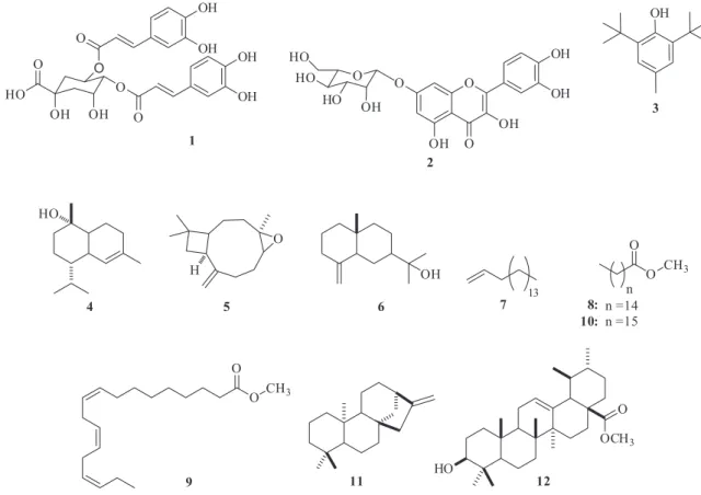
Hyptis marrubioides Epling is a native plant from Brazilian Cerrado. In this paper, the response of in vitro microplants of this species to inoculation with bacterial and fungal endophytic isolates is evaluated. HPLC-DAD analysis showed the presence of 3,4-O-(Z)-dicaffeoylquinic acid and quercetin-7-O-glucoside as the main components. GC/MS analysis demonstrated that the sesquiterpenes τ-cadinol and caryophyllene oxide were only produced in microplants inoculated with endophytic bacteria, while methyl hexadecanoate, methyl heptadecanoate and methyl (Z,Z,Z) 9,12,15-octadecatrienoate and the triterpene methyl 3β-hydroxy-urs-12-en-28-oate were overexpressed only when the microplant was treated with endophytic fungi.
Keywords:in vitro culture; Hyptis marrubioides; Lamiaceae.
INTRODUCTION
Currently, many mechanisms are employed for the induction of resistance responses in plants. These methods involve the acti-vation of latent defense mechanisms, which increase the ability of the plant to either prevent or delay the establishment of pathogenic microorganisms.1 Post-formed defenses can be triggered by exposure to inducing agents, which act as elicitors of the defense response.2 Resistance induction often involves the accumulation of secondary metabolites in plant tissues and the production of phytoalexins with antimicrobial properties by the plant itself.3
Elicitors could have a chemical or biological nature, such as non-pathogenic microorganisms,4 avirulent forms or incompatible strains of a pathogen, or even virulent pathogens. Fungal culture filtrate,5 yeast extract,6 and polysaccharides from microorganism walls7 can also be utilized. The major bacterial determinants for systemic resistance induction elicitors are the lipopolysaccharides (LPS) present in their membrane, siderophores, and salicylic and jasmonic acids.8
Plant-endophyte association is a type of symbiotic relationship with a similar or higher degree of complexity than a phytopatho-genic microorganism. Plant–endophytic association involves fungi and bacteria living inside plant tissues without causing any visible disease symptoms and sometimes interacting mutualistically with their hosts.9 Endophyte-host interactions involve a balance of antagonism and exhibit stronger phenotypic plasticity compared to the interactions of plant pathogens.10 Currently, the signals rele-ased by the two partners and their roles remain largely unknown. Researchers have tried to clarify the molecular mechanisms involved during the establishment of endophyte colonization.11 Yet there is little literature about the secondary metabolism of plants induced by its endophytes.12
Tissue culturing is a useful tool for the reproduction of explants with desirable characteristics, such as resistance to pests and other
stress conditions, high productivity, and high yield of active substan-ces of interest.13In vitro elicitation has been proven as an efficient biotechnological process for the attainment of secondary metabolites from plant cells or tissues. Chong and coworkers have used yeast extract, jasmonic acid, a mycelia homogenate of Aspergillus niger and Aspergillus flavus, chitosan, and glucan in cell suspensions of Morinda elliptica (Hook. f.) Ridl.6 These elicitors had different effects on anthraquinone production. Naoumkina and coworkers have reported that the use of yeast extract in cell cultures of Medicago truncatula induced the accumulation of flavonoids, triterpenes, and other defense-related molecules.14
Several species of the genus (Lamiaceae) occur in the Brazilian savannah (Cerrado biome), some of which have several medicinal properties and great pharmaceutical relevance.15 The species Hyptis marrubioides Epling is a medicinal plant native to this region,16 with no scientific data on its in vitro elicitation, even though this plant has drawn researchers’ attention due to the remarkable concentra-tion of terpenoids in its essential oils (e.g., cariofila-4(14),8(15)-dien-5β-ol, eudesma-4(15),7-dien-1β-ol, caryophyllene oxide, and β-caryophyllene).17 The roots of H. marrubioides are colonized by bacteria and endophytic fungi18 with different functional traits important to promoting plant growth.19
In this study, we have evaluated the phytochemical profile of H. marrubioides microplants exposed to inoculation with endophytic isolates.
EXPERIMENTAL
Plant material selection, in vitro establishment, and maintenance
with ethanol 70% (v/v) for 30 sec and sodium hypochlorite (2.5% of active chlorine) for 20 min, and inoculated in Murashige & Skoog (MS) medium (50% salt concentration).20 Seedlings were cut into small pieces, using stalk segments with a height of approximately 1 cm as explants, and kept in 50% MS. The cultures were incubated in a growth chamber for 45 days at 0.5 µmol m-2 s-1 photosynthetically active radiation (PAR), with an average temperature of 25 °C and a photoperiod of 16 hours.
Inoculum preparation
The endophytic microorganism lineages were obtained from H. marrubioides roots used in a previously mentioned study.18
The following isolates were selected as bacterial inoculants: RG8 (simple coccus, Gram-positive, catalase negative); RG12 (coccoba-cilli, positive, catalase negative); RF10 (coccoba(coccoba-cilli, Gram-positive, catalase negative); and RF11 (large bacillus, Gram-Gram-positive, catalase negative). These isolates were grown in nutrient broth (3 g beef extract, 5 g peptone, and H2O qs 1 L) overnight at 30 °C, under 150 rpm agitation. Subsequently, 15 µL aliquots of cultures were inoculated near the roots of H. marrubioides microplants growing in 50% MS medium (T2). Axenic nutrient broth inoculum (15 µL) was inoculated into microplant control (T3).
The fungal lineages used were RG29 (Trichoderma sp.), RG31 (Papulaspora sp.), and RG32 (Fusarium sp.). These fungi were grown on solid PDA (infusion of 200 g potato, 20 g dextrose, 15 g agar, and H2O qs 1 L) and their colonies were fragmented using the bottom of 1000 µL tips. This fragmentation generated disks with a diameter of 5 mm, which were then inoculated near the base of H. marrubioides Epling microplants grown in 50% MS medium. PDA discs without the presence of mycelium were inoculated in the microplants control (T8).
Culture conditions
The Hyptis marrubioides microplants were inoculated with the elicitors after 45 days of establishment in 50% MS. After inoculation, the flasks containing the cultures were kept in a BOD incubator with 15 µmol m-2 s-1 PAR, at an average temperature of 30 °C, and with a photoperiod of 16 hours. The plants were evaluated on a daily basis for the appearance of disease symptoms, and the ones subjected to inoculation with the bacterial isolates had their shoots collected on the tenth day after inoculation. The plants subjected to treatment with fungal isolates were collected on the fifth day after inoculation. These shoots, as well as the H. marrubioides plants grown ex vitro (T1), were dried in a forced-air oven at 35 °C until a constant weight was reached.
Chemical analysis of plants grown ex vitro to obtain standards
Extraction and purification
Hyptis marrubioides shoots grown ex vitro were collected in the Seedling Nursery of the Laboratory Tissue Culture Instituto Federal de Educação, Ciência e Tecnologia Goiano in Rio Verde, Goiás (S17°48’15.9” W50°54’19,5”), on April 20th, 2011. A voucher spe-cimen (HRV71) has been deposited in the Herbarium of that Institute (Herbarium HRV).
The air-dried H. marrubioides shoots (502 g) were powdered and exhaustively extracted by maceration with 96% ethanol. After concentration in a rotary evaporator, an ethanolic crude extract (26.10 g) was obtained. A sample (20 g) of this extract was dissolved in a MeOH:H2O mixture (2:8 v/v) and partitioned successively with n--hexane, ethyl acetate, and n-butanol (n-BuOH). After removal of
the solvent under reduced pressure, the following fractions were obtained: n-hexane (H1, 4.01 g), ethyl acetate (H2, 3.67 g), n-BuOH (H3, 6.28 g), and hydroalcoholic (H4, 2.16 g).
The ethyl acetate fraction (H2, 3.67 g) was initially chromato-graphed on a silica gel column (Merck silica 100 g, 30 cmx3.5 cm) using an n-hexane/ethyl acetate (EtOAc) and EtOAc/MeOH gradient solvent system as eluent, which yielded 66 fractions. Fraction 65 (1.61 g, MeOH) was purified over Sephadex LH-20® (Sigma-Aldrich, 75 g, 37.5x3.0 cm) eluted with a MeOH and MeOH/H2O (1:1 v/v) mixture, from which 74 fractions were obtained. Fraction 33 (46 mg, MeOH) was subsequently submitted to Preparative Thin Layer Chromatography (Sigma-Aldrich PTLC, 20x20x0,5 cm) using CHCl3/ MeOH/H2O (43:37:20 v/v/v) as the eluting system. This procedure afforded results 1 (8 mg) and 2 (10 mg), which were used as standards for the quantitative analysis performed by the High-Performance Liquid Chromatography with Diode-Array Detection (HPLC-DAD) on the microplants inoculated with endophytic microorganisms as well as the control microplants. These results were compared with those of ex vitroH. marrubioides plants.
Chemical general experimental procedures
The Nuclear Magnetic Resonance (1H NMR and 13C NMR) spec-tra were registered on a Varian 500 instrument using tespec-tramethylsilane (TMS) as the internal standard (1%). The spectra of 1 were obtained in CD3OD (Sigma-Aldrich
®), and the spectra of 2 were acquired in DMSO-d6 (Sigma-Aldrich
®).
The quantitative analysis by HPLC-DAD was carried out on a Shimadzu LC-6AD system equipped with a degasser DGU-20-A5, a diode array detector Prominence SPD-M20A series, and a com-munication bus mode CBM-20A. The analyses were conducted on a Phenomenex ODS column (250x4.6 mm, 5 µm) equipped with a pre-column of the same material and with the following parameters: injection volume: 20 µL; flow rate: 1.0 mL min-1; mobile phase: CH3OH/H2O/HOAc (40:59.9:0.1, v/v/v) linear gradient until 100% CH3OH in 30 min, 10 min at 100% CH3OH, and 15 min to return to the initial condition; UV detection at 230, 254, and 325 nm. Quantitative HPLC-DAD analysis
Pulverized air-dried H. marrubioides microplants (50 mg) were extracted with 3 mL HPLC grade methanol, using an ultrasound bath (20 min). The samples had been previously filtered through a filter with 0.2 µm pore size prior to the injection. Extractions were accomplished in triplicate.
The chlorogenic acid (1) and flavonoid (2) solutions were pre-pared in HPLC grade MeOH (2 mg mL-1). Stock solution dilutions were done in HPLC grade MeOH, to obtain solutions containing 0.102, 0.203, 0.406, 0.813, and 1.626 mg mL-1 of 1, and 0.05, 0.109, 0.218, 0.436, and 0.872 mg mL-1 of 2. Each standard solution was injected in triplicate. A four-point calibration curve was constructed to determine the linearity of the method. The calibration curve was determined by plotting the peak area versus the concentration of the substance in mg mL-1.
Gas chromatography mass spectrometry (GC/MS) analysis Sample preparation for GC/MS analysis
GC/MS operating conditions
The methylated extracts of H. marrubioides microplants by means of different treatments were analyzed by GC/MS (Shimadzu model QP-2010) (low resolution). A capillary column Rtx-5MS (30 m × 0.25 mm i.d. × 0.25 µm of film) was employed and a He carrier gas flow of 1.1 mLmin-1, split 1:10, was utilized. The injector temperature was set at 250 °C, and the oven was heated from 100 to 290 °C at 3 °C per minute; the temperature was maintained at 290 °C for an additional 30 min. The mass spectra obtained at 70 eV were compared with the Wiley 7, NIST 08, and FFNSC 1.2 libraries using GCMS Solution software (Shimadzu), which furnished the similarity index (SI) expressed as a percentage. The retention indices relative to a homologous series of n-alkanes (C8-C40) measured on a Rtx-5MS capillary column under the same operating conditions were also provided. Acid compounds present in the extracts were identified as their corresponding methyl esters.
Experimental design
The experiment was carried out with a completely randomized design involving 11 treatments (T1 to T11) T1 was the ex vitro plant control; T2 the microplant control; T3 the bacterial elicitation control; T4 to T7 microplants with bacterial inoculation; T8 fungal elicitation control and T9 to T11 microplants with fungal inoculation (Table 1). Each culture bottle was considered a sampling unit, and each flask contained four microplants. The samples were analyzed in triplicate. Averages for the emergence of symptoms, as well as for the 3,4-O-(Z)-dicaffeoylquinic acid (1) and flavonoid quercetin-7- O--glucoside (2) quantification were obtained by HPLC-DAD analysis and subjected to the Tukey test (5%) with the support of the SISVAR statistical software.
RESULTS AND DISCUSSION
The interaction between plant and endophyte was evaluated on a daily basis. For the statistical analysis, we included the last ten and five evaluations performed on plants inoculated with bacteria and fungi, respectively. Despite strains of fungi and bacteria being obtained from healthy roots of H. marrubioides, the effect of ino-culation with endophyte in aseptic in vitro plants varied according
to the strain used. These factors disturbed the equilibrium between microorganism virulence and plant defense that is important for asymptomatic colonization.11 The same effect was reported for Arabidopsis thaliana where only three of eight strains of fungi colonized the root system asymptomatically in vitro.22 The bacteria RF11 and RG8 were pathogenic in this work, leading to wilting, leaf--descending death, and darkness in the roots. More specifically, RF11 resulted in wilting of 100% of the inoculated microplants (Table 1). The incidence of disease symptoms has been previously described as typical bacterial symptoms.23 In the presence of the bacteria RF10 and RG12, the plants exhibited no obvious pathology signs (Figure 1). These observations demonstrated that those endophytic bacteria had the same endophytic behavior in in vitro conditions. Although the inner tissues of the H. marrubioides have not been evaluated, the penetration of these lineages into the root’s host may have occurred.
On the other hand, in the same context, bacteria RF11 and RG8, as well as the fungi Trichoderma sp., Papulaspora sp., and Fusarium sp. (isolated from the roots of H. marrubioides in the in vitro system) started to act as pathogens despite being originally endophytic strains. This behavior was evidenced by the appearance of visual symptoms in the affected plants. The wilting symptom is often related to vascular diseases caused by the colonization of xylem vessels by pathogenic microorganisms and presents with associated leaf fall. It is possible that bacteria RF11, RG8 as well as the fungi, Trichoderma sp., Papulaspora sp., and Fusarium sp. colonized the xylem conductor system in the H. marrubioides roots causing the observed invitro wilting.24
Although the fungus Papulaspora sp. caused widespread wilting in the microplants, it did not trigger leaf drop, as in the case of the other tested fungal species. The successful symbiosis of endophytic organisms is directly related to environmental conditions and the physiological state of the host, and under adverse conditions, they may manifest themselves as pathogens. In this case, the endophytic organism can be seen as a pathogen under in vitro conditions.25 The experimental conditions used were possibly a stress factor for the plants; additionally, the MS agar culture medium improved the conditions for growth of the endophyte.
Trichoderma, Papulaspora, and Fusarium are common genera in endophytic symbiosis.26 However, there are few reports on the
Table 1. Symptoms of pathogenicity caused by endophytic bacteria (at the 10th inoculation day) and fungi (at the 5th inoculation day) used as elicitors in H.
marrubioides microplants
Treatment Plants with Wilting (%) Plants with Falling Leaves (%) Color of the Roots
Plant
T1 Ex vitro plant (control) - -
-T2 Microplant (control ) 0da 0b Light
Bacterial Elicitation
T3 Microplant + nutritive broth (control) 0d 0b Light
T4 Microplant + RF11 100a 80a Dark
T5 Microplant + RG8 80b 80a Dark
T6 Microplant + RF10 0d 0b Light
T7 Microplant + RG12 0d 0b Light
Fungal Elicitation
T8 Microplant + PDA (control) 0d 0b Light
T9 Microplant + Trichoderma sp. 30c 0b Light
T10 Microplant + Papulaspora sp. 100a 0b Light
T11 Microplant + Fusarium sp. 30c 0b Light
pathogenicity of species from these genera in in vitro systems.27 Trichoderma species have been used in the biological control of plant pathogens, and this control can be manifested through mycoparasitism or antibiosis.28 The genus Papulaspora has also been shown to dis-play antimicrobial activity29 and potential for secondary metabolites production.30 On the other hand, the genus Fusarium is known to trigger wilting, or Fusariosis, characterized by xylem hypertrophy31 and wilting followed by death in affected plants.32 These symptoms were detected in plants submitted to treatment T7 (RG32), which underwent inoculation with Fusarium (Figure 1). These data de-monstrated that this endophytic organism had pathogenic behavior in the in vitro systems.
The chromatographic purification of the ethyl acetate fraction led to the isolation of compounds 1 and 2. Compound 1 presented three absorption maxima at 194, 220, and 329 nm. The 1H and 13C NMR spectra, together with the two-dimensional HMBC experiment, revealed the presence of two caffeoyl units, as well as signals related to quinic acid. The positions of the caffeoyl groups were determined on the basis of the chemical shifts and coupling constants obtained for the hydrogens H-3 (δ 5.62, m), H-4 (δ 5.13, dd, 8.6 Hz; 2.9 Hz) and H-5 (δ 4.42, m). The coupling constant value for H-4 indicated the axial-equatorial relationship between H-4 and H-5, according to the 3,4-diequatorial conformation proposal for the caffeoyl groups. Comparing our results with literature data for the isomeric 3,4-; 3,5- and 4,5-O-dicaffeoylquinic acids, the identity of compound 1 was confirmed as 3,4-O-(Z)-dicaffeoylquinic acid (Figure 2 ).33
The response of the UV detector at 325 nm was linear from 0.102 to 1.626 mg mL-1 for 1 and from 0.05 to 0.872 mg mL-1 for 2. The obtained regression equations were y = 4.0 × 10⁷ × –217572 and y = 3.0 × 10⁷ × –409470 with a correlation coefficient (r2) of 0.9995 and 0.9989, respectively.
In this study, the quantification of chlorogenic acid evidenced that T11 (i.e., in which Fusarium sp. was employed as elicitor) doubled the amount of acid that was produced (128.74 mg gDW-1, as compared to the control treatment without fungi (T8, 60.57 mg gDW-1) (Table 2).
The endophyte–plant interaction leads to qualitative and quantitative alterations in the phytochemical profile of the host. 34Neotyphodium lolii, endophyte fungus, establishes a symbiotic relationship with their host Lolium perenne and this relationship alters the metabolic profiles of L. perenne dramatically.35 This alteration is characterized by the production of a range of chemically unrelated alkaloids such as ergovaline, lolitrem B, and peramine that have never been detected in endophyte-free L. perenne. Neotyphodium lolii infection is found to enhance the level of chlorogenic acid in L. perenne, and also increases the concentrations of polyphenol biosynthesis precursors, quinic and shikimic acids.35,36
The production of 3,4-O-(Z)-dicaffeoylquinic (1) by elicited microplants can be considered a defensive response to the patho-genic behavior that this endophyte assumed in the in vitro system. Chlorogenic acid has been described as being responsible for plant resistance against attack by pests such as caterpillars, beetles, lea-fhoppers, aphids, fungi, and bacteria.37 Baptista and coworkers have inoculated Eucalyptus urophylla plants in vitro with two Pisolithus tinctorius ectomycorrhizal isolates and they found that the roots had a significantly higher chlorogenic acid level 96 hours after inoculation as compared to a control.38
On the other hand, the UV spectrum of compound 2 displayed absorption bands at 197, 255, and 354 nm, typical of flavonoids. The 1H and 13C NMR spectra exhibited the characteristic signals of a flavonoid skeleton, whose ring B coupling pattern suggested a quercetin core. Diagnostic signals for a monoglycoside unit were also observed in this spectrum. The downfield-shift of ortho carbon signals C-6 (δ 98.9) and C-8 (δ 94.9) and para carbon signal C-10 (δ 104.7), together with upfield-shift of C-7 (δ 162.8) suggesting the 7-O-glycosilation of the aglycone quercetin. The structure of compound 2 is proposed on the basis of the set of 1H and 13C NMR spectral data and on the heteronuclear, multiple-bond correlation spectroscopy (HMBC), which were consistent with those reported in the literature for the flavonoid quercetin-7-O-glucoside.39
The quercetin-7-O-glucoside (2) quantification results obtained for H. marrubioides microplants inoculated with endophytic isolates, both bacterial and fungal, showed that all the microplants, elicited or control, contained low concentrations of this compound as com-pared to the amount found in the ex vitro plant (19.02 mg g-1 dry weight) (Table 2). In contrast, other studies have shown increased synthesis of flavonoids in vitro with or without elicitors, for example, quercetin-3-O-glucoside has been identified as a secondary metabolite obtained from the ethanol extract of in vitroDrosera binata cultures,40 while Xu and coworkers have verified increased flavone synthesis in Scutellaria baicalensis Georgii suspension cultures using methyl jasmonate as an elicitor.41
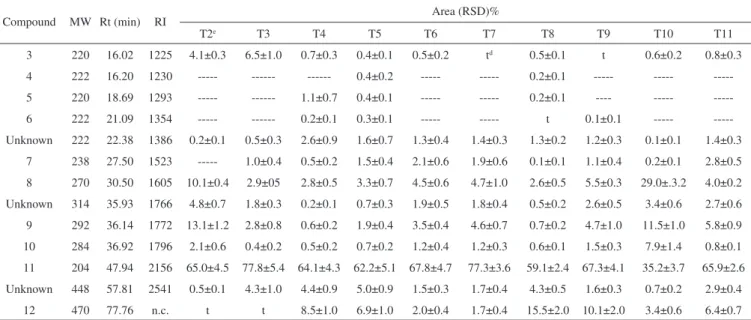
GC/MS analysis of the extracts obtained from the 10 treatments undergone by H. marrubioides revealed the presence of one phenolic compound (3, C15H24O), three sesquiterpenes (τ-cadinol, 4, C15H26O; caryophyllene oxide, 5, C15H24O; and eudesm-4(14)-en-11-ol, 6, C15H26O), one diterpene (kaur-16-ene, 11, C20H32), one triterpene (me-thyl 3β-hydroxy-urs-12-en-28-oate, 12, C31H50O3), one unsaturated hydrocarbon (heptadec-1-ene, 7, C17H26), and three fatty acid methyl esters (methyl hexadecanoate, 8, C17H34O2; methyl (Z,Z,Z )9,12,15-octadecatrienoate, 9, C19H32O2; and methyl heptadecanoate, 10, C18H36O2), as well as two unknown compounds (Figure 2). The diterpene kaur-16-ene was the major biosynthesized constituent in all the treatments, with the best results being attained in the case of treatments T3 and T7 (Table 3).
A significant amount of the fatty acid methyl esters 9 (13.1% and 2.8%) and 8 (10.1% and 2.9%) were detected in treatments T2 and T3, respectively. Concerning microplant inoculation with bacteria RF11, RG8, RG12, and RF10, the sesquiterpenes caryophyllene Figure 1. H. marrubioides microplants used in the control experiment and
oxide (5) and eudesm-4(14)-en-11-ol (6) appeared in treatments T4 and T5 while the τ-cadinol was found only in T5. The presence of the triterpene methyl 3β-hydroxy-urs-12-en-28-oate (12) was observed in higher relative proportion in treatments T4 (8.5%) and T5 (6.9%), contrasting with the intensified hydrocarbons and fatty acids contents verified for treatments T6 and T7.
Terpenoid biosynthesis induction has been investigated in many plant species. Accumulation of the terpenoid essential oil in Ocimum basilicum L. following elicitation with methyl jasmonate gave rise to a total percentage increase of the terpenoids β-caryophyllene, 1,8-ci-neole, linalool and limonene.42 Suspension-cultured cells treated with
fungal elicitors have been found to increase the terpenoids content, especially in the case of sesquiterpenoids. This is because some of them have been classified as phytoalexins, due to their induced biosyn-thesis and their potential role in plant resistance to phytopathogens.43 Comparing treatments T9, T10, and T11 with endophytic fun-gi, to the control T8, it was possible to note that the sesquiterpene eudesm-4(14)-en-11-ol (6) was present only in T9 (0.1%). The highest relative concentration of the methyl 3β-hydroxy-urs-12-en-28-oate (12) was obtained for T9 (10.1%) followed by treatment T11 (6.42%). The strains employed in T9 and T11 also increased the diterpene 11 content (67.3% and 65.9%, respectively), compared with the control T8 (59.1%). On the other hand, treatment T10 prompted a significant rise in the production of the fatty acid methyl esters 8 (29.0%), 9 (11.5%), and 10 (7.9%).
The effect of yeast extract on the accumulation of different types of triterpenoid acids has been previously described. Significant enhancement of oleanolic acid accumulation has been observed upon elicitation with the fungus Trichoderma viride homogenate in Calendula officinais L. cell suspension cultures.44
The induced accumulation of oleanolic and ursolic acids by yeast extract in Uncaria tomentosa cell suspension cultures has also been verified by Feria-Romero and coworkers.45 Triterpene production in the presence of yeast extract might be promoted as a response to cell wall damage involving plant defense mechanisms mediated by endogenous jasmonate induction.46 Another study has revealed that ursolic acid inhibits the synthesis of jasmonate-induced proteins.47
Interestingly, comparison of fatty acids production in the expe-riments conducted in the presence of bacteria and fungi showed that the predominant fatty acids were 8, 9 and 10 in all cases. Ribeiro and coworkers have demonstrated that sesame (Sesamum indicum L. - Pedaliaceae) leaf fractions contained a mixture of fatty acids, for which the major components were tetradecanoic, hexadecanoic,
Table 2. Chlorogenic acid (1) and flavonoid (2) present in H. marrubioides
Treatment 1 (mg gDW-1) 2 (mg gDW-1)
Control Plants T1b 1.91b ± 0.31 19.02ac ± 0.47d
T2 32.84b ± 8.08 1.26b ± 0.20
Bacterial Elicitation T3 34.50b ± 7.50 1.74b ± 0.44
T4 16.32b ± 4.56 0.92b ± 0.06
T5 19.13b ± 6.70 0.85b ± 0.01
T6 38.13b ± 2.23 0.94b ± 0.03
T7 29.71b ± 8.16 1.27b ± 0.17
Fungal Elicitation T8 60.57ab ± 10.8 1.59b ± 0.36
T9 58.54ab ± 8.6 2.36b ± 1.15
T10 16.57b ± 10.5 2.19b ± 0.20
T11 128.74a ± 35.20 1.00b ± 0.04
b Treatments T1 to T11 were described in table 1. c Letters compare rows Means
followed by the same letter do not differ from each other by the Tukey test (5%). d Mean standard deviation.
octadecanoic, eicosanoic, docosanoic, and 9,12,15-octadecatrienoic acids.48 These constituents were able to strongly inhibit the symbiotic fungus Leucoagaricus gongylophorus (Möller) Singer (syn. of Rozites gongylophora Möller) cultivated by the leafcutter ant Atta sexdens L. Because the in vitro culture, is a highly stressful system,49 it may have stimulated the synthesis of fatty acids by H. marrubioides Epling microplants. However, the production of these acids was higher for most of the fungi and bacteria tested as elicitors, as compared to con-trol treatments. This may represent a plant response to the presence of the microorganism.
The obtained data suggested that, in the case of the bacteria, T5 followed by T4 were the treatments that afforded greater structural diversity and a larger number of produced metabolites. As for the fungal species, T9 provided more diverse data.
CONCLUSIONS
The in vitro elicitation technique has been an important strategy for the production of active compounds from medicinal plants. The in vitro inoculation response of H. marrubioides with different strains of bacteria and fungi ranged from asymptomatic symbiotic relationships characterized by the possible endophytism of the bacteria RF10 and RG12, to symptoms of parasitic associations with bacteria RF11 and RG8 and also the fungi Trichoderma sp. Papulaspora sp. and Fusarium sp. In this study, some metabolites were only synthesized in treatments where there was elicitation, suggesting that this method could be better designed to increase the metabolic diversity of H. marrubioides elicited in vitro. Indeed, the sesquiterpenes τ-cadinol (4) and caryophyllene oxide (5) were only produced in treatments with bacteria, while the fatty acids methyl esters 8 (methyl hexadecanoate) and 9 (methyl (Z,Z,Z )9,12,15-octadecatrienoate) and the triterpene methyl 3β -hydroxy-urs-12-en-28-oate (12) were overexpressed only in treatments with fungi. These results indicated that the bacterial and fungal endophytic isolates employed as elicitors induced different metabolic respon-ses from H. marrubioides microplants. However, other biotic, or even abiotic, elicitors should be tested, in order to obtain more metabolites of interest.
ACKNOWLEDGEMENTS
The authors would like to thank the Research Support Foundation of the Goiás State (FAPEG), Brazilian Federal Agency for Support and Evaluation of Graduate Education (CAPES), São Paulo Research Foundation (FAPESP), National Council for Scientific and Technological Development (CNPq) programs for financial support. REFERENCES
1. Alves, E.; Summa Phytopathol. 2007, 33, 154.
2. Choudhary, D. K.; Prakash, A.; Johri, B. N.; Indian J. Microbiol. 2007,
47, 289.
3. Alwis, R. D.; Fujita, K.; Ashitani, T.; Kuroda, K.; Plant Biotechnol. Rep. 2009, 3, 57.
4. Coqueiro, D. S. O.; Silva, C. N.; Cerqueira-Silva, C. B. M.; Lima, G. S. A.; Santos, A.; Oliveira, A. C.; Trop. Plant Pathol. 2011, 36, 54. 5. Hwang, J.-S.; You, Y.-H.; Bae, J.-J.; Khan, S. A.; Kim, J.-G.; Choo, Y.-S.;
J. Coast Res. 2011, 27, 544.
6. Chong, T. M.; Abdullah, M. A.; Lai, O. M.; Nor’aini, F. M.; Lajis, N. H.;
Process Biochem. 2005, 40, 3397.
7. Guzzo, S. D.; Bach, E. E.; Martins, E. M. F.; Moraes, W. B. C.; J. Phytopathol. 1993, 139, 119; Oliveira, M. C.; Simões, K.; Braga, M. R.; Rev. Brasil. Bot. 2009, 32, 509.
8. Sharma, M.; Sharma, A.; Kumar, A.; Basu, S. K.; Am. J. Plant Physiol. 2011, 6, 50.
9. Yuan, Z.-l.; Zhang, C.-l.; Lin, F.-C.; J. Plant Growth Regul. 2010, 29, 116.
10. Schulz, B.; Boyle, C.; Mycol. Res. 2005, 109, 661.
11. Sherameti, I.; Shahollari, B.; Venus, Y.; Altschmied, L.; Varma, A.; Oelmüller, R.; J. Biol. Chem. 2005, 280, 26241; Bailey, B. A.; Bae, H.; Strem, M. D.; Roberts, D. P.; Thomas, S. E.; Crozier, J.; Samuels, G. J.; Choi, I.-Y.; Holmes, K. A.; Planta 2006, 224, 1449.
12. Mucciarelli, M.; Scannerini, S.; Bertea, C.; Maffei, M.; New Phytol. 2003, 158, 579; Wang, J.; Zheng, L.; Tan, R.; Chin. J. Biotechnol. 2006,
22, 829; Hasegawa, S.; Meguro, A.; Shimizu, M.; Nishimura, T.; Kunoh, H.; Actinomycetologica 2006, 20, 72.
13. Srivastava, S.; Srivastava, A. K.; Crit. Rev. Biotechnol. 2007, 27, 29.
Table 3. GC/MS identification of the main chemical constituents of H. marrubioides microplants extracts subjected or not to elicitation by endophytic bacterial
and fungal isolates
Compound MW Rt (min) RI Area (RSD)%
T2e T3 T4 T5 T6 T7 T8 T9 T10 T11
3 220 16.02 1225 4.1±0.3 6.5±1.0 0.7±0.3 0.4±0.1 0.5±0.2 td 0.5±0.1 t 0.6±0.2 0.8±0.3
4 222 16.20 1230 --- --- --- 0.4±0.2 --- --- 0.2±0.1 --- ---
---5 220 18.69 1293 --- --- 1.1±0.7 0.4±0.1 --- --- 0.2±0.1 ---- ---
---6 222 21.09 1354 --- --- 0.2±0.1 0.3±0.1 --- --- t 0.1±0.1 ---
---Unknown 222 22.38 1386 0.2±0.1 0.5±0.3 2.6±0.9 1.6±0.7 1.3±0.4 1.4±0.3 1.3±0.2 1.2±0.3 0.1±0.1 1.4±0.3
7 238 27.50 1523 --- 1.0±0.4 0.5±0.2 1.5±0.4 2.1±0.6 1.9±0.6 0.1±0.1 1.1±0.4 0.2±0.1 2.8±0.5
8 270 30.50 1605 10.1±0.4 2.9±05 2.8±0.5 3.3±0.7 4.5±0.6 4.7±1.0 2.6±0.5 5.5±0.3 29.0±.3.2 4.0±0.2
Unknown 314 35.93 1766 4.8±0.7 1.8±0.3 0.2±0.1 0.7±0.3 1.9±0.5 1.8±0.4 0.5±0.2 2.6±0.5 3.4±0.6 2.7±0.6
9 292 36.14 1772 13.1±1.2 2.8±0.8 0.6±0.2 1.9±0.4 3.5±0.4 4.6±0.7 0.7±0.2 4.7±1.0 11.5±1.0 5.8±0.9
10 284 36.92 1796 2.1±0.6 0.4±0.2 0.5±0.2 0.7±0.2 1.2±0.4 1.2±0.3 0.6±0.1 1.5±0.3 7.9±1.4 0.8±0.1
11 204 47.94 2156 65.0±4.5 77.8±5.4 64.1±4.3 62.2±5.1 67.8±4.7 77.3±3.6 59.1±2.4 67.3±4.1 35.2±3.7 65.9±2.6
Unknown 448 57.81 2541 0.5±0.1 4.3±1.0 4.4±0.9 5.0±0.9 1.5±0.3 1.7±0.4 4.3±0.5 1.6±0.3 0.7±0.2 2.9±0.4
12 470 77.76 n.c. t t 8.5±1.0 6.9±1.0 2.0±0.4 1.7±0.4 15.5±2.0 10.1±2.0 3.4±0.6 6.4±0.7
MW: molecular weight; Rt: retention time; RI: retention index relative to a mixture of homologous series of n-alkanes (C8-C40); RSD: Relative Standard
14. Naoumkina, M. A.; He, X.; Dixon, R. A.; BMC Plant Biol. 2008, 8, 1. 15. Arrigoni-Blank, M. F.; Antoniolli, A. R.; Caetano, L. C.; Campos, D. A.;
Blank, A. F.; Alves, P. B.; Phytomedicine 2008, 15, 334; Coutinho, H. D. M.; Costa, J. G. M.; Lima, E. O.; Siqueira-Junior, J. P.; J. Photochem. Photobiol. B 2009, 96, 63.
16. Rodrigues, V. E. G.; Carvalho, D. A.; Ciênc. Agrotec. 2001, 25, 102. 17. Sales, J. F.; Pinto, J. E. B. P.; Botrel, P. P.; Oliveira, C. B. A.; Ferri, P. H.;
Paula, J. R.; Seraphin, J. C.; J. Essential Oil Res. 2007, 19, 552; McNeil, M.; Facey, P.; Porter, R.; Nat. Prod. Commun. 2011, 6, 1775.
18. Vitorino, L. C.; Silva, F. G.; Soares, M. A.; Souchie, E. L.; Afr. J. Biotechnol. 2012, 11, 12766.
19. Vitorino, L. C.; Silva, F. G.; Soares, M. A.; Souchie, E. L.; Costa, A. C.;
Int. Res. J. Biotechnol. 2012, 3, 47.
20. Murashige, T.; Skoog, F.; Physiol. Plant 1962, 15, 473.
21. Leonard, J.; Lygo, B.; Procter, G.; Advanced Practical Organic Chemistry, Champman & Hall: Oxford, 1995.
22. Junker, C.; Draeger, S.; Schulz, B.; Fungal Ecol. 2012, 5, 657. 23. Nery-Silva, F. A.; Fernandes, J. J.; Juliatti, F. C.; Melo, B.; Semina:
Ciênc. Agra. 2007, 28, 3; Zoccoli, D. M.; Tomita, C. K.; Uesugi, C. H.;
Trop. Plant Pathol. 2009, 34, 45.
24. Annis, S.; Goodwin, P.; Eur. J. Plant Pathol. 1997, 103, 1.
25. Azevedo, J. L.; Microrganismos endofíticos, EMBRAPA-CNPMA: Jaguariúna, 1998.
26. Errasti, A.; Carmáran, C. C.; Victoria Novas, M.; Fungal Divers. 2010,
41, 29.
27. Almeida, C. V.; Yara, R.; Almeida, M.; Pesq. Agropec. Bras. 2005, 40, 467.
28. Ownley, B. H.; Gwinn, K. D.; Veeega, F. E.; BioControl 2010, 55, 113. 29. Ramos, H. P.; Braun, G. H.; Pupo, M. T.; Said, S.; Braz. Arch. Biol.
Technol. 2010, 53, 629.
30. Gallo, M. B. C.; Cavalcanti, B. C.; Barros, F. W. A.; Moraes, M. O.; Costa-Lotufo, L. V.; Pessoa, C.; Bastos, J. K.; Pupo, M. T.; Chem. Biodiver. 2010, 7, 2941.
31. Pinto, Z.; Bettiol, W.; Morandi, M. A. B.; Trop. Plant Pathol. 2010, 35, 16.
32. Costa, M. D.; Lovato, P. E.; Sete, P. B.; Pesq. Agropec. Bras. 2010, 45, 376.
33. Arbiser, J. L.; Li, X. C.; Hossain, C. F.; Nagle, D. G.; Smith, D. M.; Miller, P.; Govindaraian, B.; DiCarlo, J.; Landis-Piwowar, K. R.; Dou, Q. P.; J. Invest. Dermatol. 2005, 125, 207; Timmermann, B. N.; Hoffmann, J. J.; Jolad, S. D.; Schram, K. H.; Klenck, R. E.; Bates, R. B.; J. Nat. Prod. 1983, 46, 365; Meira, M.; David, J. M.; David, J. P.; Araújo, S. V.; Regis, T. L.; Giulietti, A. M.; Queiróz, L. P.; Quim. Nova 2008, 31, 751.
34. Qawasmeh, A.; Obied, H. K.; Raman, A.; Wheatley, W.; J. Agr. Food Chem. 2012, 60, 3381.
35. Rasmussen, S.; Parsons, A. J.; Fraser, K.; Xue, H.; Newman, J. A.; Plant Physiol. 2008, 146, 1440.
36. Rasmussen, S.; Parsons, A.; Newman, J.; Phytochem. Rev. 2009, 8, 535. 37. Leiss, K. A.; Choi, Y. H.; Verpoorte, R.; Klinkhamer, P. G. L.;
Phytochem. Rev. 2011, 10, 205.
38. Baptista, M.; Glória, B. A.; Pascholati, S. F.; Krugner, T. L.; Rev. Bras. Bot. 1999, 22, 309.
39. Clae, S. W.; Lee, S.; Kang, S. S.; Lee, H. J.; Nat. Prod. Res. 2002, 8, 141.
40. Marczak, L.; Kawiak, A.; Lojkowska, E.; Stobiecki, M.; Phytochem. Anal. 2005, 16, 143.
41. Xu, H.; Park, N. L.; Li, X.; Kim, Y. K.; Lee, S. Y.; Park, S. U.; Bioresour. Technol. 2010, 101, 9715.
42. Deschamps, C.; Simon, J.; J. Essent. Oil Res. 2006, 18, 618.
43. Liu, C.-J.; Heinstein, P.; Xiao-Ya Chen, X.-Y.; Mol. Plant. Microbe. In. 1999, 12, 1095.
44. Wiktorowska, E.; Długosz, M.; Janiszowska, W.; Enzyme Microb. Technol. 2010, 46, 14.
45. Feria-Romero, I.; Lazo, E.; Ponce-Noyola, T.; Cerda-García-Rojas, C. M.; Valdivia, A. C. R.; Biotechnol. Lett. 2005, 27, 839.
46. Mueller, M. J.; Brodschelm, W.; Spannagl, E.; Zenk, M. H.; Proc. Natl. Acad. Sci. USA 1993, 90, 7490.
47. Wasternack, C.; Atzorn, R.; Blume, B.; Leopold, J.; Parthier, B.;
Phytochemistry 1994, 35, 49.
48. Ribeiro, S. B.; Pagnocca, F. C.; Victor, S. R.; Bueno, O. C.; Hebling, M. J.; Bacci Jr., M.; Silva, O. A.; Fernandes, J. B.; Vieira, P. C.; Silva, M. F. G. F.; An. Soc. Entomol. Brasil. 1998, 27, 421.