Potent
in vitro
Cytotoxic Effect of
Gmelina
arborea
Roxb. (Verbenaceae) on Three
Human Cancer Cell lines.
David Punitha,1Department of Botany,
Providence College for women, Coonoor, The Nilgiris
Angamuthu Thandavamoorthy, Karuppanan Arumugasamy2*
Uthaman Danya2, Madathupatti Ramanathan Udhayasankar2 and Thimmayan Baluprakash2. 2
PG and Research Department of Botany
Sreenivasapuram Natarajan Suresh3 3
PG and Research Department of Biotechnology, Kongunadu arts and science college (Autonomous),
Coimbatore, Tamilnadu. E. mail: [email protected]
ABSTRACT:
Cancer is one of the most common devastating disease affecting millions of people per year. It has been estimated as the second leading cause of death in humans. In the present study, the cytotoxicity of ethanolic leaf extracts of Gmelina arborea (Verbenaceae) was tested against Colon cancer (COLO 201), Gastric cancer (HT-29) and Human oesophagel cancer (TE-2) cell lines using the thiazolyl blue test (MTT) assay. Ethanolic leaf extracts of G. arborea was exhibited a prominent inhibitory effect against COLO 201 (IC 50- 20±0.15 mg/ml), HT-29 (IC 50-12±0.32 mg/ml) and TE-2 (IC 50- 16±0.05mg/ml) under in vitro condition. From the results it could be found that G. arborea ethanolic leaf extract has potent in vitro cytotoxic activity.
Keywords: Gmelina arborea, Leaf extract, COLO 201, HT-29, TE-2, MTT assay.
INTRODUCTION
Cancers are characterized by the deregulation of cell signaling pathways at multiple steps. However, most current anticancer therapies involve the modulation of a single target. The lack of safety and high cost of mono targeted therapies have encouraged alternative approaches. Cancer is a major public health burden in both developed and developing countries. It was estimated that there were 10.9 millions new cases, 6.7 million deaths, and 24.6 million persons living with cancer around the world [1].There is a compelling evidence from epidermiological and experimental studies that highlight the importance of compounds derived from plants phytochemicals to reduce the risk of colon cancer and inhibit the development and spread of tumors in experimental animals. The advantage of using such compounds for cancer treatment is their relatively non-toxic nature and availability in an ingestive form [2]. Plants have long history of use in the treatment of cancer. Several studies have been conducted on herbs under a multitude of ethnobotanical grounds. For example, Hartwell has collected data on about 3000 plants, those of which possess anticancer properties are subsequently used as potent anticancer drugs [3]. Plant secondary metabolites and their semi-synthetic derivatives continue to play an important role in anticancer drug therapy [4]. These include vinblastine, vincristine, the camptothecin derivatives, topotecan and irinotecan, etoposide, derived from epipodophyllotoxin and paclitaxel (Taxol). Several promising new agents are in clinical development based on selective activity against cancer related molecular targets, including flavopiridol and combretastin A4 phosphate, and some agents which failed in earlier clinical studies are stimulating renewed interest. Sixty percent of currently used anticancer agents are derived in one way or another from natural sources [5].
Gmelina arborea Roxb. (Family: Verbenaceae) is locally known as ‘Gambhari’. In English it is known
as the ‘Candahar tree’ or ‘White teak’. It is a moderate sized, deciduous tree, found distributed in deciduous forests throughout the greater part of India up to an altitude 1500 meters [6].The root and bark of Gmelina
abdominal pains, burning sensations, fevers, 'tridosha' and urinary discharge. Leaf can be used for stomach disorders, can be given orally for coughs, headache, gonorrhoea, and ulcers, and can be used topically for scorpion stings and snakebites. Flowers are sweet, cooling, bitter, acrid and astringent. They are useful in leprosy and blood diseases. This woody plant part has been used as cardiotonic in traditional Indian medicine. It also has been found to prevent abortions in the early stages of pregnancy [7]. The stem bark and heartwood possess hypoglycaemic action [8]. The stem bark can be used as an antidiarrhoeal and for treatment of intermittent fevers. Wood is suitable for molding, furniture, interior wood working, ship building, plywood, pulp and paper, and can be used for carpentry and fodder. Lignans, iridoid glycosides, flavonoids, furanoresorcinol, and an isoxazole alkaloid were isolated from the heartwood. The stem bark and heartwood contain a number of chemical constituents such as lignans, gmelinol, n-hexacosnol, n-octanol along with β- sitosterol [9]. An apiose-containing coumarin glycoside was isolated from the root [10].
MATERIALS AND METHODS
Collection of plant material: The leaf of Gmelina arborea was collected during blooming season (November, 2010) from nearby sholas of Kothagiri Hills the Nilgiri District, Western Ghats, Southern India, Tamil Nadu. The plants were identified and authenticated by a plant taxonomist.
Preparation of extract: The leaf of the plant was cleaned of dirt and ground to powder, using a commercial mill. Dried powder was defatted with light petrol (60-80 ºC) and filtered. The residue was extracted with 90% ethyl alcohol by using Soxhlet extraction apparatus. Then solvent was completely removed under reduced pressure and the extract was stored in vacuum desiccators. The percentage yield of the extracts was calculated.
Cancer cell lines: Colon cancer cells (COLO 201), Gastric cancer cells (HT-29) and Human oesophagel cancer cells (TE-2) cell lines were provided by Department of Biotechnology, Anna University, Chennai, Tamilnadu.
Cytotoxicity analysis with MTT Assay
The cytotoxic effect of ethanolic leaf extract of G. arborea was evaluated by MTT assay using COLO 201, HT-29 and TE-2 cancer cell lines. This MTT assay was performed by [11]. Cytotoxicity of the plant extracts on the cell lines was determined using the MTT Proliferation Assay kits from ATCC and Sigma Aldrich. The HEp-2 and NCI-H292 cell lines were maintained in a suitable medium (Dulbecco's modified Eagle’s Minimum Essential Medium [Sigma]) with the addition of 10% fetal bovine serum (Sigma) and 1% L-glutamine (200 mM). Cell viability was determined by 0.4% Trypan blue (Merck). Cell counting was performed on a microscope using a hemocytometer. The cell suspensions were distributed in 96-well culture plates (198 μL in each well). These were incubated at 37°C and 5% humidity in an appropriate incubator. The assay is based on the conversion of yellow tetrazolium salt MTT to purple formazan crystals by metabolically active cells. Cells were seeded in 96-well flat bottom tissue culture plates at a density of approximately 1–1.2 × 10⁴ cells/well and allowed to attach for 24 hours at 37ºC. The cells were then incubated with (0, 2, 4, 8, 16 and 32 µg/mL) plant extracts for 24 hours. Control cultures received 100 μL of medium, and blank wells without cells contained 100
μL of medium. After the drug exposure period, the cells were grown for additional 24 hours in extract-free fresh medium. A volume of 10μL of the MTT reagent was then added to each well, and the plate was incubated for 4 h at 37ºC. The MTT crystals were then solubilized overnight with 100 μL of the MTT detergent reagent. Absorbance measurements were made at 570nm using a Spectrophotometer. Cytotoxicity was expressed as the percentage of cells surviving relative to untreated cultures. Percentage inhibitions [100 - (absorbance of test wells/absorbance of control wells) x 100] were calculated and plotted against the concentrations used to calculate the IC50 values [12]. The experiments were performed in triplicate.
Statistical analysis: The data were expressed as means ± S.E. Significant differences among the groups were determined by one-way analysis of variance using the DMRT statistical analysis program.
RESULT
value of 15mg/ml. The various concentrations and percent of inhibition of cell proliferation of three cell lines that is Colon cancer cell – COLO 201, Gastric cancer cell – HT-29 and Human Oesophagal cancer cells TE-2 (Table 1, 2 & 3 and Fig. 1, 2 & 3) are evaluated. The maximum cell proliferation percent inhibition was found to be 62% in Colon cancer cell- COLO 201, 80% in Gastric cancer cell – HT-29 and 70% in Human Oesophagal cancer cells TE-2.
DISCUSSION
Plant substances continue to serve as viable source of drugs for the world population and several plant-based drugs are in extensive clinical use [13]. Agents capable of inhibiting cell proliferation, inducing apoptosis or modulating signal transduction are currently used for the treatment of cancer [14]. The use of multiple chemo-preventive agents or agents with multiple targets on cancer cells are considered to be more effective in cancer treatment [15]. Medicinal plants are playing an important role in the health care since time immemorial. Activities of medicinal plants were due to the safe, compared with costly synthetic drugs that have adverse effects.
As seen from the results of the MTT assay, G. arborea leaf extracts induced varying levels of cytotoxicity on different cell lines. The maximum cell proliferation percent inhibition was found to be in Gastric cancer cell – HT-29 (80%) and next level of inhibition found to be in Human Oesophagal cancer cells TE-2 (70%). In the present study, the cytotoxic effect of ethanolic G. arborea leafextract on COLO 201, HT- 29 and TE-2 cells were evaluated by MTT assay. MTT assay is a well-established in vitro method for cytotoxicity against cancer cell lines, and here it was utilized to determine the selective activity of the extract. Different dilutions of extract were treated and IC50 values were calculated. In a parallel study, it was investigated for the antiproliferative activity against other cancer cell lines, namely COLO 201, HT-29 and TE-2. In these cell lines, at the concentration of 32 µg/ml, the extract was not toxic and the proliferation rates of the cells were not significantly different from that of the controls (untreated cells). The toxicity of the extract on normal culture and the exact determination of IC50 value was another point to be further assessed in order to clarify their safety.
Different cytotoxicity of the tested G. arborea leaf extract on various cell lines were based on the phytochemical constituents and relative content of biologically active substance. The literature data provided the presence of flavonoids, steroids, glycosides, terpenoids, saponins and phenolic acids in the family Verbenaceae [16]. These phytochemicals are responsible for anti-proliferative effects. Flavonoids have been shown to possess antimutagenic and antimalignant effects [17]. Further more, flavonoids have a chemopreventive role in cancer through their effects on signal transduction in cell proliferation [18] and angiogenesis [19].
Chitra et al.,[20] evaluate the antitumour activity of the ethanolic extract of leaves of Vitex negundo has been evaluated against Dalton’s ascitic lymphoma in Swiss albino mice at the dose of 250 & 500 mg/kg, body weight. The experimental parameters used were tumour volume, tumour cell count, viable tumour cell count, mean survival time and increase in life span to assess antitumour activity. The extract administered orally for 14 consecutive days to tumor bearing group of animals. The extract increase the life span of Dalton’s ascitic lymphoma treated mice and restore the hematological parameters as compared with the Dalton’s ascitic lymphoma bearing mice in dose dependant manner. The ethanolic extract of leaves of V. negundo showed significant antitumour activity in tested animal models. The ethanolic extract of leaves of V. negundo was found to be cytotoxic to mouse lung fibroblast (L-929) cells in long term chemosensitive cytotoxic assay.
Premna herbacea Roxb. or Pygmaeopremna herbacea (Roxb.) (Verbenaceae) is used for treatment of
cancer and rheumatism in Thailand [21]. Phenolic compounds, including flavonoids are especially promising candidates for cancer prevention [22]. Much information is available on the reported inhibitory effects of specific plant phenolic compounds and extracts on mutagenesis and carcinogenesis [23]. Plant extracts containing catechin, epicatechin, quercetin, kaempferol, rutin etc, have shown to decrease proliferation of breast, pancreatic, prostate and other cancer cell lines [24].
the anticancer drug armamentarium is the class of clinically-active agents derived from camptothecin, which is isolated from the Chinese ornamental tree Camptotheca acuminate. Topotecin and irinotecan, originally developed by Japanese company, YAKUH Honsha, are now in clinical use. These are used for the treatment of ovarian lung cancer and colorectal cancers [24]. The two clinically active agents, etoposide and teniposide which are semisynthetic derivatives of the natural product, epipodophyllotaxin may be considered as being more closely linked to a plant, Podophyllum species used for the treatment of cancer. P. peltatum and P. emodii from India have a long history of medicinal use, including the treatment of skin cancers and warts. The major active constituent of this plant is Podophyllotaxin. With the identification of an increasing number of molecule targets associated with particular cancers; anticancer drug discovery is now based on high throughput screening of compounds against a range of such target [25]. Natural products discovered from medicinal plants have played an important role in the treatment of cancer. Based on the present results of G. arborea, is anticipated that this plant could provide potential bioactive compounds for the development of new ‘leads’ to combat cancer diseases.
REFERENCES
[1] D.M. Parkin, F. Bray, J. Ferlay, P. Pisani, et al. Global cancer statistics,CA Cancer J Clin., 2005, 55: 74-108.
[2] A. Amin, H. Gali-Muhtasib, M. Ocker, R. Scchneider-stock, et al. Overview of major classes of plant derived anticancer drugs.
International journal of Biomedical science., 2009, 5(1): 1-11.
[3] G.M. Cragg and D.J. Newman. Natural product scaffolds as leads to drugs. Future Med Chem., 2009, 1(8): 1415-1427.
[4] L. Pan, H. Chai, A.D. Kinghorn, et al. The continuing search for anti-tumor agents from higher plants. Phytochem Lett., 2010, 3(1): 1-8.
[5] G.M. Cragg, P.G. Grothaus, D.J. Newman, et al. Impact of natural products on developing new anti-cancer agents. Chem Rev., 2009, 109(7): 3012-3043.
[6] Controller of Publication, The Ayurvedic Pharmacopoeia of India, Govt. of India, Ministry of Health & Family welfare, Dept. of ISM & H., Delhi, 2003, 3: 53-54.
[7] K.M. Nadkarni, Indian Material Medica, Bombay Popular Prakashan, Mumbai, 1994, 1: 584-585.
[8] Khare C.P., Encyclopedia of Indian Medicinal Plants, Springer-Verlag Berlin-Heidelberg, Germany, 2004, 236-237.
[9] K.P. Unnikrishnan, S. Raja, A.B. Remashree, I. Balachandran, et al. Occurance of gmelinol in stem bark of Gmelina arborea Roxb.,
Aryavaidyan., 2007, 21: 34-37.
[10] S. Falah, T. Katayama, T. Suzuki, et al. Chemical constituents from Gmelina arborea bark and their antioxidant activity. J. Wood Sci., 2008, 54:483–489.
[11] T. Mosmann. Rapid colorimetric assay for cellular growth and survival application to proliferation and cytotoxicity assays. J. Immunol. Methods., 1983, 65: 55-63.
[12] R.I. Freshney. Culture of animal cells, A manual of basic technique, Wiley‐Liss; 5th edition, 200‐1, 209‐11, 213‐4, 251,328‐32, 335‐8, 359‐70, 508.
[13] M. Heinrich and P. Bremner. Ethnobotany and ethnopharmacy – their role for anti-cancer drug development. Curr. Drug Targets., 2006, 7: 239- 245.[14]. S. De Flora and L.R. Ferguson. Overview of mechanisms of cancer chemopreventive agents. Mutat. Res., 2005, 591: 8-15.
[14] L.M. Howells and M.M. Manson. Prospects for plant-derived chemopreventive agents exhibiting multiple mechanisms of action.
Curr. Med. Chem. Anticancer Agents., 2005, 5: 201- 213.
[15] T. Satyanarayana, B.M. Katyayani, E. Hema Latha, K.V. Rothu, Y. Durga Prasad, et al. Phytochemical studies on root of Gmelina asiatica Linn. Phcog. Mag., 2007, 3 (11): 156-158.
[16] T. Hirano, K. Oka, M. Akiba, et al. Antiproliferative effect of synthetic and naturally occurring flavonoids on tumor cells of human breast carcinoma cell lines, ZR-75-1. Research in Community Pathology and Pharmacology., 1989, 64: 69-78.
[17] G. Weber, F. Shen, N. Prajda, Y.A. Yeh, H. Yang, et al. Increased signal transduction activity and down regulation in human cancer cells. Anticancer Research., 1996, 16: 3271-3282.
[18] T. Fotsis, M.S. Pepper, E. Aktas, S. Breit, S. Rasku, H. Adlercreutz, et al. Flavonoids, dietary-derived inhibitors of cell proliferation and in vitro angiogenesis. Cancer Research., 1997, 57: 2916- 2921.
[19] V. Chitra, S. Sharma, N. Kayande, et al. Evaluation of Anticancer Activity of Vitex negundo in Experimental Animals: An In Vitro &
In Vivo Study. International Journal of Pharm Tech Research., 2009.1(4): 1485-1489.
[20] A. Itharat, P. Singchangchai, P. Ratanasuwan, et al. Wisdom of Southern Thai traditional doctors. pp. 126 Research Report of Prince of Songkla University, Songkla. 1998.
[21] L. Bravo. Polyphenols: Chemistry, dietary sources, metabolism and nutritional significance. Nutr.Rev., 1998, 56: 317-333.
[22] K.J. Meyers, C.B. Watkins, M.P. Pritts, R.H. Lin. et al. Antioxidant and antiproliferative activities of strawberries. J. Agric. Food Chem., 2003, 51: 6887-6892.
[23] Kampa M, Nifli AP, Notas G, Castanas E. Polyphenols and Cancer cell growth. Rev.Physiol. Bicohem. Exp.Pharmacol, 2007; 159: 79-113.
0 10 20 30 40 50 60 70
1 2 3 4 5 6
Concentration of the extract
%
o
f
in
hi
bi
ti
o
n
Concentration % of inhibition
Fig.1. Cell proliferation percent inhibition of ethanolic leaf extract of Gmelina arborea on human Colon cancer cell line (COLO 201)
0 10 20 30 40 50 60 70 80 90
1 2 3 4 5 6
Concentration of the extract
%
o
f
in
hi
bi
ti
o
n
Concentration % of inhibition
0 10 20 30 40 50 60 70 80
1 2 3 4 5 6
Concentration of the extract
%
o
f
in
hi
bi
ti
o
n
Concentration % of inhibition
Fig.3. Cell proliferation percent inhibition of ethanolic leaf extract of Gmelina arborea on human oesophagal cancer cell line (TE 2)
0 5 10 15 20 25
COLO 201 HT 29 TE 2
Cancer cell lines
IC 50 Value mg / ml
Fig.4. IC 50 values calculated from dose response curves of three human cancer cell lines (COLO 201, HT 29 & TE 2)
Table 1. Self proliferation percent inhibition of ethanolic extract of Gmelina arborea on celline: Colon cancer cells-COLO 201.
S. N0 Concentration
(mg/ml) % of inhibition
1 0 0
2 2 12
3 4 20
4 8 32
5 16 48
Table 2. Self proliferation percent inhibition of ethanolic extract of Gmelina arborea on celline: Gastric cancer cells-HT-29.
S. N0 Concentration
(mg/ml) % of inhibition
1 0 0
2 2 12
3 4 22
4 8 38
5 16 70
6 32 80
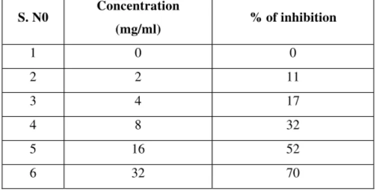
Table 3. Self proliferation percent inhibition of ethanolic extract of Gmelina arborea on celline: human Esophagel cancer cells-TE 2.
S. N0 Concentration
(mg/ml) % of inhibition
1 0 0
2 2 11
3 4 17
4 8 32
5 16 52
6 32 70
Table 4. IC 50 values calculated from dose response curves.
S. No Cell lines IC 50 Value (mg/ml)
1 Colon Cancer Cells COLO 201 20±0.15
2 Gastric Cancer Cells HT 29 12±0.32